Anatomy - The Liver and Gallbladder Flashcards
Give 4 functions of the liver.
- Synthesis of bile. 2. Glycogen storage. 3. Clotting factor production. 4. Detoxification of blood.
What is the liver an embryological derivative of and in which mesentery does it develop?
Derivative of the foregut. Develops in the ventral mesentery.
What structure(s) lie anterior to the liver?
The rib cage and the anterior abdominal wall.
What structure(s) lie superior to the liver?
The diaphragm.
What structure(s) lie posterior to the liver?
Oesophagus, stomach, gall bladder, first part of duodenum. (All are foregut derived organs).
What aspects of the liver does the diaphragmatic surface refer to?
The anterior superior aspects. This surface is smooth and convex.
What aspects of the liver does the visceral surface refer to?
The posterior inferior aspects. It is moulded by the shape of surrounding organs and so is irregular.
Name the 5 liver ligaments.
- The falciform ligament. 2. The right and left coronary ligaments. 3. The right and left triangular ligaments.
What is the function of the falciform ligament?
To attach the liver to the anterior abdominal wall.
What is found in the free edge of this ligament?
The ligamentum teres (remnant of the umbilical vein).
What is the function of the coronary and triangular ligaments?
They attach the superior surface of the diaphragm to the liver.
Name the 4 lobes of the liver.
- Right. 2. Left. 3. Caudate. 4. Quadrate.
What structures bind the Caudate lobe and where is it located?
The IVC and a fossa produced by the ligamentum venosum. It is located on the upper aspect of the right lobe on the visceral surface.
What structures bind the Quadrate lobe and where is it located?
The gall bladder and a fossa produced by the ligamentum teres. It is located on the lower aspect of the right lobe on the visceral surface.
What divides the liver into the right and left lobes?
The falciform ligament.
What vein supplies the liver with dexoygenated blood?
The hepatic portal vein.
What vein supplies the liver with oxygenated blood?
The hepatic artery proper.
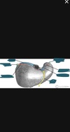
What is the function of the gall bladder?
A temporary storage for bile.
What is the storage capacity of the gall bladder?
30-50ml.
What are the 3 divisions of the gall bladder?
Fundus, body and neck. The neck is where the gall bladder tapers and becomes continuous with the cystic duct.
Briefly describe the biliary tree.
Left and right hepatic ducts = common hepatic duct. Common hepatic duct + cystic duct = common bile duct. Common bile duct + pancreatic duct = hepatopancreatic ampulla of Vater.
What is it called where the common bile duct and pancreatic duct meet?
The hepatopancreatic ampulla of Vater.
What is the orifice called where bile empties into the duodenum?
The major duodenal papilla.
What sphincter regulates the emptying of bile into the duodenum?
The sphincter of Oddi.
What artery supplies blood to the gall bladder?
The cystic artery.
Does parasympathetic or sympathetic innervation result in contraction of the gall bladder and the secretion of bile?
Parasympathetic.
What is the arrangement anterior to posterior of the ducts, artery and portal vein at the porta hepatis?
Anterior: Common bile duct, hepatic artery, hepatic portal vein. (DAV)
What is Calot’s triangle?
An anatomic space bounded by the liver superiorly, the cystic duct laterally and the common hepatic duct medially.
What is the cystic artery a branch of?
The right hepatic artery - hepatic artery proper - common hepatic artery - coeliac trunk.
Where is the bare area of the liver?
Under the tendinous part of the right hemi-diaphragm. This area has no peritoneal covering.
What is the surface marking for the gall bladder?
The tip of the 9th costal cartilage. Where the lateral rectus sheath joins the costal margin.
Where do the hepatic veins drain?
Into the IVC.
Where in the abdomen is the Liver located?
right hypochondria and epigastric areas, extends into the left hypochondria
Describes the diaphragmatic surface of the liver?
- anteriosuperior Surface of the liver
- Smooth and can be vexed fitting underneath the curvature of the diaphragm
- posterior aspect is not covered by visceral peritoneal, it is in direct contact with the diaphragm itself ( known as the bare area)
Described the visceral surface of the liver?
- Posterioinferior surface of the liver
- except for the fossa of the gallbladder and porta hepatis, it is covered in peritoneum
- Irregular shaped due to surrounding organs
- lies in contact with the right kidney, right adrenal gland, right colic flexure, transverse colon, first part of the duodenum, gallbladder, oesophagus and the stomach
What is the falciform ligament?
- Attaches the anterior surface of the liver to the anterior abdominal wall
- forms a natural anatomical division between the left and right lobes of the liver
- the free edge contains the ligamentum teres, a remnant of the umbilical vein
What is the Coronary ligament?
- Attached is the superior surface of the liver to the inferior surface of the diaphragm
- marks the bare area of the liver
- divided into the anterior and posterior folds
- folds unite to form a triangular ligament on the right/left lobe of the liver
What is the triangular ligament?
- Formed by the union of the anterior and posterior layers of the coronary ligament at the apex of the liver
- attaches the left/left lobes of the liver to the diaphragm
What relevance is the lesser omentum to the liver?
- Attaches delivered to the lesser curvature of the stomach and the first part of the duodenum
- consists of:
- hepatoduodenal ligament ( extends from the duodenum to the liver), Surrounds the portal triad
- hepatogastric ligament (Extends from the stomach to the liver)
How is the posterior surface of the liver secured to the inferior vena cava?
by 4 large hepatic veins
What is a hepatic recess?
- anatomical space between the liver and surrounding structures
- clinically important as infections may collect in these areas to form abscesses
Where are the sub-phrenic spaces?
- Between the diaphragm and the anterior and superior aspects of the liver
- divided Into the right and left by the falciform ligament
Where is the sub-hepatic space?
- a subdivision of the supracolic compartment (above the transverse mesocolon)
- peritoneal space located between the inferior surface of the liver and the transverse colon
Where is Morison’s Pouch?
- a potential space between the visceral surface of the liver and the right kidney
- deepest part of the peritoneal cavity when lying supine
- Pathological abdominal fluid is more likely to be collected in this region
Name the fibrous layer that covers the liver? What divides it into left and right?
- Glisson’s capsule
- the falciform ligament
Name and describe the 2 accessory lobes that arise from the right lobe on the visceral surface of the liver?
- caudate lobe - On the upper aspect of the visceral surface, lies between the inferior Vena Cava and the Fossa produced by the ligamentum venosum (Remnants of the fetal ductus venosus)
- quadrate lobe - located on the lower aspect of the visible surface, lies between the gallbladder and the fossa produced by the ligamentum teres
Name the deep transverse fissure that separates these accessory lobes? What is its function?
- porta hepatis
- transmits all the vessels, nerves and ducts entering or leaving the liver with the exception of the hepatic veins
How are hepatocytes arranged in the liver?
- in lobules, hexagonal - shaped and drained by a central vein
- at the periphery of the lobules are three structures collectively known as the portal triad
What is contained within the portal triad at the periphery of the lobule?
- arteriole - a branch of the hepatic artery entering the liver
- venule - a branch of the hepatic portal vein entering the liver
- bile - a branch of the bile duct leaving the liver
- also contains lymphatic vessels and the vagus nerve (parasympathetic)
Describe the unique dual blood supply to the liver?
- hepatic artery proper (25%) - supplies the non-parenchymal structures of the liver with arterial blood, Derived from the coeliac trunk
- hepatic portal vein (75%) - supplies the liver with partially deoxygenated blood, carrying nutrients absorbed from the small intestines
- dominant blood supply to the liver parenchyma, allowing the liver to perform it’s gut-related functions such as detoxification
How is blood drained from the liver?
- through hepatic veins
- the central vein of the hepatic lobule forms a collecting vein which combines to form multiple hepatic veins
- These then open up into the inferior vena cava
Describe the innervation of the liver?
- The parenchyma is innervated by the hepatic plexus
- contains sympathetic ( coeliac plexus) and the parasympathetic (vagus nerve)
- enter the liver at the porta hepatis
- Glisson’s capsule is innervated by the branches of the lower intercostal nerves,
- sharp well-localised pain
Describe the lymphatic drainage of the liver?
- lymphatic vessels of the anterior aspect of the liver drain into the hepatic lymph nodes
- lie along the hepatic vessels and ducts in the lesser omentum
- Empty into the colic lymph nodes which in turn drain into the cisterna chyli
- lymphatic vessels of the posterior aspect of the liver drain into the phrenic and posterior mediastinal nodes
- join the lymphatics and the thoracic duct
What are the three important structures are transported in the porta hepatis in and out of the liver?
- hepatic artery proper
- hepatic portal vein
- common bile duct
- ( also nerves and lymphatics)
Why might the liver become enlarged?
- if blood flow through the hepatic portal vein is impeded
- conditions such as congestive heart failure, cirrhosis, tumours or viral Infections like hepatitis
What is the clinical relevance of hepatic Endocepalopathy?
- If the liver is diseased, blood will bypass it through the portosystemic shunt
- causing the patient to develop a neurological disease
- or if the short-chain proteins are broken down by the liver they can act as neurotransmitters


What region of the abdomen would you find the gallbladder?
right hypochondrial region
What is the primary function of the gallbladder?
to concentrate and store bile which is produced in the liver
When is stored bile released from the gallbladder?
- as part of the gustatory response, in response to cholecystokinin
- also, a small amount released by the Parasympathetic stimulation causing a contraction of the gallbladder, and secretion of bile into the cystic duct due to the relaxation of the sphincter of Oddi
Describe what is on the anterior, superior, posterior and inferior borders of the gallbladder?
- entirely surrounded by peritoneum
- direct relation to the visible surface of the liver
- anteriorly and superiorly - inferior border of the liver and the anterior abdominal wall
- posteriorly - transverse colon and the proximal duodenum
- inferiorly - biliary tree and the remaining part of the duodenum
What is a storage capacity of the gallbladder?
- 30-50ml
Describe the three parts that the gallbladder is typically divided into?
- fundus - the rounded distal portion of the gallbladder, projects into the inferior surface of the liver in the mid-clavicular line
- Body - largest part, lies adjacent to the posteroinferior aspect of the liver, transverse colon and superior part of the duodenum
- Neck - tapers to become continuous with the cystic duct, Leading to the biliary tree
- contains mucosal folds known as Hartmann’s pouch
- common location for gallstones to become lodged causing cholestasis
- contains mucosal folds known as Hartmann’s pouch
What is the biliary tree? Describe its function?
A series of gastrointestinal ducks allowing newly synthesised bile from the liver to be concentrated and stored in the gallbladder
Describe where bile is initially produced and its journey to the gallbladder?
- initially secreted from hepatocytes
- drain from both lobes of the liver into the left and right hepatic ducts
- Ducts combine to form the common hepatic duct ( runs alongside the hepatic vein)
- the cystic duct joins the hepatic duct, allowing bile to flow in and out of the gallbladder for storage and release
- Becomes the common bile duct
Describe the journey of bile through the common bile duct?
- Common bile duct ascends and passes posteriorly to the first part of the duodenum and the head of the pancreas
- joins the main pancreatic duct, forming the hepatopancreatic ampulla (ampulla of Vater)
- empties into the duodenum via the major duodenal papilla
- regulated by a muscular valve called the sphincter of Oddi
Describe the arterial supply of the gallbladder?
- via the cystic artery
- Usually a branch of the right hepatic artery ( derived from the common hepatic artery one of the major branches of the coeliac trunk)
- Can be a branch of the left hepatic or the hepatic artery
- Usually a branch of the right hepatic artery ( derived from the common hepatic artery one of the major branches of the coeliac trunk)
Describe the venous drainage of the neck of the gallbladder? How does this differ from venous drainage of the fundus and body of the gallbladder?
- Neck - via the cystic veins, which drain directly into the hepatic portal vein
- body and fundus - Flows into the hepatic sinusoid
Describe the innervation of the gallbladder?
- sympathetic and sensory - coeliac plexus T5,6,7,8 and 9
- parasympathetic - vagus nerve
Describe lymph drainage from the gallbladder?
- drains into the cystic lymph nodes, in the gallbladder’s neck
- cystic lymph node then drain into hepatic lymph nodes and ultimately the coeliac lymph nodes
What are gallstones formed of?
bile components mainly cholesterol, bile salts and bilirubin
What is the clinical relevance of gallstones?
- often asymptomatic
- biliary colic pain is when they obstruct Cystic duct outflow Or the common bile duct
- cramping like pain
What is the clinical relevance of cholecystitis?
- infection caused by obstruction of the outflow
- the gallbladder becomes infected
- Can lead to complications like pancreatitis and jaundice
- pain is felt in three places:
- via the greater splanchnic nerve (foregut), dull vague pain in the epigastric region)
- via the parietal peritoneum, pain fell accurately over the area of contact as a sharp well-localised pain
- via the phrenic nerve, if the gallbladder comes in contact with the diaphragmatic layer of the parietal peritoneum (C345), pain is felt on the right shoulder
What is the spiral valve?
- honeycomb appearance of the mucosa of the gallbladder
- spiral folds of the mucosa running down the cystic duct
What is the surface marking of the fundus of the gallbladder?
- Tip of the 9th costal cartilage, on the mid-clavicular line (also transplyoric line)
- or the most lateral attachment of the rectus-abdominis