Anatomy - Retroperitoneum (kidneys) Flashcards
What are the functions of the kidneys?
They act to filter and excrete waste products from the blood. They are also responsible for H2O and electrolyte balance.
What are the posterior relations to the kidneys?
Diaphragm, psoas major, quadratus lumborum and transversus abdominis.
What are the anterior relations to the right kidney?
Liver and duodenum, coils of intestine.
What are the anterior relations to the left kidney?
Stomach, spleen, pancreas, coils of intestine.
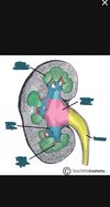
What is found at the renal hilum?
Renal arteries and veins, ureter, nerves and lymphatics.
Which kidney is often positioned lower in the abdomen and why?
The right kidney, this is due to the presence of the liver.
What are the renal arteries a branch of?
The abdominal aorta just below the SMA.
Which renal artery is longer?
The right renal artery, this is due to the position of the aorta being slightly to the left.
Which artery crosses the IVC posteriorly?
The right renal artery.
Describe the branching of the renal artery.
Renal artery -> interlobar artery -> arcuate artery -> interlobular artery -> afferent arteriole.
Where do the renal veins drain?
Into the IVC.
How would you describe the shape of the: a) right adrenal gland? b) left adrenal gland?
a) Tetrahedral. b) Crescent-shaped.
Which adrenal gland does the IVC lie anteriorly to?
The right adrenal gland.
What are the 3 main layers of an adrenal gland?
- An outer connective tissue capsule. 2. A cortex. 3. A medulla at the centre.
Adrenal glands: What are the 3 layers of the cortex?
- Zona glomerulus. 2. Zona fasciculata. 3. Zona reticularis.
Adrenal glands: What cells are contained within the medulla and what do they secrete?
Chromaffin cells - they secrete catecholamines e.g. adrenaline. This is a ‘fight or flight’ response.
What are the 3 arteries that supply the adrenal glands?
- Superior adrenal artery. 2. Middle adrenal artery. 3. Inferior adrenal artery.
What is the superior adrenal artery a branch of?
The inferior phrenic artery.
What is the middle adrenal artery a branch of?
The abdominal aorta.
What is the inferior adrenal artery a branch of?
The renal arteries.
What does the right adrenal vein drain into?
The IVC.
What does the left adrenal vein drain into?
The left renal vein (and then into the IVC).
What section of the spine is found in the posterior abdominal wall?
The lumbar section.
What muscle lies superficially to psoas major?
Quadratus lumborum.
What is the origin of quadratus lumborum?
Iliac crest and the iliolumbar ligament.
What is the insertion of quadratus lumborum?
The transverse processes of L1 to L4 and the 12th rib.
What is the action of quadratus lumborum?
Extension and flexion of the vertebral column.
What is the innervation of quadratus lumborum?
T12-L4 nerves.
What is the origin of psoas major?
T12-L5 vertebrae.
What is the insertion of psoas major?
The lesser trochanter of the femus.
What is the action of psoas major?
Flexion of the hip.
What is the innervation of psoas major?
L1-3 nerves.
What nerves lies on the anterior surface of psoas major?
The genitofemoral nerve.
Where does the femoral branch of the genitofemoral nerve go?
It passes under the inguinal ligament and supplies sensory innervation to the inner thigh.
Where does the genital branch of the genitofemoral nerve go?
It passes through the deep inguinal ring to enter the inguinal canal. It supplies the cremaster muscle.
Why can renal tumours become very large before invading adjacent structures?
The tumour has to grow through multiple tough layers: fibrous capsule, perirenal fat, renal fascia and pararenal fat.
What vein does the right gonadal vein drain into?
The IVC.
What vein does the left gonadal vein drain into?
The left renal vein (and then the IVC).
Where would you palpate an abdominal aortic aneurysm?
In the epigastrium, above the umbilicus.
What is the origin of psoas major?
T12-L5.
What is the insertion of psoas major and iliacus muscles?
Lesser trochanter of femur.
What is the origin of iliacus muscle?
Iliac fossa and anterior inferior iliac spine.
What is the innervation of iliacus muscle?
Femoral nerve (L2-4).
What nerve lies in the groove between the iliacus and psoas major?
The femoral nerve.
What nerve emerges from psoas major medially?
Obturator nerve.
What does the femoral nerve give motor innervation to?
The anterior thigh muscles that flex the hip and extend the knee.
What is the action of iliacus?
Flexion of the hip.
What does the femoral nerve give sensory innervation to?
Cutaneous branches pass to the antero-medial thigh. The terminal branch, saphenous nerve, supplies the medial side of the leg and foot.
What are the nerve roots for the femoral and obtruator nerves?
L2-L4.
What does the obtruator nerve give motor innervation to?
The medial thigh muscles inolved in adduction.
What does the obtruator nerve give sensory innervation to?
The skin of the medial thigh.
What are the sciatic nerve roots?
L4 - S3 (lumbosacral plexus).
What is the largest nerve in the body?
The sciatic nerve.
What does the sciatic nerve give motor innervation to?
Muscles of the posterior thigh and hamstring of adductor magnus.
When the sciatic nerve terminates it bifurcates into 2 nerves. What are they?
- Tibial. - Common fibular.
What are the walls of ureters composed of and why?
Smooth muscle walls. When the muscle contracts it produces peristaltic waves that propels the urine into the bladder.
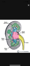
What is the name of the point at which the renal pelvis narrows?
The ureteropelvic junction.
What is the significance of the ureters piercing the bladder obliquely?
It creates a physiological valve that acts to prevent the back flow of urine.
Do the ureters cross the ovarian arteries and the ductus deferens anteriorly or posteriorly?
Posteriorly. (Water (ureters) under the bridge (artery)).
At what vertebral levels are the kidneys?
- T12 to L3
- often right is lower due to the liver
What muscles are on the posterior abdominal wall behind the kidneys?
- diaphrapgm
- psoas major
- quadratus lumborum
- transversus abdominalis
Describe the vasculation of the adrenal glands?
- 3 arteries:
- superior (from inferior phrenic artery)
- middle (from abdominal aorta)
- inferior adrenal arteries (from renal arteries)
- 2 veins: right adrenal vein drains into IVC and left drains into left renal rein
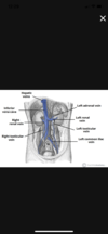
What is the difference between the right venous drainage and left venous drainage of the adrenal glands, kidneys and gonads?
- right: all drain separately into the inferior vena cava.
- Left: all join to form a single vein that passes in front of the aorta to enter the right-sided inferior vena cava
At what level does the aorta bifurcate? What does it bifurcate into?
- L4/5 (umbilicus)
- into right and left common carotid
- (they both then divide into external (lower limbs) and internal (pelvic organs) iliac arteries
What vertebra do the hypogastric and ilioinguinal nerves arise from?
- hypogastric: T12
- ilioinguinal: L1
Renal tumours can become very large before they invade adjacent structures. What anatomical features explain this?
- Fibrous capsule (inner layer) closely applied to the renal substance, perirenal fat, renal fascia (encloses the kidney and adrenal glands together), pararenal fat.
- So the tumour has to grow through multiple tough layers before invading adjacent structures.
Describe two common variations in the anatomy of the renal pelvis and ureter?
Duplex system, pelvoureteric junction obstruction.
What veins do the right and left gonadal veins drain into?
Right into the inferior vena cava, left into the left renal vein.
What is a polar artery? Explain why polar arteries exist
- An accessory renal artery usually supplying the lower pole of the kidney.
- The kidney develops as a number of separate kidneys which fuse together during development, if this fusion is incomplete duplex kidney can occur or part of the kidney can have a separate blood supply
Where would you palpate an abdominal aortic aneurysm?
In the epigastrium (above the umbilicaus).
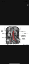
Where are the narrow parts of the ureter where a stone may get stuck?
The junction between the pelvis of the kidney and top of the ureter, where the ureter passes over the iliac vessels, where the ureter passes into the bladder.
Describe the relationship of the ureter to bony landmarks of the abdomen and pelvis (useful for finding the ureter on an X-ray).
The ureter runs anterior to the tips of the transverse processes on the lower three lumbar vertebrae, over the sacroiliac joints and just medial to the lateral pelvic side wall