Anatomy - The Abdominal Wall Flashcards
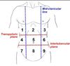
What 2 vertical lines divide the abdomen into it’s 9 nine regions?
Mid-clavicular lines that extend to the mid-inguinal point.
What 2 horizontal lines divide the abdomen into it’s 9 nine regions?
transpyloric: joins lower coastal margins.
Intertubercular: connects the iliac crests.
How would you draw the transpyloric plane?
Half way between the suprasternal notch and the pubic symphysis.
What vertebral level is the transpyloric plane found?
L1.
Name 5 structures found on the transpyloric plane.
- Pylorus of stomach. 2. Fundus of gall bladder. 3. Pancreas. 4. Hilum of Kidneys. 5. Duodenojejunal flexure.
How would you draw the transtubercular plane?
Joins the iliac crests of the pelvis.
What vertebral level is the transtubercular plane found at?
L4. (Same level as the bifurcation of the abdominal aorta).
What is the intercristal plane?
It joins the highest points of the pelvis at the back.
What vertebral level is the intercristal plane found at? Why is this important?
Between the L4 and L5 vertebrae. This is important for lumbar punctures and epidurals.
What is the subcostal plane and at what vertebral level does it lie?
It joins the lower points of the costal margins and lies at the L2 level.
What can the subcostal plane (L2) be a marker for?
- The end of the spinal cord. - The superior mesenteric artery.
What is McBurney’s point?
2/3 of the way along a line extending from the umbilicus to the right anterior superior iliac spine.
What is the significance of McBurney’s point?
Marks the base of the appendix and can act as a guide for the location of the caecum.
Name the 3 flat muscles of the Abdominal wall.
- External Oblique. 2. Internal Oblique. 3. Transversus Abdominis.
In what direction do the fibres of the flat muscles run in?
- External oblique - inferiorly and medially (down and in). 2. Internal oblique - superiorly and medially (up and in). 3. Transversus abdominis - transversely.
What is the origin of external oblique?
Lower 8 ribs and thoraco-lumbar fascia.
What is the insertion of external oblique?
Pubic crest, pubic tubercle, iliac crest and linea alba.
What is the origin of internal oblique?
Thoraco-lumbar fascia, iliac crest, lateral 1/2 of the inguinal ligament.
What is the insertion of internal oblique?
Linea alba, pubic tubercle.
What is the origin of transversus abdominis?
Thoraco-lumbar fascia, iliac crest, lateral 1/3 of the inguinal ligament.
What is the insertion of transversus abdominis?
Linea alba, pubic tubercle.
What are the fibrous intersections of the rectus abdominis called?
Tendinous intersections.
What are the attachments of rectus abdominis?
Rectus sheath, pubis, costal cartilages.
Name 5 structures contained within the rectus sheath.
- Rectus Abdominis. 2. Sup and inf epigastric arteries. 3. Sup and inf epigastric veins. 4. Nerves. 5. Lymphatics.
What is the inferior epigastric artery a branch of?
The external iliac artery.
What is the internal thoracic artery a branch of? And what does it supply?
It is a branch of the subclavian artery and it supplies the anterior chest and abdominal wall and also the breasts.
Define dermatome.
An area of skin supplied by nerves from a single spinal root.
What are the dermatomes of the abdominal wall?
T7 to L1.
If a disease process involves the peritoneum of the abdominal wall, then the pain is localised. Why is this?
The peritoneum covering the inside of the abdominal wall has the same sensory nerve supply as the overlying skin.
What are the four main functions of the abdominal wall?
- firm yet flexible boundary, that keeps the abdominal viscera in anatomical positions against gravity
- protect the abdominal viscera from injury
- assist enforceable exploration by pushing the abdominal viscera inwards
- involved in any action that increases the intra-abdominal pressure eg coughing
Name the four main layers of the abdominal wall (externally to internally)?
- skin
- superficial fascia
- muscles and associated fascia
- parietal peritoneum
Describe the difference in the superficial fascia Above the umbilicus below the umbilicus?
- above - single sheet connective tissue continuous with other regions
- below - divided into two layers: fatty superficial layer (camper’s fascia) and membranous deep layer (scarpa’s fascia)
- superficial vessels and nerves run in between these two layers
Describe the two Muscle groups on the anterior lateral abdominal wall?
- Flat muscles - three situated laterally on either side of the abdomen
- vertical muscles - two situated near the midline of the body
Describe how the three flat must are positioned?
- fibres running different directions crossing each other
- strengthening the wall and decreasing the risk of abdominal contents her negating through
Where is the abdominal aponeurosis formed?
- broad flat tendon
- on the anterior medial aspect of the abdominal wall each muscle forms an aponeurosis
- Covers the vertical rectus abdominis muscle
What occurs when the aponeurosis intertwine?
- entwine at the midline
- forms linea alba
- fibrous structure that extends from the xiphoid process of the stone into the pubic symphysis
Describe the external oblique muscle? attachments function and innervation?
- largest most superficial flat muscle
- run inferomedially ( hands in pocket)
- originates from ribs 5 to 12, inserts of the iliac crest and pubic tubercle
- Contralateral rotation of the torso
- innervated by T7-T11 and T12
Describe the internal oblique muscle? Attachments function innervation?
- Lies deep to the external oblique, smaller and thinner
- Runs superomedially
- Originates from the inguinal ligament, iliac crest and lumbodorsal fascia
- Insert into ribs 10 to 12
- bilateral contraction compresses the abdomen, while unilateral contraction ipsilaterally rotates the torso
- Innervation is T7-T11 and T12 and branches of the lumbar plexus
Describe the transversus abdominis Muscle? attachments function and innervation?
- deepest muscle, runs transversely
- deep to this is a well formed layer of fascia, called transversalis fascia
- Originates from the inguinal ligament, costal cartilages 7-12, the iliac crest and thoracolumbar fascia
- inserts onto the conjoint tendon, xiphoid process, linea alba and pubic crest
- Compression of the abdominal contents
- Innervation by T7-11, T12 and branches of the lumbar plexus
Describe the vertical abdominal muscle rectus abdominis?
- paired muscle found each side of the midline (4 in total)
- Split into by the linea alba
- create surface markings known as linea semilunaris
- Intersected by fibrous strips, known as tendinous intersections
- Originates from the crest of the pubis before inserting into xiphoid process of the sternum and costal cartilages of ribs 5-7
- Compresses the abdominal viscera, stabilises the pelvis during walking and depresses the ribs
- innervation from T7-11
Describe the vertical abdominal muscle pyrimidalis?
- small triangular muscle found superficially to rectus abdominis
- Located inferiorly with base on the pubis bone, apex of the triangle attaches to the linea alba
- originates from the pubic crest and pubic symphysis for inserting into the linea alba
- axed to tense the linea alba
- T12
Describe the location of the umbilicus?
- roughly halfway between the xiphoid process and pubic symphysis
What indicates the lateral and medial borders of the rectus abdominis?
- linea semilunaris
- curved line running from the ninth rib to the pubic tubercle
- linea alba
- Visible as a virtual groove extending inferiorly from the xiphoid process
Describe the 2 horizontal and 2 vertical planes that split the abdomen into nine regions?
- horizontal planes:
- transpyloric - halfway between the jugular notch and pubic symphysis ( at the level of L1)
- the gallbladder, the pancreas, the pylorus of the stomach and duodenal-jejunal flexure all lines plain
- intertubercular - runs between the superior aspect of the right and left iliac crest (Bifurcates the abdominal aorta)
- (Sub coastal plane - lowest point the costal margin)
- (Intercristal plane - Lies across the highest point of the pelvis)
- transpyloric - halfway between the jugular notch and pubic symphysis ( at the level of L1)
- vertical planes
- mid-clavicular to mid inguinal points
Describe the rectus sheath?
- the fascial covering of the rectus abdominis and pyramidalis muscles
- formed by aponeuroses of the three flat muscles
- Anterior wall is formed by - external oblique and half of internal oblique
- posterior wall is formed by - half of internal oblique and transversus abdomis
- halfway between the umbilicus and pubic symphysis, all muscle is the anterior wall
- at this point there is no posterior wall so the rectus abdominis is in direct contract with the transversalis fascia
- this is called the arcuate line
What is aponeurosis?
- Takes place of a tendon in a flat muscle that has a wide area attachment (sheet)
Describe the inguinal ligament? What is found just above it?
- the lowest most extent of the external oblique muscle inferiorly is attached to the anterior superior iliac spine laterally and the pubic tubercle medially
- inguinal canal, Transmits all structures to and from the testes
Name the six bony landmarks of the abdomen?
- xiphisternum
- Coastal margin
- iliac crest
- Anterior superior iliac spine
- pubic tubercle
- pubic symphysis
Describe McBurney’s point?
- Perpendicular line, point is two thirds of the way along between the umbilicus and right anterior superior iliac spine
- base of the appendix
- a guide to the position of the caecum
- Splits the fibres of the muscle without cutting them
Describe the positioning of the umbilicus?
- often unreliable
- around the level of L3
What is the upper extent of the abdominal cavity?
- anteriorly the under surface of the diaphragm reaches the 5th intercostal space

