Test 3: Mara Rendi review Flashcards
What is the general definition of arteriosclerosis?
hardening and thickening of blood vessels
Name 3 arterioscelreosis diseases
Mockeberg’s arteriosclerosis
arteriolosclerosis
atherosclerosis
What is this?

Mockeberg’s arteriosclerosis
It is the calcification of the media

Does Mockeberg’s arterioscleosis obstruct arterial flow?
Nope
What is arteriolosclerosis?
hyaline thickening or proliferation of small vessels. The type of thickening depends on the type of arteriolosclerosis
What are the two types of arteriolosclerosis?
Hyaline arteriolosclerosis
hyperplastic arteriolosclerosis
What is this?

hyaline arteriolosclerosis

Does hyaline arteriolosclerosis obstruct flow?
Yes!
What is this?

hyperplastic arteriolosclerosis

Does hyperplastic arteriolosclerosis obstruct flow?
Yes!
What is atherosclerosis?
The formation of atheromas within the INTIMA
What are the components of a atheroma-plaque?
central core
fibrous cap

What is in the central core?
cholesterol
foam cells (macrophages with lipid inside of them)
necrotic debris
calcium
What is in the fibrous cap?
collagen
fibrin
smooth muscle
foam cells
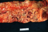
What are these pictures trying to show?

fatty streaks
What is this trying show show?

More fatty streaks
Why are fatty streaks important to know about?
They are the first steps in becoming an atheroma-plaque
What is this?

a god damn fatty streak!
What is this?

A atheroma-plaque.
Notice the fibrous cap and the necrotic core

What is this?

early atherosclerosis
What is this slide? What is the arrow showing?

Atherosclerosis
the arrow is pointing to calcification

What is this? What are the “red chunks”?

late atherosclerosis
thrombi that are forming due to the ruptured plaques
What are the “needle” shapes in the central core?

Cholesterol needles
Name the components


What is the difference between these two pictures?

A - Atheroma-plaue
B- Ruptured atheroma-plaque
What are the major complications of atherosclerosis?
Ruptured plaques result in thrombus formation which can lead to emboli. These emboli, depending on where the thrombus formation occured, can cause occlusion which is bad.
Atheroscleorsis can also lead to aneurysms, which are also bad

What are the modifiable and non-modifiable risk factors for atherosclerosis?
Non-Modifiable:
advanced age, male sex, family history, genetics
Modifiable:
smoker, diabetes, hypertension, hyperlipidemia,
Is atherosclerosis an inflammatory process?
Yes
Where does venous thrombosis occur?
Usually in the deep veins of the legs
What three factors make you at high risk for a DVT?
stasis of blood flow
endothelial injury
hypercoagulability

What is this? Bonus: Where is this?

embolus
Bonus: in the lungs
What is the difference between ischemia and hypoxia?
ischemia is the lack of blood flow
hypoxia is the lack of oxygen
What is this picture showing? What would happen is you started to increase physical exertion?

atherosclerosis of a coronary artery
Ischemia leading to angina
This is a slide of a coronary artery. What is going on here?

Ruptured plaque that resulted in thrombus formation. This is a STEMI

Evolution of MI
What changes can bee seen grossly and histologically within 12 hours?
no gross changes
not much histologic changes
Evolution of MI
What changes can bee seen grossly and histologically within 12 - 24 hours?
- Grossly: blue-gray discoloration caused by stagnant blood
- Histologically: coagulative necrosis and contraction bands
How old is this MI? What is the arrow pointing to?

This is between 12-24 hours
You can see coagulative necrosis (notice there are no nuclei) and you can see contraction bands indicated by the arrow

How old is this infarction?

12-24 hours
Myocardium on the right is normal while the myocardium on the left is undergoing coagulative necrosis

Evolution of MI
What changes can bee seen grossly and histologically within 1 - 4 days?
- Grossly: blue-gray discoloration caused by stagnant blood
- Histologically: neutrophils within the necrotic myocardium
How old is this infarct?

1 - 4 day old
Notice the presence of neutrophils

Evolution of MI
What changes can bee seen grossly and histologically within 4-10 days?
- Grossly: tan-yellow and soft at the area of infarction with hyperemic boarders (red due to recruitment of blood vessels)
- Histologically: macrophages arrive to clean up the scene
Why is the 4-10 period a very danegrous time for the patient?
The area of infarction is the point of maximal weakness and is susceptible for rupture
Where is the point of maximal weakness?
How old is this infarct?

4 - 10 days
How old is this infarct?

4 - 10 days
notice the macrophages and the nearly complete removal of the necrotic caridomyocytes

How old is this infarct?

4 - 10 days
notice the macrophages and the nearly complete removal of the necrotic caridomyocytes

Evolution of MI
What changes can bee seen grossly and histologically within 10 days - 3 weeks?
- Grossly: mottled yellowish tan and gray depressed area
- Histologically: granulation tissue present
How old is this infarct?

10 day to 3 weeks
Note: grossly, it would be VERY difficult to distinguish between 4-10 days and 10 days - 3 weeks, which is why you would want to look at it histologically
How old is this infarct?

10 days - 3 weeks old
Granulation tissue characterized by loose connective tissue and abundant capillaries.
Evolution of MI
What changes can bee seen grossly and histologically within 3 - 8 weeks?
Grossly: white scar
Histologically: dense scar tissue
How old is this infarct?

3 - 8 weeks
notice the white scar

How old is this infarct?

3 - 8 weeks
notice the dense fibrous connective tissue and residual myocardiocytes

How old is this infarct?

3 - 8 weeks
notice the dense fibrous connective tissue and residual myocardiocytes

How old is this infarct?

1 - 4 days
What’s going on here?

Reperfusion causes hemorrhage in area of infarction

What are 5 complications of MI?
arrhythmias
heart failure
tamponade (4-10 days)
mural thrombosis
ventricular aneurysm
What is this?


What is this?


What is this?


What is this?


What is this?


What is this?


What is this?


What are aneurysms?
Abnormal dialtions of arteries and veins that can rupture
What are the 3 causes of aneurysms that we should know for this test?
hypertenstion
atherosclerosis
marfans syndrome
What is this?

ruptured abdominal aortic aneurysm
What is this?

ascending aortic aneurysm

What is this?


What is patent foramen ovale?
The foramen ovale is not closed, but the pressures from the valve keep the flap closed
What is a paradoxical embolization?
It is an embolism that forms on in the venous system but can travel through a septal defect into the arterial system

What is a VSD?
hole in the ventricular septum
What is patent ductus arteriosus? What can it lead to?
failure of the ductus arteriosus to close
pulmonary hypertention

What is coarctation of the aorta?
constriction of the aorta
Who develops coarctation of the aorta?
men>women
turner’s syndrome (female with one X)
What is this?

coarctation of the aorta
What is this?

patent ductus arteriosus
Do patients with coarctation of the aorta have higher blood pressure in the upper extermities?
yes
What is tetralogy of fallot? Is this a left to right flow?
- Sub-pulmonary Stenosis (obstruction of right ventricular outflow tract)
- Right ventricular hypertrophy
- Overriding aorta
- Ventricular Septal Defect
NO! this is a right to left flow
What is transposition of the great arteries?
The reveral of pumlonary trunk and aorta

is transposition of the great vessels a cyanotic disease? How can a fetus survive this?
yes
they need a large VSD or a patent ductus arteriosus