Sudden Death in Lambs Flashcards
what are reasons for sudden death in growing lambs
Clostridial diseases
Pasteurellosis —> septicaemia (M. hemolytica, P. Multocida), systemic (Pasteurella trehalosi)
Acidosis (grain overload) vs cerebrocortical necrosis (CCN)
Acute liver fluke
Others
what are clostridial diseases and the bacteria that cause them (7)
- lamb dysentery (clostridium perfringens type B)
- pulpy kidney (cl. perfringens type D)
- black leg (cl. chauvoei)
- tetanus (cl. tetani)
- black disease (cl. novyi type B)
- braxy (cl. septicum)
- abomasitis (cl. sordellii)
what are the clinical features of pulpy kidney
Enterotoxemia
what does pulpy kidney cause
Sudden death in non-immune lambs
what age of lamb does pulpy kidney commonly affect
4 weeks to 8 month old lambs
Often the bigger, better lambs
what could be seen in pulpy kidney if the lambs are alive
Severe depression
Abdominal pain
Grinding teeth
Neurological signs (seizures, opisthotonus)
describe the pathogenesis of pulpy kidney 12
- organism is present in gut
- reaches high # in presence of undigested CHO (high milk intake or excess food in weaned)
- in presence of trypsin and absence of antibody the protoxin is cleaved to the active ε toxin
- ε is 3rd most lethal C toxin
- ε produced and enters bood
- increased intestinal permeability
- toxin absorbed and fixes to cells in liver, kidney and brain
- in brain causes liquefactive necrosis, perivascular edema and hemorrhage (focal symmetrical encephalomalacia)
- in brain the cell tight junctions degenerate, increased permeability with fluid loss followed by elevated intracranial pressure
- mobilization of hepatic glycogen stores
- hyperglycemia, glycosuria, nervous changes
- death
how is pulpy kidney diagnosed
history of recent move to rich feeding
PM
C. perfringens type D in smears by gram stain, by culture and toxin gene PCR
toxin detectable in ELISA
what is the pathology of pulpy kidney
Good condition, some fecal staining
No gross lesions, clear fluid in body cavities
Small petechial hemorrhages on lungs and epicardium
Pulpy kidneys and glycosuria
Small intestinal contents in fluid
Focal symmetrical hemorrhages in basal ganglia of brain and later focal symmetrical encephalomalacia
C. perfringens type D and its toxin in small intestinal contents
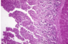
what pathology is shown here

pulpy kidney
how does clostridium chauvoei enter
via skin wounds
ex. shearing, castration
what does blackleg affect
skeletal and cardiac muscle
how do blackleg lambs present
sudden death
or
if alive
dullness
febrile (<41C)
toxic mucous membranes
severe lameness with edema and emphysema
what toxins does c chauvoei produce
α, β, γ, δ
where are C tetani spores found
ubiquitous in soil
when is c tetani commonly associated
docking
castration wounds
what is responsible for the clinical signs in C tetani
production of potent neurotoxin tetanospasmin
what are the clinical signs of C tetani
generalized stiffness
rigidity of limbs
unable to swallow or eructate
progress to convulsions
how are early cases of C tetani treated
tetanus antitoxin
penicillin
what age does black disease affect
sheep of all ages
what does black disease affect
liver
what is C novyi associated with
migration of immature liver fluke in the liver
when is C novyi commonly seen
late autumn/winter
what does C novyi cause
sudden death
what toxins do C novyi produce
α & β
what are the clinical features of Braxy
Rapidly fatal abomasitis
what sheep does Braxy commonly affect
Sudden death of young non-immune sheep eating frosted forage
Usually store lambs and ewe lambs
what are the clinical signs of Braxy if the lambs are alive
Sudden onset of illness with weakness, anorexia
Inability to keep up with groups
May be fevered (to 42ºC)
what is the pathogenesis of C septicum 3
- Frosted kale, grass or other forage damages the abomasal wall
- Infection of the wall of the abomasum, invasion by C. septicum which produces α toxin to cause local and systemic effects
- α toxin of C. septicum is a pore forming toxin — inserts into cells and lyses them resulting in cell death — causes release of cell potassium and hemoglobin
what toxins does C septicum produce
Toxins produced α, β, γ, δ
how is c septicum diagnosed
- History of frost and absence of vaccination
- PM
- Presence of C. septicum in profuse culture — direct detection by gram stain of mucosal lesions and heart blood
- Detection by immunohistochemistry in tissue
- Demonstration of its toxin in tissue or body cavity fluids
what is the pathology of C septicum
Abomasitis
Abomasal, duodenal walls thickened edematous, hemorrhage and necrosis (mucosa intensely inflamed or blackened)
Excess turbid peritoneal fluid
Some epicardial petechiae
where is C sordellii found
Primary pathogen of sheep intestine
what age of lambs does C sordellii affect
- Acute abomasitis young lambs 3-10 weeks
- Sudden death and abomasitis in finishing 6-12 months
- Rotten lambs post vaginal prolapse in pregnant ewes?
what is the pathology for C sordellii
C. sordellii morphologically distinctive (hairpin cells)
Distinct colonial morphology
Two toxins, hemolysin and lethal toxin — antigenically similar to C. difficile toxins A and B
Lesions include single abomasal ulcers which ruptures or diffuse enteritis which may be necrotic or emphysematous
what is shown here

C sordellii enteritis in sheep jejunum
how are clostridial diseases treated
Sensitive to penicillin (44, 000 IU/kg)
Treatment for blackleg (penicillin) and black disease (fluke control)
how are clostridial diseases prevented
Vaccination!
Introducing to rich feed gradually
Remove affected groups to pasture, housing to prevent access to frosted grass?
when should ewes be vaccinated with clostridial vaccines
booster 4-6 weeks pre lambing to provide protective colostrum
how long to maternal clostridial antibodies last
up to 10 weeks
when should lambs be vaccinated for clostridial disease
10 weeks old
booster 4-6 weeks later
when should lambs of non immune ewes be vaccinated for clostridium diseases
2 weeks
booster 4-6 weeks later
what is the pathogen that causes septicemic pasteurellosis
mannheimia hemolyica
what age does septicemic pasteurellosis affect lambs
sudden death in lambs up to 12 weeks old
best lambs of group
how do septicemic pasteurellosis lambs present
Depressed
Febrile (>41ºC)
Toxic mucous membranes
Dyspnea
what causes systemic pasteurellosis
Pasteurella trehalosi
when does Pasteurella trehalosi affect lambs
sudden death in recently weaned lambs
when does systemic pasteurellosis appear
stress conditions
how does systemic pasteurellosis present
Depressed
Febrile (>41ºC)
Toxic mucous membranes
Dyspnea
how is septicemic and systemic pasteurellosis diagnosed
- history
- clincal signs
- pm
what is the gross pathology of septicemic and systemic pasteurellosis
Lungs edematous and congested
Enlarged, hemorrhagic lymph nodes
Widespread petechiae, serosal surfaces, epicardium, kidneys
Focal necrotizing hepatitis
Upper alimentary tract erosions
Pleurisy and pericarditis
what is the histology of septicemic and systemic pasteurellosis
Widespread bacterial emboli in arterioles
Multifocal necrotizing hepatitis
Pulmonary congestion and edema
how is Systemic and septicemic pasteurellosis treated
oxytetracyline?
how is Systemic and septicemic pasteurellosis prevented
With clostridial vaccine (Ovivac and Heptavac P plus)
Ovipast plus
what causes acidosis
Sudden and unaccustomed intake of high quantities of rapidly fermentable carbohydrate (grain overload)
when does acidocis occur
Sheep (lambs) turned to graze on grain stubble with high quantities spilled grains
Sudden introduction of concentrates
what are the two types of acidosis
rumenal and metabolic (L and D-lactate)
what are the clinical signs of acidosis
Sudden death
- Within 24 hours from sudden introduction
Anorexia and bruxism
Distended abdomen and ruminal stasis
Severe metabolic acidosis
- Tachypnea
- Hyperpnea
Diarrhea
Dehydration and toxic mucous membranes
how is acidosis diagnosed
History
Clinical signs (if alive)
PM
what can be found in PM exam with acidosis
Rumen contents milky grey (porridge like)
Rancid odour
Rumen epithelium sloughing off
Rumen pH <5.5
how is acidosis prevented
Gradually introduce to grain concentrate feeding over 3 weeks before ad-lib 100g/day per lamb recommended (as introduction)
Good quality roughage available
Closed feed storage
how is acidosis treated
In severe emergency consider rumenectomy
Correct ruminal and metabolic acidosis and dehydration (sodium bicarbonate per os and IV)
Encourage feeding on forage
Rumen function stimulants (Pro-rumen)
Transfer of rumen contents from healthy sheep
what age is polioencephalomalacia seen in
adult sheep and weaned lambs
what is polioencephalomalacia preceded by
dietary change (concentrates)
disruption to normal feeding in prev 2 weeks (anthelmintic treatment?)
what is polioencephalomalacia caused by
Caused by induced thiamine (vit B12) deficiency (ex. by overgrowth of thiaminase-producing bacteria in rumen —> altered glucose metabolism —> necrosis superficial cerebral grey matter —> generalized cerebral edema)
Occasional outbreaks also associated with high levels of dietary sulphur
what are clinical signs of polioencephaomalacia (CCN)
Typical of diffuse cerebral lesion:
- Blind, wandering, “stargazing”
- High stepping gait
- Head pressing
- Dorso-medial strabismus in some cases
- Progression to recumbency, backwards flexion of neck
- Hyperaesthesia
- Convulsions
what are some ddx of polioencephaomalacia
Pregnancy toxemia
Listeriosis
Focal symmetrical encephalomalacia
Acute coenurosis
how is polioencephaomalacia diagnosed
Decrease erythrocyte transketolase and increase thiaminase activity in feces, rumen fluid, blood (rarely used)
Usually based on clinical signs and response to treatment
PM
what is seen on PM with polioencephaomalacia
Fluorescence of cerebral cortex under UV light
Histology: bilateral laminar necrosis
how is POLIOENCEPHALOMALACIA treated
Early cases most likely to respond
- IV vitamin B1 (10-20mg/kg) and dexamethasone (1mg/kg)
- Continue vit B1 IM BID for 3 days
- Check adequate levels of thiamine in ‘multivitamin’ injectables
- Usually improve within 24 hours
- May remain blind for 2-3 weeks
when is obstructive urolithiasis seen
Male animals on high concentrate diets
Tups & fattening lambs
what are causes of obstructive urolithiasis
Low calcium high phosphate/magnesium
what are the crystals seen obstructive urolithiasis
struvite crystals
what are clinical signs of obstructive urolithiasis
Hematuria
Dribbling urine
Abdominal pain
Tail flicking
End stages
- Depression
- Possibly prepucal swelling