Physics, Artifact, M-mode Flashcards
What are the types of cavitation?
- Squeeze flow
- acceleration of blood as occluder approaches stop causing drop in pressure
- Vortex flow
- turbulence at edge of rapidly moving occluder
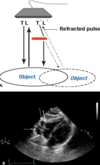
What is velocity (propagation speed) error artifact?
- sound propogates through some structures at velocity other than 1540 m/sec
- takes longer to travel through a structure - which is assumed by the ultrasound machine
What is specular reflection?
- mirror-like reflection of waves from a surface, in which waves from a single incoming direction (a ray) is reflected into a single outgoing direction.
- angle of reflection equals angle of incidence for objects > 1 wavelength diameter (~0.5mm)
What is cavitation?
- rapid formation of vaporous microbubbles in a fluid due to a local reduction in pressure
What are High Intensity Transient Signals (HITS)?
- under some conditions (vortices), vaporous microbubbles can coalesce into larger bubbles
- degassing: removal of gasses (principally CO2 in blood) from liquid medium
- larger bubbles persist and ar visible on echo
- no demonstrated neurologic sequelae from HITS microbubbles
Why are bubbles such good reflectors? Why do we see them with prosthetics?
- they have a very low acoustic impedance
- acoustic impedance (velocity x density)
- interface between materials with dissimilar acoustic impedances (velocity x density) reflects sound
- 23 (air) vs. 1465 (water)
To reduce aliasing on color-flow Doppler, a sonographer should perform this action?
- Changing the baseline shift
- allows velocities to be displayed up to twice th original Nyquist limit
What is pulse repetition frequency (PRF)?
- number of pulses of a repeating signal per unit time
- inversely related to PRP (PRF = 1 / PRP)
- inversely related to imaging depth (PRF x 1/d)
What environment can sound waves not travel through?
- vacuum
- pressure waves can only be transmitted through physical media consisting of molecules that interact with each other
What is the upper limit of human hearing?
20,000 Hz or 20 kHz
The frequency of a sound wave is measured in Hz as the:
- number of times particles vibrate each second in the direction of wave propogation
- 1/s
- the number of times a particle in a conducting medium vibrates per unit time
Ultrasound imaging is usually performed in what frequency range?
- 1-30 MHz
- lower frequencies (greater penetration) –> larger organs or deeper structures
- higher frequencies (better spatial resolution) –> smaller, more superficial structures
Define wavelength
the distance a wave travels during a single cycle
What type of tissue results in the fastest loss of ultrasound wave strenght?
- Lung
- because of the high content of air and the abundance of highly reflective tissue/air interfaces, the sound waves dissipate in the lung so fast that the lungs are virtually opaque to ultrasound
What is the main goal of the gel used during ultrasound imaging?
- Improve contact between the transducer surface and the skin
- eliminates any tissue/air interfaces which are highly reflective and thus prevent ultrasound transmission
What are Piezoelectric crystals?
materials that respond to electric signals by vibrating and generating acoustic waves and vice versa
To better assess rapidly moving structures, a sonographer should perform this action?
- Narrow scan sector width and decrease imaging depth = increased frame rate
- Higher frame rate is required to increase temporal resolution
- some machines enable direct manipulation of frame rate, others can be adjusted by sector width and imaging depth
- Higher frame rate is required to increase temporal resolution
How do Piezoelectric crystals generate ultrasound images?
- transmit waves by “exciting” the crystals in the transducer by an electrical stimulus,
- then receiving the ultrasound waves reflected by structures inside the body,
- translating them back into electrical signals that are used to form an image of the reflecting structures
Define Doppler shift?
- change in the frequency of a sound wave reflected by a moving target
- negative / red shift
- object moving away
- wavelength made longer (increased)
- frequency decreased
- positive / blue shift
- object moving toward transducer
- frequency incerased
- wavelength decreased
- negative / red shift

Define doppler angle?
- angle between:
- the direction of flow
- ultrasound beam
With time gain compensation, the machine is generally preset to perform this action?
- Decrease signal in near field, increase signal in far field
- increasing depth = increased attenuation
- TGC accounts for this signal loss by effectively incresing gain in parallel with increase in depth
- many modern systems automatically account for this so the knobs should be left in neutral position to start with then adjusted as needed
Define Harmonic imaging?
Echocardiogaphic images are formed from returning echoes at twice the insonifying frequency (second harmonic)
When does aliasing occur?
when doppler shift of high-velocity flow exceeds the Nyquist limit (1/2 the Pulse Repetition Frequency)
What are the two forms of “harmonics” or image enhancement currently used in diagnostic ultrasound?
- native tissue harmonic imaging
- contrast harmonics
Define contrast harmonics
- occurs when tiny bubles are insonified at a lower frequency than their natural frequency of vibration
- the vibrating bubbles then radiate back significant amounts of energy at the second harmonic
- requires a high-pass filter on the ultrasonic receiver to eliminate signal at the fundamental frequency
- the second harmonic image will show the echo contrast with significantly enhanced intensity relative to the surrounding tissue, which does not reflect the harmonic frequency
The energy of the transmitted ultrasound wave can be changed by adjusting which of the following?
- Power
- increasing the power increases the energy / heat delivered to the tissues
- increased gain will not do this
- visually this can create similar changes compared to adjusting the gain, which increases the amplitude of the signal
- increasing the power increases the energy / heat delivered to the tissues
Define spatial resolution of an ultrasound image?
- smallest distance between two objects that allows distinction between them
- also determines the size of the smallest object that can be visualized
Image resolution for a region of interest can be improved by this action?
Increasing the write zoom
What does time gain compensation do?
- part of postprocessing of the reflections designed to correct for beam attenuation as it travels through the body
- aims to provide a correction for the loss of intensity (or attenuation) by all these different mechanisms (scattering, absorption, reflection)
- based on assumption that acoustic properties of surrouding tissues are the same
The dynamic range of echoes displayed on the screen is adjusted by this?
- Compression control
- used to include or suppress weak echoes
When does time gain compensation become inaccurate?
- large differences in acoustic properties between adjacent tissues
- contrast agents that show much stronger attenuation
- This is the reason why acoustic shadowing artifacts are frequently seen distal to contrast filled blood pools, such as ventricles or atria
Attenuation is the combined result of these:
- scattering
- absorption
- reflection
The strength of the transmitted ultrasound wave is controlled by adjusting this?
Power control
What does Gain Control do?
determines to what extent the received signal is amplified
What is Range ambiguity artifact
- Deeper structures –> appear closer to the transducer than true location
- Occurs with high Pulse Repetition Frequency (PRF):
- when a second pulse is sent out, before the first signal along the same scan line is received
- Pulse repetition period determines maximum depth imaged
- US does not “disappear” beyond this depth and may be strong enough to return a signal
- Machine assumes all returning signals are result of most recent pulse
If a rapidly moving structure such as a cardiac valve appears to be moving in slow motion, what may be set too high?
- Persistence
- images can be averaged together to create a smoothing effect by increasing persistence
- lower persistence maintains temporal resolution and can keep a structure from appearing as though it were moving in slow motion
What does compression control do?
determines the dynamic range of received signals that are used to create the image
What is the difference between read and write zoom?
- Read zoom only magnifies the image without a change in resolution
- Write zoom increases line density and number of pixels in a given area
What does a positive doppler shift indicate?
indicates that the reflector is moving so that the angle between the transmitted beam and the direction of flow is > 90 degrees
What does a Doppler shift of zero indicate?
- indicates that the reflector is stationary or moving in a direction perpendicular to the beam
- Doppler angle = 90 degrees –> flow is neither toward nor away but perpendicular to the beam
Filtering eliminates “ghosting” artifact by removing this?
- Low-velocity signals
- when imaging higher-velocity regions, movement of cardiac structures produces low-velocity signals that appear on the screen as color and make it harder to interpret the area of interest
What is Pulsed repetition period (PRP)?
- time between pulses
- directly related to imaging depth
Increasing scan line density with a fixed sector width results in this?
Increase in spatial resolution
- increased resolution at the cost of decrease in frame rate and temporal resolution
The spatial resolution of an ultrasound image is equal to this?
- size of a pixel in the relevant direction
- also resolution is directly related to wavelength
Define temporal resolution
- shortest time between two events that allows distinction between them
- determines the shortest duration of an event that can be detected but “with confidence”
Define contrast resolution
- Minimal difference in the parameter displayed in the image as distinct pixel intensities
- reflection intensity in US images
What can be done to increase image resolution?
- increase write zoom
- reducing sector width (while maintaing scan lines –> increased spatial resolution)
- changing focal point on display
The temporal resolution of a sequence of ultrasound images is equal to this?
directly related to frame rate
- increased FR = improved TR
- decreased FR = decreased TR
What does compression control do?
- used to adjust the dynamic range of echoes displayed on the screen
- can be used to include or suppress weak echoes
Define duty factor
the fraction of time the transducer is sending compared to the time it is receiving
Describe how PW doppler works
- transducer sends out packets of US waves in a single pulse
- transducer waits for waves to interact with the subject and return to the transducer where frequency shift is measured
- transducer will only send out another pulse after it receives the preceding pulse
- PW doppler can determine the velocity of blood cells at a very specific spatial location
- because the time it takes for the beam to return is a known constant (velocity of US in tissue = 1540 msec)
What are the positives/negatives of PW doppler when compared to CW doppler?
- Positive
- spatial localization better
- Negative
- there is a maximum velocity that can be measured
- can only sample at a defined pulse rate frequency (PRF)
- PRF is the time it takes to send out and receive a signal pulse
What is the Nyquist Limit?
- Maximum velocity that can be sampled without aliasing
- equal to = PRF/2
- Synonymous with aliasing
What are ways to reduce aliasing (and thereby increase the Nyquist Limit)?
- change the baseline
- increase the PRF
- decrease the imaging depth
- decrease the frequency of the transducer
- decrease the size of the sampling volume
What are the positives/negatives of CW doppler when compared to PW doppler?
- Positive
- Can sample much higher velocities (because it is constantly sending/receiving pulses)
- like those seen in the LVOT / aorta
- not limited by PRF
- Can sample much higher velocities (because it is constantly sending/receiving pulses)
- Negative
- inability to localize velocity to a specific location
- samples all frequency shifts along a given sample line

Myocardial Speckle tracking
- speckles are formed from interface patterns between ultrasound and the myocardium
What are the 2 main assumptions when working with the Bernoulli equation?
- Linear acceleration
- Negligent viscous friction
What changes will result in improved near field spatial resolution?
- changing to a high frequency probe (shorter wavelength)
What are two ways to increase temporal resolution?
- decreasing depth
- narrowing the sector width
What is one way to avoid Range Ambiguity artifact?
decreasing PRF when scanning deeper
In a patient with A-fib and calcific aortic disease, what adjustment needs to be made when obtaining continuous wave (CW) Doppler in order to capture the best representation of the aortic velocity in one image?
- Lowering the sweep speed
- Patients with A-fib will have varying RR intervals resulting in variable peak velocities across a stenotic AV due to differing stroke volumes
- important to calculate an average of 10 cardiac cycles
- lower sweep speed allows you to see more cardiac cycles per frame, allowing for calculation of the average velocity
Describe the findings

Reverberation artifact
- appears as parallel equally spaced lines extending from the object away from the transducer
- at increasing depths
- and more distant than the true object

Define reverberation artifact
- occurs when an ultrasound beam encounters two strong parallel reflectors
- ultrasound waves will reflect back and forth between the reflectors (“reverberates”), the US transducer interprets the sound waves returning as deeper structures since it took longer for the wave to return to the transducer
- amount of reflected energy is proportional to the difference in the impedance of the two media: the larger the impedance mismatch, the higher the probability of reflection to occur
What objects typically cause reverberation artifact?
- metallic valves
- calcified structures
How can you eliminate reverberation artifact?
- Decrease Gain
- Alternative imaging windows / angle of insonation
- Change:
- depth of transducer
- frequency of transducer
Describe the findings

“comet-tail” artifact
- type of reverberation artifact
- multiple reverberations from very closely spaced reflectors merge
- appears as a single, long, hyperechoic echo, located parallel to the beam’s main axis
- Commonly seen with mechanical heart valves, cholesterol crystals in adenomyomatosis
- not to be confused with “ring-down” artifact which is a series of reverberations behind trapped air bubbles (not commonly seen in Echo)

Describe the findings

Comet-tail artifact
Describe the findings

Reverberation artifact
Define shadowing artifact?
What causes these artifacts?
How can you reduce or eliminate this artifact?
- acoustic shadowing that results in the absence of echoes behind a strong reflector
- strong reflector prevents wave propagation distally
- Causes: mechanical valve, calcification, bioprosthetic ring’s
- Reduce: alternative windows, increase gain or TGC
Describe the findings

Enhancement Artifact
- Opposite of shadowing artifact
- Echo enhancement of images that occurs distal to weak reflectors (cyst, bladder)
- medium through which the US wave traveled has a lower attenuation rate than soft tissue, beam is attenuated less than normal
- Echoes below the weak reflector are enhanced and those structures appear to be brigher or hyperechoic
- Error of TGC - overcompensates through fluid-filled structures causing deeper structures to be brighter
How does mirror image artifact occur?
- created when
- sound is reflected off a strong reflector, which acts like a mirror
- sound is redirected toward a second structure which is closer to the transducer than the reflector
- secondary structures reflect the wave back to the strong reflector –> back to the transducer
- Appears as a symmetric signal of less intensity than the true signal in the opposite site of the baseline
- “Image on two sides of the reflector”
What assumptions are violated in mirror image artifact?
- sound travels in straight lines
- sound travels directly to the reflector and back
What can be used to compensate for image losses due to attenuation?
- Increasing Gain
- amplifies the return signal
- can come at the expense of greater noise
What is the most common strong reflector causing mirror image artifact?
the lung
Give an example of a common mirror image artifact?
- double-barreled aorta
- seen in suprasternal notch window

What is one way to correct for mirror image artifact?
- 2D Echo –> change scanning angle
- Spectral Doppler –> decrease power output or gain
Describe the findings

Mirror image artifact
- strong reflector is the pericardium
- replica of mitral valve below the pericardium at equal distance to the from pericardium to the true mitral valve
- displayed under a strong reflector at equal distance between the strong reflector and the true object
- appears as a symmetric signal of less intensity in the opposite site of the baseline
- It can be seen in two-dimensional, color, and spectram echocardiography (cross talk artifact)
Describe the findings

- Mirror image artifact on Color Doppler
- descending aorta with appearance of a double lumen
Define pulse repetition frequency
number of pulses per unit time (pulses per second)
- decreased PRF –> pulses are more spread out
What is the best way to avoid / improve mirror image artifact?
Decrease Gain
What conditions will result in paradoxical septal motion of the IVS?
Characaterized by early systolic anterior (rightward) motion of the septum
- LBBB
- RV pacing
- RV volume overload
- severe TR
- ASD
- Aortic valve replacement
Aortic insufficiency will have normal septal motion (posteriorly or leftward in systole)
What is one advantage of M-mode vs. 2D Echo?
Superior temporal resolution
- much higher sampling rate than 2D Echo
What is the most specific sign to suggest cardiac tamponade on M-mode?
RV diastolic collapse
Define pseudodyskinesis
When is the associated condition?
- Diastolic flattening of the inferior / inferolateral wall
- Advanced liver disease
What is the sampling rate in M-mode?
> 1,000 samples / second
- excellent temporal resolution
- usefuly for structures that move rapidly
Describe the findings

- A
- atrial contraction
- B
- C
- represents the end of diastole and closure of MV
- D
- represents the beginning of diastole, when MV leaflets open
- E
- early passive filling of the LV, maximal opening of the valve occurs
- corresponds to E-wave of the mitral inflow (obtained by PW doppler)
- F
- valve closes in mid-diastole

Describe the findings and diagnosis

Hypertrophic cardiomyopathy with SAM

Describe the findings and diagnosis

MVP of the posterior mitral valve leaflet during systole

Describe the findings and diagnosis

Severe MS
- loss of the M-configuration
- flattening of the E-F slope (blue arrow)
- more flat –> more severe

Describe the findings and diagnosis in regards to his volume status
- middle aged man with dyspnea
- BP 120/90mmHg

Stroke Volume is low
- patient with idiopathic dilated cardiomyopathy
- marked LV dilation, with an LVEDD ~ 6cm and LVESD ~ 5.5cm
-
e-point septal separation
- large separation between the anterior leaflet of the mitral valve and the septum
- peak anterior position of the anterior leaflet is known as the e point in M-mode parlance
- This sign is associated with a low forward stroke volume
- LV dilation by itself does not lead to an abnormal e-point septal separation

What effect does imaging depth have on fram rate?
- Decrease depth –> increase frame rate = better resolution
- Increase depth –> decrease frame rate = worsening resolution
What effect does decreasing / narrowing sector width have on:
- frame rate
- temporal resolution
- scan lines per pixel
increased frame rate
increased temporal resolution
decreased scan lines per pixel
Continuous spectral Doppler image displays the strength of each velocity component by assigning to them:
Different gray scale levels
- each vertical line represents a power spectrum of the Doppler signal at one time point
- brigthness of each point indicates how predominant the specific velocity is at that moment
Color pattern characterizing turbulent flow in color-flow Doppler
Mosaic
Describe how phased-aray transducers work?
What is the benefit?
- use differences in phase of pulses transmitted by individual elements
- to steer the US beam in different directions
- and thus scan a “slice” rather than a single line
The main cause of acoustic shadowing artifact is the inability of the imaging system to accurately compensate for this:
Increased attenuation by structures such as contrast-filled ventricular cavity
What is one way to reduce shadowing artifacts?
using less contrast material
What has been adjusted to result in greater contrast between the images?

Compression
- also known as Dynamic range
- describes the difference between the highest and lowest amplitude received signals that can be displayed
- increasing dynamic range –>
- allows echoes of higher and lower intensity to be displayed
- may be helpful in differentiating endocardial borders
- decreasing dynamic range –>
- more black and white high contrast is produced

What has been adjusted to improve the resolution of:
- LV apex (A)
- mitral valve (B)

Focus

Describe the findings

Doppler flow pattern with Low-velocity filter turned on
- velocities between 0 and 20 cm/s are not visible –> low velocity filter on
- also known as wall filter

What happened to the sweep speed in the image?

Decreased
- Sweep speed does not impact the US beam itself but rather the speed with which the Doppler spectrum moves across the screen
- Higher sweep speeds
- useful in making detailed time estimates
- Lower sweep speeds
- better appreciation of respiratory variation or other muticycle events

What is one requirement in tissue harmonic imaging in regards to depth?
Need to travel sufficient depth to generate harmonics and thus images with less noise and better clarity
- harmonic imaging is dependent upon sound waves being distorted as they pass through tissue –> generating higher frequency harmonics
Define smoothing
- refers to the process of averaging adjacent pixels to create a “smoother” image
- can be used to reduce the pixelated appearance
What is the most useful tool to distinguish ascending aortic artifacts from intimal flaps?
M-mode during TEE
- useful in showing that intimal flaps have independent motion
What causes linear artifacts in the ascending aorta?
Reverberation
When will a consistent linear image (artifact) in the aorta be visualized on TEE?
Aortic diameter > LA diameter
Where are TEE artifacts in the ascending aorta usually created?
posterior aortic wall interface with the left atrium
Describe the findings and diagnosis

Comet-tail artifact
- artifact is located parallell to the sound’s beam and is a violation of time of flight and speed of sound assumpctions

Describe beam width artifact
also called section thickness or slice thickness
- occur distal to the focal zone where lateral resolution is least optimal
- Two types:
- related to slice thickness with superimposition of images from different planes
- related to suboptimal lateral resolution distal to the focal zone of the beam
- Two types:
- Similar to side lobe artifacts, they can appear as flaps in the aorta, catheters in the wrong position, thrombi in the LAA
- occurs when the assumption that sound waves are infinitely thin throughout is violated
- sound waves are 3D, so structures that are adjacent to the imaging plane but still within the imaging cone can still be displayed in the imaging plane

What are several examples of beam width artifact (section / slice thickness)?
- Similar to side lobe artifacts, they can appear as:
- flaps in the aorta
- catheters in the wrong position
- thrombi in the LAA
What is one way to avoid / improve beam width artifact?
Focusing
- adjustment of the focal zone of the beam narrows the beam width and the lateral resolution improves
Define Snell’s law
Requires information on the angle of incidence of the US beam and speed of propogation of US in two different media
- describes the principle by which refraction of US occurs and contributes to the development of refraction type artifacts
- Under normal conditions if the speed of US is the same in two media, the transmission angle will equal the incident angle
Describe the findings

Side lobe artifact - in ascending aorta/aortic root

What is the most common strong reflector that causes mirror image artifact?
lung
Define side lobe artifact
Image will appear at the wrong location, lateral to the true object location
- most of the energy emitted by the US machine is concentrated along a central beam
- not all energy remains within central beam
- some energy of the mechanical array transducer is also directed to the sides of the central beam (side lobes) that will return to the transducer
- transducer assumes these have been generated from the central beam

What is one way to avoid / improve side lobe artifact?
Decreasing gain
and
Applying Color Doppler
What is the best way to avoid / improve refraction artifact?
Transducer:
- repositioning
- change in angle
Describe refraction artifact
- Produced when the transmitted US beam is deviated from its straight path line (change in angle of incidence) as it crosses the boundary between two media with different propagation velocities
- Sound beam bends –> artifact displaying a “duplicated” structure
- Refracted beam is reflectet back to the transducer –> image in the wrong location
- Examples: double aortic valve or double LV

What is another name for mirror-image artifact?
crosstalk
What is one way to help distinguish a LV thrombus from a near-field clutter artifact?
Changing from fundamental to harmonic imaging
- helpful in reducing several types of artifacts
- side lobe, grating lobe, reverberation, near-field clutter
When should mechanical index be decreased?
when using contrast –> to avoid excessive destruction of the bubbles
What are artifact types that can be seen in the LV?
- Near field clutter
- Reverberation (comet tail)
- Range ambiguity
- Shadowing (attenuation)
A mirror-image artifact in two-dimensional echocardiography develops when:
- A structure is located in front of a highly reflective surface
- resulting in a mirror-image copy of the structure
- in a deeper position
Describe the findings and artifact

Range ambiguity artifact
- Resolved after increasing the depth

Describe the findings and artifact

Near field clutter
- Created from high-amplitude oscillations of the piezoelectric elements

What can be done to improve / resolve near field clutter?
Improving near field resolution
- Increase frequency (high frequency transducers)
- Decrease depth
- Change views
- Contrast imaging
- Harmonic imaging
Describe the findings and artifact

Beam width artifact (slice thickness)
- secondary to a calcified aortic valve/ascending aorta that is out of plane
- All reflected signals encountered at any position or plane along the main two-dimensional ultrasound beam are displayed as if they originate from the same central and relatively thin plane
- Strong signals at the margins of the beam or slightly out of plane create echoes that will be interpreted by the machine as arising from a point along the central beam, and the images will be superimposed

Describe the findings

Reverberation artifact
- Type B artifact
- twice the distance from the LA posterior wall to the aortic posterior wall dimension
- movement of artifact by M-mode parallels the movement of the aortic posterior wall and therefore does not have independent motion

Describe the multiple artifacts present

- Comet tail
- multiple comet tail artifacts caused by strong reflectors at the border of the diaphragm and the heart
- Attenuation
- Enhancement

Describe the findings

ASD occludder in place - “figure-of-eight” artifact from the device

Describe the findings and artifact

Ghosting
- refers to color Doppler that is distorted beyond anatomic borders due to multiple reflections
- can be seen as brief flashes that are inconsistent with physiologic or pathologic jets of regurgitation

Describe the findings and type of artifact

Reverberation
- artifact in the LAA (appears to be thrombus) caused by prominent coumadin ridge

Describe the findings

color Doppler artifact from - inflow cannula of an LVAD
- common artifact from continuous flow inflow cannulas related to the degradation of the color Doppler US singal
- Absence of this type of artifact is a sign of pump dysfunction
- Improve artifact: modified views

What artifacts demonstrate:
- artifact farther away from true object
- Reverberation
- Mirror image
- Comet Tail
- Ring Down
What artifacts demonstrate:
- artifact to the side of the true object
- Refraction
- Side Lobe
- Grating Lobe
- Beam Width
What artifacts demonstrate:
- artifact closer than the true object
Range ambiguity
What artifacts demonstrate:
- brighter
Enhancement
What artifacts demonstrate:
- artifact less bright
Shadowing
What assumption is violated:
- Beam Width
-
Narrow Beam
- reflections return from a narrow transmit beam
-
Central Axis
- Reflections return form objects located on the beam’s central axis
What assumption is violated:
- Side lobe
- Grating lobe
-
Central Axis
- Reflections return from objects located on the beam’s central axis
What assumption is violated:
- Refraction
-
Central Axis
- Reflections return from objects located on the beam’s central axis
-
Straight beam
- sound waves travel in straight line
-
Speed of Sound
- sound travels at a constant speed in tissue (1,540 m/s)
What assumption is violated:
- Propagation speed
- Comet tail
-
Speed of Sound
- sound travels at a constant speed in tissue (1,540 m/s)
-
Time of flight
- sound takes a single round trip to a reflector and back
What assumption is violated:
- Shadowing
- Enhancement
-
Proportional brightness
- reflections from an object are related directly to the reflective strength of that object
What assumption is violated:
- Reverberation
- Near Field Clutter
- Range Ambiguity
- Mirror Image
-
Time of flight
- sound takes a single round trip to a reflector and back
What assumption is violated:
- Comet Tail
-
Time of flight
- sound takes a single round trip to a reflector and back
-
Speed of Sound
- sound travels at a constant speed in tissue (1,540 m/s)
Describe the findings

- Mirror image artifact below the heart
- duplicate mitral valve (deeper) appears in the echolucent region –> confirms that this is not related to an effusion or other pathologies

Describe the findings

-
Comet-tail / reverberation and Side-lobe artifact
- Pacemaker wire in the RA
- comet tail reverberation (arrowhead) below the wire and
- side-lobe artifact extending in the radial direction

Describe the findings

Reverberation artifact
- reverberation artifact –> mimicking a mass in the LA

Describe the findings

Reverberation artifact
- “stepladder” of reverberations (full arrowheads)
- below a “multilayered” aortic calcification (arrow)
- comet-tail artifact below strongly reflecting pericardium (empty arrows)

Describe the findings

-
Side-lobe artifact
- linear side-lobe artifact in the ascending aorta (arrow) due to a calcified sinotubular junction (arrowhead)

Describe the findings

-
Side-lobe artifact
- strongly reflecting pericardium –> side-lobe artifact in the LA (arrow)

Describe the findings

-
Acoustic shadowing
- acoustic shadowing (asterisk) distal from an implanted MitraClip device (arrow)

Describe the findings

-
Reverberation artifact
- TEE images of LA
- transseptal guiding catheter during pulmonary vein isolation procedure presenting with a series of closely spaced reverberations (arrowheads) due to reflections at the upper and lower side of the (hollow) catheter and one reverberation at twice the distance to the probe due to reflection at the transducer itself

Describe the findings

-
Side-lobe artifact
- Defibrillator wire in the RV (arrow)
- linear arc-like side-lobe artifact crossing anatomic borders (IVS).
- should not be misinterpreted as dislocated (perforated) wire into the LV
