Diastology Flashcards
Define diastolic dysfunction
- Inability of the ventricle to fill to an adequate end-diastolic volume at normal pressure
- abnormality of LV diastolic:
- distensibility
- filling
- relaxation
LV diastolic dysfunction is usually the result of this:
- impaired LV relaxation with or without reduced restoring forces (and early diastolic suction)
- increased LV chamber stiffness
Both of which lead to increased cardiac filling pressures
Describe the algorithm differentiating CP and Restrictive Cardiomyopathy?

How will pulmonary venous Doppler flow pattern immediately change in the case of left atrial stunning (e.g. after cardioversion for persistent A-fib)?
A decrease of the systolic filling fraction, particularly S1

Define LV filling pressure
Can refer to:
- mean PCWP (which is an indirect estimate of LV diastolic pressures)
- mean left atrial pressure (LAP)
- LV pre-A pressure
- mean LV diastolic pressure
- LV end-diastolic pressure (LVEDP)
What is the diagnosis in patient with:
- Normal systolic function
- PCWP: significant V-waves
- Echo: no evidence of MR
Loss of left atrial reservoir function
or
severely decreased left atrial compliance
61 year old male with PMH HTN with complaints of exercise intolerance.
- LFT’s normal
- HR 60 bpm
- LVEF normal, mild LVH
What is the diagnosis? Next step?

-
Diastolic stress test
- Normal myocardial relaxation –> E/e’ will remain unchanged because both E and e’ velocities increase proportionally
- Impaired myocardial relaxation –> increase in e’ is much less than that of E –> E/’e increases

When performing PW Doppler imaging in the A4C view to acquire mitral annular velocities, where should the sample volume be positioned?
At or 1 cm within the septal and lateral insertion sites of the mitral leaflets
- Should be adjusted as necessary (usually 5-10 mm) to cover the longitudinal excusion of the mitral annulus in both systole and diastole
What are supportive findings of CP with mixed mitral medial e’ (6-8 cm/s) in assessment of CP vs. RC?
-
Annulus reversus
- Mitral lateral e’ < medial e’
- Most likely constriction if present
- Hepatic vein expiratory end-diastolic reversal velocity / forward flow velocity = > 0.8
- definitely constriction if present
What are the elements of a basic diastolic function assessment?
- Left atrial volume index
- Mitral inflow Doppler
- Mitral annular tissue doppler (medial and lateral)
- medial is sufficient in most instances, also easier to align
- Right ventricular systolic pressure
What is two echo machine adjustment that should be made when obtaining annular velocities?
- Doppler spectral gain settings
- usually automatic
- velocity scale should be set at ~ 20 cm/s above and below the zero-velocity baseline
- lower settings may be needed in severe LV dysfunction
- Minimal angulation - < 20 degrees
Describe the findings of Grade II diastolic dysfunction
- LV relaxation - impaired
- LAP - Elevated
- Mitral E/A ratio - > 0.8 - < 2
- Average E/e’ ratio - 10-14
- Peak TR velocity (m/s) - > 2.8
- LAVI - Increased

What is the best way to estimate LV filling pressures in A-fib?
What indicates elevated LV filling pressures?
- E/e’ ratio
- E/e’ > 11 –> LVEDP > 15 mmHg
Define LV untwisting
-
Major determinant of the isovolumic relaxation time (IVRT)
- measurable manifestation of elastic recoil
- energy generated by helically oriented fibers and stored in the heart’s elastic tissue during systole is released before end-systole
- creating early diastolic suction
- filling the LV for the next cardiac cycle
What is second step in assessment of CP vs. RC?
Ventricular interdependence (with respiration)
- No –> suspicion still high –> further imaging or cardiac cath
- Yes –> Mitral medial e’
What mitral deceleration time is associated with elevated LV filling pressures?
< 150 ms in the presence of LV dysfunction
What is the best two-dimensional (2D) and doppler echo finding to differentiate restrictive cardiomyopathy from constrictive pericarditis?
Early diastolic mitral annular velocity
- mitral medial e’ velocity > 8 cm/s
- normal mitral e’ velocity (in patient with heart failure) –> CP
What is the optimal sample volume size in PW Doppler assessment:
- Mitral valve inflow
- Pulmonary vein doppler flow
- 1-3 mm at mitral valve tips
- 2-3 mm placed > 0.5 cm into pulmonary vein
What is first step in assessment of CP vs. RC?
Mitral inflow E/A > 0.8
+
Dilated IVC
- No –> Constriction / Restriction unlikely
- Yes –> Next step
Describe the findings in Grade III diastolic dysfunction
- LV relaxation - impaired
- LAP - Elevated
- Mitral E/A ratio - > 2
- Average E/e’ ratio - > 14
- Peak TR velocity (m/s) - > 2.8
- LAVI - Increased

Describe the diagnosis

Restrictive cardiomyopathy

What deceleration time of the pulmonary venous diastolic velocity indicates elevated LV filling pressures?
< 220 ms
Describe pulmonary venous Doppler flow pattern:
AR wave

- AR wave
- atrial flow reversal velocity and duration
- influencenced by LV late diastolic pressures, atrial preload, LA contractility

What are supportive findings of RC in assessment of CP vs. RC?
- DT < 150 ms
- IVRT < 50 ms
- PV Systolic Fraction < 40%
- E/e’ > 15
- LAVI > 48 mL/m2
Describe pulmonary venous Doppler flow pattern:
S1 and S2

- S1
- related to atrial relaxation
- most noticeable with prolonged PR
- influenced by changes in LAP (relaxation and contratction)
- S2
- related to SV, pulse wave propagation in pulmonary arterial tree
- should be used to compute the ratio of peak systolic to peak diastolic velocity
***A-fib or atrial stunning –> blunted S wave, mainly due to a loss of S1 with a decreased systolic fraction and absence of AR velocity

What is third step in assessment of CP vs. RC?
Mitral medial e’
- > 8 cm/s –> CP
- 6 - 8 cm/s –> Mixed constriction/restriction
- < 6 cm/s –> RC
Describe findings of Grade I diastolic dysfunction
- LV relaxation - impaired
- LAP - low or normal
- Mitral E/A ratio - < 0.8
- Average E/e’ ratio - < 10
- Peak TR velocity (m/s) - < 2.8
- LAVI - Normal or increased

Define E/A wave ratio of Mitral inflow measurements
represents the ratio of:
- E wave: peak velocity blood flow from gravity in early diastole
- to
- A wave: peak velocity flow in late diastole caused by atrial contraction
Describe pulmonary venous Doppler flow pattern:
D wave

D wave
- diastolic velocity
- influenced by changes in LV filling and compliance
- related to LV relaxation
- young individuals can exhibit large D waves
- changes in parallel with mitral E velocity

Describe the diagnosis

Constrictive pericarditis

What are comorbidities that confound assessment of diastolic function?
- Tachycardia (causes E-A fusion)
- A-fib
- AR
- MR
- Mitral annular calcification
- Mitral stenosis
- Ventricular pacing
- Prior surgery / procedure
- MAZE procedure
- Pulmonary vein isolation
- Mitral valve surgery
- LVAD
- Cardiac transplant
What mitral annular velocity values (usually) indicate normal diastolic function?
- septal (E’) = ≤ 7
- lateral (E’) = ≤ 10
What LA volume values (usually) indicate normal diastolic function?
LA < 34 ml/m2
What does mitral inflow pattern provide?
insight into the pressure gradient between the left atrium and the left ventricle
How do you differentiate between a normal and pseudonormal mitral inflow pattern?
EF
- If EF is impaired –> diastolic dysfunction is present and can classify based on pattern
What are the first two parameters to check in the assessment of diastolic function?
- LA volume indexed (LAVI)
- Doppler Tissue Imaging –> E’ (septal and lateral)
*** if both normal then most likely normal diastolic function
What is an easy way to determine normal LA pressure when assessing Left Atrial Inflow or Pulmonary Vein Flow (PVF) in assessment of diastolic function?
S > D
- 85% have normal LA pressure
What is another method for determining normal vs. Pseudonormal filling pattern for diastolic dysfunction?
Valsalva maneuver
In the assessment of diastolic function:
- How is the valsalva response obtained/measured?
- What does it determine?
- How can the Valsalva maneuver help to distinguish between normal/pseudonormal?
- Continuously recording mitral inflow (PW doppler) for 10s during the straining phase of the maneuver
- whether restrictive LV filling is reversible or not
- decrease in E/A ratio of ≥ 50%, not caused by E and A velocities fusion, is highly specific for increased LV filling pressures (supporting presence of diastolic dysfunction)
***Continuous recording of mitral inflow during standardized Valsalva maneuever for 10s –> decrease in E/A ratio with straining, which is consistent with elevated LV filling pressures

What pulmonary vein doppler flow atiral reversal velocity is usually consistent with elevated LV filling pressures particularly at end diastole?
≥ 35 cm/s
In the assessment of diastolic function, explain the relation of the S and D wave in PVF or LAI?
- S wave
- related to LV contractility, atrial function, atrial pressure and mitral regurgitation
- D wave
- related to LV relaxation
- young, healthy individuals can exhibit large D waves indicating forceful elastic recoil of the LV rather than high left atrial pressure
When are Mitral and Pulmonary inflow patterns reliable for the assessment of LV filling pressures?
Only with a reduced LV function
Pulmonary venous atrial reversal wave can be obtained in what percentage of patients?
> 70%
What is the most accurate method to quantify the left atrial size and provide useful prognostic data?
- direct measurement of the left atrial volume using the biplane method of disks
- measured in A2C and A4C views
- LA volume should preferably be indexed
What is the movement on TTE to transition for PLA to a better view of the ascending aorta?
shift the probe up one interspace without changing the location of the index marker (notch)
What is the main utility of the assessement of diastolic function?
to predict filling pressures
What does fractional shortening tell you?
How is it obtained?
What is the equation for FS?
- Gives a rough estimate of LV systolic function using linear dimensions
- PLA at or below the level of the mitral leaflet tips using 2D or M-mode –> linear internal measurements of the LV at end-diastole and end-systole are obtained
- FS% = (LVIDd - LVIDs) / LVIDd x 100
- normal values 25-45%
- 10% is consistent with severe LV dysfunction
- normal values 25-45%
In assessment of the AV/Aorta which measurements should be measured:
- leading edge to leading edge
- inner edge to inner edge
- Leading-to-leading edge (during mid-systole)
- LVOT diameter
- Aortic valve annulus
- Inner-to-inner edge
- sinuses of Valsalva
- sinotubular junction
- ascending aorta
What conduction disturbances must be ruled out prior to initiatiation of flecainide therapy?
- 2nd or 3rd degree AV block
- LBBB
- RBBB + left hemiblock
- NSVT (asymptomatic)
What is a major contraindication to flecainide therapy?
structural heart disease
How long should a patient be monitored after initiation of Flecainide?
What should be monitored?
- 8 hours (check local practices)
- QRS duration
When should Flecainide be dc or dose decreased?
- increase in QRS duration > 25%
- potential risk of proarrhythmia
What is height of the E wave correspond to?
- transmitral pressure gradient
- does not necessarily convey information about the mean LA (or PCWP)
What does the magnitude of the A wave (in transmitral flow) correspond to?
- force of atrial stystole
- in turn, is related to atrial contractility
Describe the pathophysiology in Grade 1 (mild diastolic dysfunction)
- abnormally slow LV relaxation
- reduced early transmitral (E wave) veolocity
- coupled with a compensatory increase in velocity associated with atrial systole
What are transmitral (E and A) flow velocities dependent on?
When are they not useful?
- transmitral pressure gradients and indirectly on the integrity of ventricular relaxation
- Preserved EF
- patterns of E and A waves have limited ability to predict filling pressures
Recording the velocity of the mitral annulus in early diastole using TDI (e’) in both the septal and lateral annuli of the mitral valve gives insight into what?
ventricular relaxation
***relatively insensitive to preload
**** particularly in patients with heart disease
(Transmitral) E / e’ (TDI)
can be used to reliabiably predict what?
mean PCWP
What affect dose LVEF have on transmitral E velocity?
- Reduced LVEF (dilated cardiomyopathy)
- correlate better with LV filling pressures, functional class, and prognosis than LVEF
- Normal EF
- correlates poorly with LV filling pressures
What are additional limitations to transmitral E velocity?
- more challenging to apply with arrhythmias
- affected by alterations in LV volumes and elastic recoil
- age dependent (decreasing with age)
What are the four principal measurements in assessing presence of diastolic function in a patient with normal LVEF?
- E/e’ ratio > 14
- e’ averaged between septal and lateral annulus
- Tissue Doppler velocity (medial and lateral annuli) - e’
- medial < 7 cm/s
- lateral < 10 cm/s
- Peak continuous wave TR velocity > 2.8 m/s
- LA volume index > 34 cc/m2
**diastolic dysfunction present if > 50% positive
***indeterminate if 50% positive
****nromal diastolic function if < 50% positive

What are three special populations in which grading diastolic function is very difficult?
- Mitral annular calcification
- Atrial Fibrillation
- Coexistent mitral regurgitation
What are the three functions of the left atrium throughout the cardiac cycle?
-
Reservoir function during ventricular systole and isovolumic relaxation
- reflected by the pulmonary venous S wave
- Conduit phase from the moment the mitral valve opens until onset of atrial contraction
- reflected by the pulmonary venous D wave
- Contractile phase during atrial systole
- reflected by pulmonary venous AR wave and mitral A wave
What populations of patient’s should grading of diastolic dysfunction occur?
- reduced LVEF
- heart disease (known) and normal LVEF
Describe the initial step in the grading of diastolic dysfunction
(in patients with depressed LVEF’s and patients with myocardial disease and normal EF)?
- Transmitral Inflow ratio
- E/A ≤ 0.8 + E ≤ 50 cm/s
- –> normal LAP, Grade I DD
- if symptomatic –> consider CAD, or diastolic stress test
- E/A ≤ 0.8 + E ≤ 50 cm/s
- E/A ≤ 0.8 + E > 50 cm/s or E/A > 0.8 - < 2
- –> 3 criteria to be evaluated
- E/A ≥ 2
- –> increase LAP, Grade III DD

Describe the algorithm for grading diastolic dysfunction (in patients with impaired LV or normal LV with known CAD)

Describe the parameters to evaluated in grading diastolic function for those with:
E/A ≤ 0.8 + E > 50 cm/s or E/A > 0.8 - < 2
- Average E/e’ > 14
- TR velocity > 2.8 m/s
- LAVI > 34 ml/m2
Describe the second step in determining diastolic function grade when all 3 parameters are known?
- 2/3 or 3/3 positive –> increased LAP or Grade 2 diastolic dysfunction
- 2/3 or 3/3 negative –> normal LAP or Grade I diastolic dysfunction
Describe the second step in determining diastolic function grade when only 2 parameters are known?
- 2 positive –> increased LAP or Grade 2 diastolic dysfunction
- 1 positive/1 negative –> indeterminate
- 2 negative –> normal LAP or Grade I diastolic dysfunction
Describe the findings
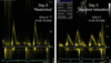
LA stunning after cardioversion
- Day 0 (cardioversion) = markedly reduced mitral A velocity and apparent “restrictive filling” pattern (basis of E/A ratio)
- Day 3 = LA function improves with increased A velocity and a decreased E/A ratio consistent with impaired LV relaxation but normal LV filling pressures
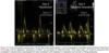
Color Doppler M-mode (CMM) echocardiography provides infomration on flow propagation (Vp) which is unique in that it is relatively independent of this?
Loading conditions

What is one of the key limitations of Vp?
- Predictive ability regarding filling pressures is predominantly in systolic dysfunction
- E / Vp > 2.5 –> predices PCWP > 15 mmHg
What is a normal Vp?
> 50 cm/s
Describe the findings

Color M-mode with white line tracing Vp
What is the strongest determinant of mitral decelration time?
LV operating stiffness
- changes in LV compliance (relationship between LV pressure and volume) and also ventricular relaxation (or early diastolic pressures)
In patients with dilated cardiomyopathy, PW Doppler mitral flow velocity variables and filling patterns correlate with these variables to what degree when compared to LV function?
- Cardiac filling pressure
- Functional Class
- Prognosis
PW Doppler mitral flow velocity > > > LVEF
in regards to filling pressure, functional class and prognosis
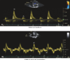
Describe the diastolic filling pattern

Grade 3-4 diastolic dysfunction
Restrictive

Describe the diastolic filling pattern?

Grade 2 diastolic dysfunction
Pseudonormal

What patterns of diastolic dysfunction are most commonly associated with acute myocardial infarction?
Pseudonormal
or
Restrictive
What finding on Valsalva maneuver, in the assessment of diastolic dysfunction, will indicate increased LV filling pressures?
decrease E/A ratio ≥ 50%
What are two findings that indicate an adequate Valsalva maneuver?
- Forceful expiration - generating 40 mmHg of pressure
- Decrease mitral PV ≥ 20 cm/s
- in patients without restrictive filling
When is Valsalva maneuver utilized in the assessment of diastolic function?
- Stage 2 diastolic dysfunction
- differentiating normal vs. pseudonormal
Patient with Cardiac Amyloidosis, which stage of disease will A-fib be most problematic?

- A - Impaired relaxation (may be asymptomatic at rest or with mild exercise) –> LV has become more dependent on atrial contraction (low E/A ratio)
- most likely to feel a change in symptoms with sudde onset of A-fib due to loss of atrial kick
****Atrial contraction hardly contributes to LV filling in most advanced stages of diastolic dysfunction (diminutive A wave in restrictive filling)

Describe the diastolic filling pattern?

Normal diastolic filling pattern
Describe the findings and diagnosis

Markedly delayed relaxation –>
preload reduction will reveal stage 1 diastolic dysfunction
- L-wave: transmitral flow during diastasis

Describe mitral “L-wave”
- L-wave: transmitral flow during diastasis
- triphasic mitral inflow pattern that can be seen in patients without structural heart disease
- Associated with bradycardia
- represents advanced diastolic dysfunction (elevated filling pressures + loss of compliance + very delayed relaxation)

Describe the diastolic filling pattern?

Grade I diastolic dysfunction
Impaired relaxation

What is one way to unmask underlying relaxation abnormalities in diastolic function assessment?
- Preload reduction
- decreases LAP and decreases operating stiffness of the LV
Patient with severe LV dysfunction due to long-standing untreated hypertension is referred for medical therapy. Based on Doppler findings:
- What is the diagnosis?
- What medical therapy should one be cautious of starting? Why?

- Grade 3-4 diastolic dysfunction
-
Beta-blockers
- long term prognosis better with BB (need careful titration)
- But, operating stiffness of heart is very high –> CO is dependent on HR (impossible to augment SV without HR)
Patient with Doppler tracing of AR, presents with SOB. What is the cause?
- SBP 154/78 mmHg

LVEDP is in the normal range - consider alternative etiology
- LVEDP = DBP - 4Vend diastolic velocity2
- LVEDP = 78 - 4 (4)2
- LVEDP = 78 - 64 = 14 mmHg
61 year old woman with ischemic cardiomyopathy is referred for cardiac resynchronization therapy. TTE prior to implantation (A) and post-implantation (B) with Doppler findings. What is the conclusion?

LV stiffness has increased
- Increased E/e’ and E/Vp ratio –> increased LV filling pressures
- Shorter DT –> increased LV operating stiffness
- Decreased A-wave velocity –> reduced LA contractility
- M-mode Vp and tissue Doppler e’ are unchanged

Describe the findings and diagnosis

60 year old man with hypertensive heart disease
- Large difference ( > 30 ms) between duration of mitral A-wave velocity and duration of the late diastolic pulmonary venous flow reversal, AR –> Elevated end-diastolic LV filling pressures
- usually seen in patients with grade 2 or 3 diastolic dysfunction
What is the effect of A-fib on mitral A waves?
they will be absent
What is the effect of MR on the pulmonary vein flow S wave?
reversal or at least blunting of the S wave (with significant MR)
Mitral inflow pattern by itself is suggestive of elevated filling pressures if:

Reduced LVEF

What will effect changes in the mitral inflow pattern, in patients with reduced EF and diastolic dysfunction?
changes in preload
- volume overload, changes in medical therapy
In the presence of moderate-severe MR and diastolic dysfunction, what are the effects on E velocity and DT?
- Increased peak E velocity
- Normal DT
What are the four components of hepatic vein Doppler velocities?
- Systolic forward flow (S)
- Diastolic forward flow (D)
- Systolic flow reversal (VR)
- Atrial flow reversal (AR)
What two conditions are diastolic flow reversal seen most commonly?
How do you differentiate between the two (with flow reversal alone)?
- CP and Pulmonary hypertension
- Respiratiory variation –> CP
- augmentation of diastolic flow reversal

48 year old male with newly diagnosed NICM, what can you conclude about LV compliance?
- ECG: ST - 108 bpm
- BP 90 / 50 mmHg
- Audible S3
- BNP 1530

Decreased LV compliance
- Mitral Inflow
- Appears to have fused E/A wave but more likely increased E wave with shortened DT
- Pulmonary vein flow
- Blunted systolic to diastolic flow with a large AR confirms –> E wave with restrictive physiology, decreased LV compliance and elevated LV filling pressures
- Decreased annular velocity and delayed Vp confirms impaired relaxation
- E/e’ = 17 and E/Vp = 5 –> both confirm elevated LV filling pressure

48 year old male with newly diagnosed NICM, what can you conclude after 1 week of intensive therapy?
- ECG: ST - 108 bpm
- BP 90 / 50 mmHg
- Audible S3
- BNP 1530

LV filling pressures are now normal
- E/e’ is now < 10 –> suggesting that LV filling pressures have been lowered

What is one way to correlate or determine which wave is present on mitral inflow?
- Pulmonary vein flow
- positive relationship of mitral E wave and pulmonary vein D wave
- larger the E wave, the larger the D wave
What are confirmatory measures of elevated LV filling pressures (in patients with reduced EF and diastolic function)?
- E/e’ > 15
- E/Vp > 2
What are the hallmarks of restrictive cardiomyopathy?
- Advanced diastolic dysfunction
- Atrial dilatation
****In spite of normal LV size and function
48 year old male presents with SOB, abdominal swelling, lower extremity edema over the past 6 months. Recurrent admissions over the last 2 years with syncope and A-fib (on Holter)
- EKG: ST - 100 bpm
- BP: 100/74 mmHg
- JVD - 8 cm
- PE: pitting edema of bilateral lower extremities
What is the diagnosis?
What can explain the Doppler/Echo findings?

- Restrictive cardiomyopathy
- Progressive myocardial stiffening
- Restrictive physiology
- shortened mitral E wave DT
- E/A ratio markedly increased ~ 20
- Grade 3 diastolic filling with normal systolic function
- Ventricular filling is notable for decreased LV compliance
What is the pathophysiology of restrictive cardiomyopathy?
- As LAP increases with disease progression
- MV opens at a higher pressure –> decrease in the IVRT
- In addition, there is increased transmitral pressure gradient –>
- increased E velocity
- decreased systolic pulmonary venous flow velocity
- systolic/diastolic ratio < 1
Patient with known CAD, presented with abnormal stress test, given SL NTG –> junctional bradycardia (HR = 40 bpm) and hypotension (BP 75/43 mmHg)
- What is the grade of diastolic dysfunction?

Extreme Grade 1 diastolic dysfunction
- Mitral E/A < 1
- severely decreased mitral E wave
- Vp is reduced (50 cm/s)
- Septal E’ < 7 cm/s
- E/e’ < 8
- filling pressures are likely not increased
Why do nitrates in patients with (extreme grade 1) diastolic dysfunction result in hemodynamic abnormalities?
-
Decreased preload:
- causes reduced end-diastolic volume –>
- patients are dependent on atrial contractility –>
- reduced SV/CO
What are the Doppler findings in Grade 1 (extreme) diastolic dysfunction?
- Mitral E velocity < 50 cm/s
- DT > 200 ms
- Mitral A velocity increased
- E/A < 1
- IVRT increased
- e’ usually < 7 cm/s
- Vp < 50 cm/s
What finding on TTE is associated with worsening prognosis in amyloidosis patients?
Short DT - < 150 ms
Increased E/A > 2.1 (also with decreased survival at 1 year)
What is one finding which can differentiate amyloidosis from other causes of LV hypertrophy?
“Apical sparing” longintudinal strain pattern
What is a desirable result after AV optimization?

Grade 1 LV diastolic filling pattern

What is one variable that remains constant in alteration of paced AV delay (in AV optimization)?
Heart rate
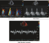
Describe the findings and grade

Pulmonary vein Doppler flow
Normal

Describe the findings and grade

Pulmonary vein doppler flow
Grade 1 - Impaired relaxation

Describe the findings and grade

Pulmonary vein doppler flow
Grade 2 - Pseudonormal

Describe the findings and grade

Pulmonary vein doppler flow
Grade 3 - Restrictive

Describe the findings and grade

TDI
Normal

Describe the findings and grade

TDI
Grade 1 - Impaired relaxation

Describe the findings and diagnosis

TDI
Grade 2 - Psuedonormal

Describe the findings and diagnosis

TDI
Grade 3 - Restrictive

