Thyroid anatomy, hormones and iodine balance Flashcards
Describe the normal embryonic development of the thyroid.
The thyroid gland originates as a invagination in the pharyngeal floor, at the foramen cecum of the tongue. From the foramen cecum a tubular structure termed ‘thyroglossal duct’ descends anterior to the trachea and bifurcates, forming two lateral lobes.

What are 2 possible congenital defects that could be observed with the thyroid?
Retrosternal thyroid - if thyroglossal duct travels too far down before growing 2 lobes
Thyroglossal cysts - fluid filled mass in neck that develops from cells and tissue remaining after formation of the thyroid gland during embryonic development


What is a goiter and why can it be dangerous to a person’s health?
Goiter = englarged thryoid that protrudes from neck

What is the epithelium of the thyroid gland?
Simple cuboidal
[…] cells produce colloid
[…] cells produce calcitonin
Follicular
Parafollicular
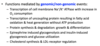
Describe the hypothalamus, pituitary, thyroid axis and the factors that regulate this axis at each level.
- Really high [I-] is what can inhibit thyroid hormone production in this image
- Deiodinases are present on peripheral tissues that will convert T4 to T3 and in this way the body can control which tissues will have more potent responses to thyroid hormone since T3 is 10x more potent than T4


A
TRH
- What signals induce its production and secretion?
- Where is this made?
- Where is it released?
- Where does it act?
- What signaling pathway does it activate?
- What are its effects on the cells it activates?
- How is its release inhibited?
- Signals: diurnal rhythm, stress (infection/starvation), temperature, metabolic state
- Parvocellular neurons in PVN of hypothalamus
- Median eminence –> hypophyseal portal system
- Thyrotropes in anterior pituitary
- GPCRs –> PLC –> IP3 and DAG –> PKC and increased Ca2+
- TSH synthesis and secretion
- T3 provides negative feedback to inhibit cells in hypothalamus from making TRH

TSH
- What signals induce production and secretion?
- Where is this made?
- Where is it released?
- Where does it act?
- What signaling pathway does it activate?
- What are its effects in the cells it activates?
- How is its release inhibited?
- TRH
- Thyrotropes anterior pituitary
- Released into systemic circulation
- Acts on thyroid epithelial cells
- GPCR –> Gas –> (+) AC –> (+) cAMP –> (+) PKA –> (+) gene transcription
- Stimulates all aspects of thyroid hormone synthesis and secretion, as well as cell growth
- T3 can feedback and inhibit anterior pituitary, also inhibited by dopamine, somatostatin, and glucocorticoids


C
Describe how the thyroid stores its hormones.
- Follicular epithelial cell synthesizes thyroglobulin (rER –> posttranslationally glycosylated in rER and Golgi –> packaged into vesicles –> secreted into the lumen of the follicle)
- Follicular epithelial cells actively transport iodide from the blood into their cytoplasm using ATPase-dependent sodium/iodide symporters (NIS). Iodide ions then diffuse rapidly toward the apical cell membrane. From here, iodide ions are transported to the lumen of the follicle by the iodide/chloride transporter called pendrin located in the apical cell membrane. Iodide is then immediately oxidized to iodine, the active form of iodide. This process occurs in the colloid and is catalyzed by membrane-bound thyroid peroxidase (TPO).
- One or two iodine atoms are then added to the specific tyrosine residues of thyroglobulin. This process occurs in the colloid at the microvillar surface of the follicular cells and is also catalyzed by thyroid peroxidase (TPO). Addition of one iodine atom to a single tyrosine residue forms monoiodotyrosine (MIT). Addition of a second iodine atom to the MIT residue forms a diiodotyrosine (DIT) residue.
- Thyroid hormones are formed by oxidative coupling reactions of two iodinated tyrosine residues in close proximity. For example, when neighboring DIT and MIT residues undergo a coupling reaction, T3 is formed; when two DIT residues react with each other, T4 is formed. After iodination, T4 and T3 as well as the DIT and MIT residues that are still linked to a thyroglobulin molecule are stored as the colloid within the lumen of the follicle.
- In response to TSH, follicular cells take up thyroglobulin from the colloid by a process of receptor-mediated endocytosis. After endocytosis, thyroglobulin follows at least two different intracellular pathways.
● In the lysosomal pathway, thyroglobulin is internalized and transported within endocytic vesicles to early endosomes. They eventually mature into lysosomes or fuse with existing lysosomes. Thyroglobulin is then degraded by lysosomal proteases into constituent amino acids and carbohydrates, leaving free T4, T3, DIT, and MIT molecules. Under physiologic conditions, this is the major pathway of colloid resorption.
● In the transepithelial pathway, thyroglobulin is transported intact from the apical to the basolateral surface of follicular cells. To enter this pathway, thyroglobulin binds to its receptor, megalin, which is a transmembrane protein expressed at the apical surface of follicular epithelial cells directly facing colloid. Thyroglobulin internalized by megalin avoids the lysosomal pathway, and endocytic vesicles are delivered to the basolateral membrane of follicular cells. In pathologic conditions of high TSH or TSH-like stimulation, megalin expression is increased, and large amounts of thyroglobulin follow the transepithelial pathway. This pathway may reduce the extent of T4 and T3 release by diverting thyroglobulin away from the lysosomal pathway. Individuals with Graves disease and other thyroid diseases have detectable amounts of circulating thyroglobulin that contains portions of megalin receptor.
- Release of T4 and T3 from follicular cells into the circulation. Follicular cells predominately produce T4 in a T4 to T3 ratio of 20:1. Most of the T4 and T3 produced is liberated from thyroglobulin in the lysosomal pathway, and only negligible amounts of T4 and T3 are released bound to thyroglobulin. Both T4 and T3 cross the basal membrane and enter the blood and lymphatic capillaries. Most of the released hormones are immediately bound to a specific plasma protein, thyroxine-binding globulin (TBG) (~70%), or a protein called transthyretin (~20%). T4 has a stronger bond to TBG, whereas T3 has a stronger bond to transthyretin. Less than approximately 10% of released hormones are bound to a nonspecific fraction of albumin, leaving only small amounts (~1%) of free circulating hormones that are metabolically active. One-third of circulating T4 is converted to T3 in peripheral organs, such as the kidney, liver, and heart. T3 is five times more potent than T4 and is mainly responsible for biologic activity by binding to the thyroid nuclear receptors in the target cells.

What are the 3 thyroid hormones to know?
What are their relative proportions in thyroid secretions?
T4 –> 90%
T3 –> 10%
rT3 –> 1%
Iodide transport into follicular cells is regulated by what?

What drugs inhibit TPO?
Propylthiouracil
Where is TPO located?
Integral membrane protein in apical membrane of follicular cells
What is the Wolff-Chaikoff effect?

What enzyme catalyzes the coupling of adjancent MIT and DIT to make T3 and T4?
TPO (also responsible for catalyzing production of MIT and DIT residues on TG prior to coupling reactions)

Does the majority of T3 come from its synthesis?
No –> comes from conversion of T4 to T3 in peripheral tissues
- What are the types of deiodinases in peripheral tissue?
- What reaction do they catalyze?
- What tissues express each type?
Type 1
- T4 –> T3
- Liver, kidney, skeletal muscle, thyroid
Type 2
- T4 –> T3
- CNS, glia, female reproductive tract
Type 3
- T4 –> rT3
- Peripheral tissues

What would be the effect of hypothyroidism on type 1 and type 2 deiodinases?
Cause their expression to be elevated

A
What are some dietary sources of iodine?

Discuss how iodine is stored and secreted in the body.
Like many ions, we secrete the same amount that we ingest when we are in homeostatic balance. The thyroid releases about 80 micrograms of iodine a day, so it takes in that much iodine from blood daily to replace iodine that is released at thyroid hormones. Iodine that is released as thyroid hormones are metabolized and lost through the stool and urine.

When do we see goiters develop?
When iodine intake < 50 micrograms/day
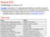
What are the consequences of iodine deficiency in:
- Fetus
- Neonates
- Children
- Adults


A - recall that TSH stimulates cell growth

TSH – high
fT4 – low
fT3 – normal (due to increased conversion of T4 to T3 by deiodinases)
serum Tg – high
[…] is the principal mediator and causes the morphological consequences of developing a goiter
TSH
Describe in detail what the Wolff-Chaikoff effect is.

Explain in detail what the Jod-Basedow effect is

The thyroid gland has about […] to […] supply of hormones ready at any given time.
2 to 3 months
[…] is the receptor that thyroglobulin binds to in order to enter the cell in an endosome for release of thyroid hormones.
Megalin
Some MIT and DIT released during TG proteolysis is rapidly deiodinated within the follicular cell by the enzyme […]
intra-thyroidal deiodinase
Hashimoto’s disease is caused by auto-antibodies against […]
TPO
What are the 3 proteins that thyroid hormones can bind to and circulate in the blood?
Thyroid binding protein
Transthyretin
Albumin
What are the effects of free T4 and T3 in the blood?
Enter cells, bind to specific receptors for hormone action, feedback inhibition at thyrotroph cells in anterior pituitary

Normally only ~ […] of TBG hormone binding sites are occupied
1/3
What is the role of TBG binding to thyroid hormones?

Deiodination reactions that occur in periphery require […] as a co-factor.
Selenium
How do thyroid hormones enter the cell?
They require transporters to allow them to cross the membrane. The transporters show specificity for T4 vs. T3 and cell-specific expression
Describe the binding of thyroid hormones to their receptor once they’ve entered the cell.

What are the general cellular functions that are mediated by thyroid hormones binding to their receptor?
What are the effects of thyroid hormones on the cardiovascular system?
Ionotropic effects (increase CO and contractility) and chronotropic effects (increase HR)

What effect do thyroid hormones have on BMR?
Increased expression of mitochondrial uncoupling proteins –> increases production of heat without production of energy –> increases body temp –> increase in BT is moderated via increase in heat loss thorugh thyroid hormone mediated increase in blood flow, sweating and ventilation

What effect do thyroid hormones have on Metabolism?

What are the effects of thyroid hormones on respiratory system?

What effect do thyroid hormones have on growth and maturation?

What effect do thyroid hormones have on MSK system?

What effect do thyroid hormones have on skin?

What effect do thyroid hormones have on nervous system?

What effect do thyroid hormones have on reproductive and endocrine systems?
Increased thyroid hormone –> increases sex hormone binding globulin –> has greater affinity for androgens –> increases estrogen : androgen ratios in men

What effect do thyroid hormones have on GI system?

What effect do thyroid hormones have on kidneys?

What effect do thyroid hormones have on blood?

Total (free + bound) thyroid hormone in plasma is dependent on both […] and […]
Thyroid function
Concentrations of binding proteins
- Increased thyroid binding proteins will have what effect on free and bound hormone levels?
- Decreased thyroid binding proteins will have what effect on free and bound hormone levels?
- Temporary fall in free hormone due to increased BP in blood, but TSH will be stimulated by decreased free hormone and this will restore free hormone levels to normal
- Temporary increase in free hormone due to decreased BP in blood, but TSH will be inhibited by increased free hormone and this will restore free hormone levels to normal
Change in total hormones but normal free hormone levels
What factors can cause an increase in thyroid binding protein levels?
What factors can cause a decrease in thyroid binding protein levels?

How do we conduct thyroid function tests?
Assays to detect TSH and free T3 and T4 in blood.
With hypothyroidism, levels will be low. With hyperthyroidism levels will be high.
What effect can drugs have on thyroid function tests?
Some drugs can displace thyroid hormones from binding proteins
If plasma TSH is high and plasma free T4 is high, this indicates a […]
TSH secreting tumor
If plasma TSH is high and plasma free T4 is normal, this indicates a […]
Borderline hypothyroidism
If plasma TSH is high and plasma free T4 is low, this indicates a […]
Hypothyroidism
If plasma TSH is normal and plasma free T4 is high, this indicates a […]
Thyroid hormone resistance
If plasma TSH is normal and plasma free T4 is normal, this indicates a […]
Normal thyroid
If plasma TSH is normal and plasma free T4 is low, this indicates a […]
Hypopituitarism
If plasma TSH is low and plasma free T4 is high, this indicates a […]
Hyperthyroidism
If plasma TSH is low and plasma free T4 is normal, this indicates a […]
T3 toxicosis
If plasma TSH is low and plasma free T4 is low, this indicates a […]
Hypopituitarism
In Graves’ disease, a person has autoantibodies against […]
TSH receptor
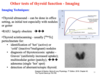
How can imaging be used to examine the thyroid?
What is the difference between primary hypothyroidism and secondary hypothyroidism?

Thyroid hormone dysfunction results from what 3 factors?
Alterations in circulating levels
Impaired peripheral hormonal metabolism
Tissue resistance to hormonal activity
