Mineralocorticoids Flashcards
(24 cards)
What factors stimulate aldosterone synthesis in the Zona Glomerulosa of the adrenal cortex?
Angiotensin 2
Hyperkalemia
Leptin
What factors inhibit aldosterone synthesis in the Zona Glomerulosa of the adrenal cortex?
ANP
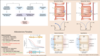
- Fill in the missing proteins/enzymes and the locations where these parts of aldosterone synthesis are occurring.
- For the missing proteins / enzymes, what factors stimulate their activity?

- See image
- STaR = ACTH, Angiotensin 2, hyperkalemia
- Aldo Synthase = Angiotensin 2, hyperkalemia

Describe the signaling pathway that angiotensin 2 uses in adrenal cortex cells to induce aldosterone production.
Ang2 binds GPCR –> Gaq –> PLC cleaves PIP2 to IP3 and DAG –> DAG inhibits K+ leaving cell –> depolarization –> stimulates voltage gated Ca++ channels –> increased Ca++ –> IP3 also binds to receptor on SR to lead to efflux Ca++ from SR –> large increase intracellular Ca++ leads to activation of Ca-dependent kinases that stimulate transcription of genes for AldoSynthase and STaR leading to increased aldosterone production.

Describe the signaling pathway that hyperkalemia uses in adrenal cortex cells to induce aldosterone production.
Increased K+ in ECF inhibits K+ leaving cell –> depolarization –> stimulates voltage gated Ca++ channels –> increased Ca++ –> IP3 also binds to receptor on SR to lead to efflux Ca++ from SR –> large increase intracellular Ca++ leads to activation of Ca-dependent kinases that stimulate transcription of genes for AldoSynthase and STaR leading to increased aldosterone production.
Describe the signaling pathway that ANP uses in adrenal cortex cells to inhibit aldosterone production.
ANP binds to receptor (RTK) –> increased cGMP –> results in stimulation of K+ leaving cell –> hyperpolarization –> inhibits release of Ca++ –> inhibits transcription of genes for aldosynthase and star proteins needed to make aldosterone
- How do cells achieve specificity for mineralocorticoids over glucocorticoids, considering that GCs can bind to MCR?
- If a person has high cortisol (cushings) what can happen to mineralocorticoid sensitive cells?
- They express 11betaHSD2 which converts cortisol to cortisone, which cannot bind to the MCR.
- 11betaHSD2 can be saturated, and if that happens then cortisol can bind to MCR and mimic effects of aldosterone

What tissues have aldosterone sensitive epithelium?
What is the net effect of aldosterone on these tissues?

True/false: Epithelial cells of the early distal tubule are directly sensitive to aldosterone.
False - They are sensitive to angiotensin 2. They don’t express 11betaHSD2.

Aldosterone’s primary actions are on the principal cells of the late distal tubule and collecting duct.
- Describe the signaling pathways involved when aldosterone is acting on these cells.
- What are the cellular results of aldosterone signaling?
- Aldo diffuses across membrane, binds to MCR in cytosol, ligand/receptor complex diffuses through nuclear pore, dimerizes with other ligand/receptor complex, binds to DNA, stimulates transcription of:
- ENac channels –> increased Na+ reabsorption and positive driving potential to move K+ into lumen
- ROMK channels –> increased permeability of basolateral membrane to K+
- Na+/K+ ATPase –> increased electrochemical driving force for Na+ reabsorption
- Increased SGK1 –> prevents ubiquitination of ENac and aids in increasing ROMK expression
- CAP1 –> cleaves off part of ENac channel increasing the amount of time the channel is open

- Review the renin-angiotensin-aldosterone pathway.
- Specifically - what are the effects of angiotensin 2 binding to its receptor in the following tissues:
- Brain
- Kidney
- Adrenals
- Pituitary
- Arterioles

What are the actions of ANP?
Opposite of angiotensin 2

Describe the signaling pathways that result from ANP binding to its receptor on aldosterone sensitive cells in the late distal tubule and collecting duct.
Inhibits ENac and Na+/K+ ATPase –> results in decreased Na+ reabsorption and decreased K+ secretion –> decreased Na+ and water reabsorption –> decreased volume

Angiotensin 2 and hyperkalemia stimulate aldosterone production and secretion by the adrenal cortex. Aldosterone then promotes Na+ reabsorption and K+ secretion by principal cells in kidney. However, this is not always the case. What are the 2 scenarios which are referred to as the “aldosterone paradox”?
The aforementioned function is true under normal function and euvolemia.
- When a person is volume depleted, their RAAS system is activated, resulting in increased Na+ reabsorption but not an increase in K+ secretion as would be expected. This works to restore Na+ balance but does not change K+ balance.
- When a person is hyperkalemic, this causes an increase in K+ excretion stimulated by aldosterone. However, you would expect a decrease in Na+ excretion (i.e. increase in Na+ reabsorption) but this is not observed. There is instead response to correct the high K+ and no change in Na+ balance.
Why don’t see increased K+ excretion in volume contraction?
Both increased aldo and increased delivery of Na+ to distal nephron are required for K+ excretion to be increased. In volume contraction, there is increased aldo, but Angiotensin 2 will stimulate Na+ reabsorption in the early nephron segments, thus reducing the amount of Na+ that reaches the distal segments in the urine. The distal segments are responsible for excreting K+, so if there is little Na+ remaining in the urine, there will be no change in K+ excretion as little Na+ is reabsorbed in the distal segments in volume contraction. Normally, the low intracellular [Na+] drives Na+ from urine into cells in distal segments. This makes the urine net (-) and favors K+ diffusion into urine via ROMK. However, when there is little Na+ in urine remaining, there is comparatively less net (-) charge that results in urine from loss of Na+ so there is no change in K+ excreted as a result. Additionally, Ag2 inhibits ROMK channels in distal nephron so there is decreased ability for K+ to be excreted.
Why don’t we see increased Na+ reabsorption with hyperkalemia?
Hyperkalemia will stimulate aldosterone production and secretion. This will result in comparatively increased Na+ reabsorption in the distal nephron that is sensitive to aldosterone and increased Na+ delivery to distal nephron. This increased delivery of Na+ to distal segments will drive the K+ excretion b/c the low intracellular [Na+] established by Na+/K+ ATPase will drive Na+ from urine into cells in distal segments. This makes the urine net (-) and favors K+ diffusion into urine via ROMK. However, because there are no signals for Na+ to be reabsorbed in any segment other than the distal nephron (aldosterone not effective on other parts of nephron and RAAS is not triggered), there will be no net change in Na+ reabsorption despite an increase in K+ excretion.

What enzymes have emerged as being important in explaining the aldosterone paradox?

What are the various causes and characteristics of hypoaldosteronism?

What are the causes and characteristics if primary hyperaldosteronism?

What are the causes and characteristics of secondary hyperaldosteronism?

What is aldosterone escape?
In primary hyperaldosteronism, there is unregulated secretion of aldosterone. However, this does not lead to unregulated Na+ retention or the development of edema. When there is an increase in aldosterone, the kidneys do respond initially by retaining Na+, however, this leads to an increase in BP which leads to an increase in blood flow through the vasa recta. In the early nephron segments this increases interstitial pressure, which decreases Na+ reabsorption. In the distal segments, this diminishes the medullary solute gradient thus decreasing the driving force for Na+ reabsorption. Thus, there is no increase in Na+ reabsorption in response to unregulated aldosterone.

Why does primary hyperaldosteronism not result in edema?
No increase in Na+ retention –> no edema
Why does secondary hyperaldosteronism result in edema?
Big thing is that you’re changing venous pressure

- In the brain, the MCR receptor is located in the […]
- In the heart, high levels of aldosterone can promote […]
- In adipose tissue, adipocytes secrete […] in proportion to body fat, which stimulates aldosterone secretion. This provides a link between […] and […].
- nucleus solitarius –> salt appetite
- myocardial fibrosis
- Leptin; Obesity; HTN
