Embryology Flashcards


At beginning of […], embryo is a trilaminar germ disc
fourth week
When do the following processes happen?
- Neurulation
- Cranial neurpore closes
- Caudal neuropore closes and primitive streak appears
All these events happen during week 4
- Neurulation = day 21
- Cranial closes = day 24
- Caudal closes = day 26
Describe closure of the neural tube.
Begins centrally and extends in both cranial and caudal directions
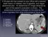
Failures of the neural tube to close most often occur in the caudal neuropore. What conditions occur if the caudal neuropore fails to close?
Many nervous system defects occur when the neural tube fails to develop properly. Spina bifida is an incomplete vertebral arch that results when the neural tube fails to sink completely below the surface, and somite sclerotome cells cannot migrate over it to complete the vertebral arch. The spinal cord may be exposed on the surface with severe functional deficits (myeloschisis); it may be completely normal in function with few visible manifestations (spina bifida occulta), or there may be a variety of intermediate conditions. In meningocele, the spinal cord is normal, but a swelling of meninges with cerebrospinal fluid projects through the defect. If part of the spinal cord is included, it is a meningomyelocele.

Although failure of the cranial neuropore to close are less common, they can still happen. What conditions arise when that happens?
Neural tube defects in the head have effects similar to those in spina bifida, but the skull and brain are involved instead of the vertebral column and spinal cord. The occipital bone (or other midline cranial bones) may fail to ossify, and meninges project through the defect with (encephalocele) or without brain tissue. An extreme neural tube defect is failure of the anterior neuropore to close, resulting in absence of most of the brain and neurocranium (anencephaly). This is the most common major malformation in stillborn fetuses.


D
Neurulation = folding process by which […] is folded into […] and […] cells form; takes place ~[…] days post-conception
neural plate
neural tube
neural crest
18-32
The adenohypophysis (anterior pituitary), lens of eye, external epithelial linings, epidermis of skin are all drived from what germ layer?
Which of these stands out as an odd exception to be in this category?
Surface ectoderm
Anterior pituitary - seems like it fits in more with those that are neuroectoderm but it’s actually surface ectoderm from the roof of the mouth
The neurohypophysis, CNS neurons, oligodendrocytes, astrocytes, ependymal cells, pineal gland are all derived from what germ layer?
Neuroectoderm
The gut tube and its derivatives (GI tract, lungs, liver, pancreas, thymus, parathyroid gland, thyroid follicular cells) all come from what germ layer?
Endoderm
The dura mater, connective tissue, muscle, bone, cardiovascular structures, lymphatics, blood, urogenital structures, peritoneum, spleen, adrenal cortex, kidneys are all derived from what germ layer?
Which of these structures stands out as being odd for this group?
Mesoderm
Spleen (fits in more with gut structures, which are endoderm) and adrenal cortex (b/c it’s different from adrenal medulla which is neural crest)
The ANS, dorsal root ganglia, cranial nerves, melanocytes, chromaffin cells of adrenal medulla, enterochromaffin cells, pia and arachnoid layers, celiac ganglia, Schwann cells, odontoblasts, parafollicular thyroid “C” cells, pharyngeal cartilage, skull bones are all derived from what germ layer?
Neural crest cells –> adrenal medulla stands out b/c different from adrenal cortex which is mesoderm
What germ layer gives rise to the nucleus pulposis of the intervertebral discs?
Notochord (induces ectoderm to form neuroectoderm)
C - Mesoderm (pointing at spleen)

A
Urogenital ridges form from […] and develop at […] weeks (using embryological notation)
mesoderm
3 weeks
Using embryological timeline, when do major and minor calices develop?
4-5 weeks
The external genetalia develop during week […] (embryological)
10
Describe the development of the kidneys.
The intermediate mesoderm differentiates into nephrogenic tissue in the nephrogenic ridge lateral to the genital ridge. From cranial to caudal it forms three successive kidneys:
- The pronephros never fully develops and quickly diminishes
- The mesonephros is the first functioning kidney, with glomeruli, mesonephric tubules, and a mesonephric duct that drains embryonic urine into the dividing cloaca
- The metanephros becomes the permanent kidney

How does the kidneys’ location change in the abdomen during development?

Because the kidneys ascend in the abdomen, there are issues that can arise with this process. What are some of them?
A kidney can fail to ascend on one side only, or a kidney can migrate to the opposite side of the body. The development of renal blood vessels is unique. Most organs “trail” their blood supply as they migrate. As the kidneys ascend, new blood vessels form at higher levels of the aorta and inferior vena cava and connect to the kidneys as lower vessels disappear. Renal arteries of pelvic kidneys originate near the bifurcation of the aorta. For normal adult kidneys, they are at the level of the superior mesenteric arteries of the midgut. Sometimes, more inferior renal vessels fail to disappear. This is the embryonic basis of multiple renal arteries and veins in the adult.

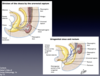
Describe the formation of the urethra and the anus from the urogenital cloaca.
The urorectal septum between the allantois and hindgut divides the cloaca in the frontal plane into an anterior urogenital sinus and posterior rectum. The septum divides the cloacal membrane into a urogenital membrane and anal membrane. The upper part of the urogenital (UG) sinus is the fusiform urinary bladder. The lower pelvic and phallic parts of the UG sinus (UG sinus proper) form the urethra and related glands and structures in each sex. The genital portion of the urogenital sinus is closely related to the genital tubercle, a swelling of somatopleure. The metanephric duct (future ureter) opens into the developing urinary bladder; the male (mesonephric) and female (paramesonephric) genital ducts shift to a more caudal position on the UG sinus.
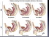
If division of the urogenical cloaca does not go smoothly, there can be pathologies that arise. What are some of these?
If the urorectal septum does not completely divide the cloaca, the rectum will connect anteriorly with urinary or genital structures derived from the urogenital sinus. The resulting fistulas are all associated with an imperforate anus. A rectoperineal fistula opens to the surface, but it is an abnormal connection anterior to the external anal sphincter (and anus) through the central tendon of the perineum (perineal body).
The urachus is the fibrous remnant of the allantois, an extension of the cloaca/urogenital sinus into the umbilical cord. It is obliterated in normal human adults. What can arise if it doesn’t get completely destroyed?
The lumen of the allantois may persist as a fistula (completely patent lumen), sinus (blind pit at either end), or cyst (enclosed swelling). These types of congenital defects may occur in any tubular primordium in the embryo that is supposed to form a fibrous cord or disappear.

What structures develop from the urogenital sinus in individuals whose genome is:
- XX
- XY

What structures develop from the mesonephric duct and tubules in individuals who are genomically:
- XX
- XY

What structures develop from the paramesonephric duct in individuals who are genomically:
- XX
- XY

What structures develop from the genital tubercle/phallus in individuals who are genomically:
- XX
- XY

What structures develop from the urogenital folds in individuals who are genomically:
- XX
- XY

What structures develop from the labioscrotal folds in individuals who are genomically:
- XX
- XY

What structures develop from the indifferent gonads in individuals who are genomically:
- XX
- XY

What structures develop from the gubernaculum in individuals who are genomically:
- XX
- XY

What is an “intersex” person?
Used to describe a variety of conditions in which human genital anatomy doesn’t match “typical” male or female
Heart development happens between days […] and […] post conception
19 and 63
When does the heart begin beating?
day 22 post conception
When is heart development complete?
End fo week 8 (post conception)
What is a persistent truncus arteriosus?

What is a patent ductus arteriosus?

What are atrial and ventricular septal defects?


B
- Lungs form from a […] from […] beginning in […] week
- Trachea and esophagus form very close together, and disruptions to their development can result in […]
- laryngotracheal diverticulum; foregut; fourth
- tracheoesophageal fistulas

During development, the fetus swallows […]
Amniotic fluid (contains urine)
Large amounts of amniotic fluid retained within the fetus indicates what?
Some kind of narrowing / blockage along the GI tract, i.e. esophageal or duodenal atresia
Small amounts of amniotic fluid retained within the fetus can indicate an issue with what?
The kidenys
What is an omphalocele?

What is Meckel’s diverticulum?
- This is the most common type of congenital gastrointestinal anomaly
- Rule of 2s: occurs in 2% of children, occurs within ~2 feet of the ileocecal vale, contains 2 types of ectopic mucosa (gastric & pancreatic), and its symptoms usually occur by age of 2


B

C
Limbs rotate to their adult locations at […]
Hip, knee, ankle, shoulder, elbow and wrist joints fully formed at […]
Bones begin to ossify at […]
7 weeks
9 weeks
10 weeks
