Posterior Leg and Ankle Joint Flashcards
Posterior Aspect of the Leg Bones


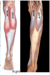
Muscles of the Posterior Compartment

7 muscles (3 superficial, 4 deep)
Superficial muscles all insert on posterior surface of calcaneous via Tendocalcaneous
Tendons of deep muscles pass behind medial malleolus to plantar surface of foot (except popliteus)

Superficial Muscles - Gastrocnemius
Attachments (Medial and Lateral head)

Medial head: Popliteal surface of the femur, superior to the medial condyle
Lateral Head: Lateral aspect of the lateral condyle of the femur

Superficial Muscles - Gastrocnemius
Innervation

Tibial nerve (S1, S2)

Superficial Muscles - Gastrocnemius
Function
Plantarflexes ankle and flexes knee

Superficial Muscles - Plantaris
Attachments

Lateral supracondylar line of femur (proximal to the lateral head of gastrocnemius)

Superficial Muscles - Plantaris
Innervation

Tibial nerve (S1, S2)

Superficial Muscles - Plantaris
Function

Assists plantarflexion of ankle

Superficial Muscles - Soleus
Attachments

Soleal line of Tibia, upper 1/3 of posterior fibula. Tendinous arch between bony attachments

Superficial Muscles - Soleus
Innervation

Tibial nerve (S1, S2)

Superficial Muscles - Soleus
Function

Plantarflexes ankle joint

Deep Muscles - Popliteus
Attachments

Lateral condyle of the femur (pit for popliteus) and lateral meniscus to the popliteal area of the tibia (above the soleal line)

Deep Muscles - Popliteus
Innervation

Tibial nerve (L4, L5, S1)

Deep Muscles - Popliteus
Function

Unlocks the knee joint by laterally rotating the femur on the fixed tibia

Deep Muscles - Flexor Digitorum Longus
Attachments

Posterior surface of Tibia to the base of the distal phalanx of digits 2-4

Deep Muscles - Flexor Digitorum Longus
Tibial nerve (L5, S1, S2)


Deep Muscles - Flexor Digitorum Longus
Function

Flexes lateral 4 digits. Weak plantarflexor of ankle.

Deep Muscles - Flexor Hallucis Longus
Attachments

Posterior surface of Fibula to the base of the distal phalanx of the hallux

Deep Muscles - Flexor Hallucis Longus
Innervation

Tibial nerve (L5, S1, S2)

Deep Muscles - Flexor Hallucis Longus
Function

Flexes Hallux and is a weak plantarflexor of the ankle

Deep Muscles - Tibialis Posterior
Attachments

Posterior surface of tibia and fibula, and the inerosseous membrane to the tuberosity of the navicular, cuneiforms, cuboid, sustentaculum tali of calcaneus, and the base of the 2nd, 3rd and 4th metatarsals

Deep Muscles - Tibialis Posterior
Innervation

Tibial nerve (L4, L5)

Deep Muscles - Tibialis Posterior
Function

Plantarflexes ankle and inverts foot

Tendons of the Deep Muscles
Tibialis Posterior

Tendon passes deep to Flexor Digitorum Longus.
Groove posterior to the medial malleolus


