Anterior and Medial Thigh Flashcards
Compartments of the Thigh


Proximal femur (anterior)


Proximal femur (posterior)


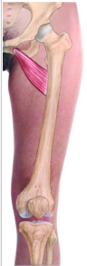
Muscles of the anterior thigh
Hip Flexors

Iliacus, Psoas major, pectineus, sartorius

Muscles of the anterior thigh
Knee extensors

Quadriceps –
Rectus femoris
Vastus medialis
Vastus lateralis
Vastus intermedius

Hip Flexors - Iliopsoas
Iliacus attachments

Iliac crest, fossa, ala of sacrum, anterior sacroiliac ligament to the psoas tendon and lesser trochanter

Hip Flexors - Iliopsoas
Iliacus innervation

Femoral nerve (L2,3)

Hip Flexors - Iliopsoas
Iliacus function

Flexes hip

Hip Flexors - Iliopsoas
Psoas major attachments

T12-L5, IV discs, lumbar transverse processes to the lesser trochanter

Hip Flexors - Iliopsoas
Psoas major innervation

Anterior rami L1-3

Hip Flexors - Iliopsoas
Psoas major function

Flex hip

Hip Flexors - Pectineus
Attachments

Superior ramus of pubis to the pectineal line of the femur

Hip Flexors - Pectineus
Innervation

Femoral nerve (L2,3) occasionally a branch from obturator

Hip Flexors - Pectineus
Function

Adducts and flexes hip, and assists with medial rotation

Hip Flexors - Sartorius
Attachments

Anterior superior iliac spine to the superior part of the medial surface of the tibia

Hip Flexors - Sartorius
Innervation

Femoral nerve (L2,3)

Hip Flexors - Sartorius
Function

Flexes, abducts, and laterally rotates hip, and flexes knee

Knee extensors - Quadriceps
Rectus femoris attachments

AIIS, ilium above acetabulum

Knee extensors - Quadriceps
Vastus lateralis attachments

Greater trochanter and lateral linea aspera

Knee extensors - Quadriceps
Vastus medialis attachments

Intertrochanteric line and medial linea aspera

Knee extensors - Quadriceps
Vastus intermedius attachments

Anterior and lateral shaft of femur

Knee extensors - Quadriceps
All quadriceps distal attachments

Quadriceps tendon then tibial tuberosity via patellar ligament

Knee extensors - Quadriceps
Function

Extend knee (rectus femoris also flexes hip)

Knee extensors - Quadriceps
Innervation

Femoral nerve (L2,3,4)