Anterior Triangle of Neck Flashcards
Triangles of the neck
Sternocleidomastoid (SCM)
Divides anterior from posterior triangle
Boundaries of the Anterior Triangle
Anterior border

Imaginary midline of neck

Boundaries of the Anterior Triangle
Posterior border

Anterior border of SCM

Boundaries of the Anterior Triangle
Base

Mandible

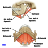
Divisions of the Anterior Triangle


Divisions of Anterior Triangle
Submental

Submental lymph nodes and beginning of anterior jugular vein

Divisions of Anterior Triangle
Submandibular

Submandibular salivary gland, lymph nodes, fascial artery and vein, carotid sheath, hypoglossal nerve etc

Divisions of Anterior Triangle
Carotid

Carotid sheath, branches of external carotid artery, internal jugular vein, hypoglossal accessory and vagus nerves etc

Divisions of Anterior Triangle
Muscular

Sternohyoid and sternothyroid form the floor beneath which lies the thyroid gland, larynx, trachea and oesophagus

Contents of Anterior Triangle


Fascia of Anterior Neck


Platysma
Attachments
From fascia above pectoralis major and clavicle to lateral neck and onto mandible
Platysma
Innervation
Facial nerve CN VII
Veins of Head/Neck


Supra-hyoid muscles
Function

Raise hyoid bone or depress mandible (mylohyoid and digastric)

Supra-hyoid muscles
Innervation

Facial nerve - posterior belly of digastric and stylohyoid
Mandibular division of trigeminal CNV3 - anterior belly of digastric and mylohyoid

Supra-hyoid muscles
Geniohyoid
Deep to mylohyoid - Innervated by C1 via hypoglossal CN XII

Infrahyoid Muscles
Function

Depress hyoid bone

Infrahyoid Muscles
Attachments

Indicated by names of muscles

Infrahyoid Muscles
Innervation

Ansa cervialis except thyrohyoid (C1 via hypoglossal CN XII)

Vasculature and Nerves


Cervical Plexus
Root value

Anterior rami C1-4

Cervical Plexus
Arrangement

Joined into loops which lie anterior to levator scapulae and scalenus medius

Cervical Plexus
Covering

Formed by pre-vertebral layer of deep cervical fascia


