Neuro HARC Lectures Flashcards
What are the three main arteries that supply the head and neck? And what are their soruces?
Source 1: Common Carotid
Artery: External and Internal Arteries
Source 2: Subclavin artery
Artery: Vertebral artery
Bifurcation of the common carotid occurs at vertebral level ___
C4
External carotid a. predominately supplies extracranial structures with one exception:_____ _______ _______, intracranial vessel supplying the meninges
middle meningeal artery
The anterior system and posterior system of the skull is joined together by?
Cerebral Arterial Circle (of Willis)
What is Anastomosis?
- connection between two normally divergent structures
The connection between anterior and posterior systems is a natural ______ _______ ____
circulatory arterial anastomosis.
What is the anterior part of the skull supplied by specifically?
- Carotid (Anterior) System
- Internal carotid artery (right and left)
What is the posterior part of the skull supplied by specifically?
Vertebrobasilar (Posterior) System
- Vertebral Arteries joining to become basilar artery.
- Vertebral arteries initially run in the transverse foramina of the first six cervical vertebrae
- Internal Carotid arteries enter the skull via the _____ _____
- Passes over the _____ _____
carotid canal
foramen lacerum
What is the carotid siphon?
The carotid siphon is a U or S-shaped part to the ICA that varies with age. It begins at the posterior bend of the cavernous part of ICA and ends at the cerebral part, at ICA bifurcation.
- Vertebral arteries run in______ _____
- Pass upwards entering the skull via the ________ _______
- Form the basilar artery on the anterior surface of the brainstem, lying on top of the ____ of the cranial base
transverse foramina
FORAMEN MAGNUM
CLIVUS
Where does the anterior cerebral artery pass through?
- Passes forward under the corpus callosum
- Progresses superiorly and posteriorly along the superior surface of the corpus callosum.
The anterior cerebral artery supplies which areas?
- Frontal lobes
- Medial aspect of parietal lobes
- Optic chiasm
- Lower limb motor and sensory cortexes
The posterior cerebral artery passes through?
- Terminal branch of basilar artery
- Passes posteriorly towards the parietal lobe
Posterior Cerebral artery supplies ?
• Supplies occipital lobe - visual cortex
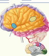
Anterior and Posterior cerebral artery
Middle Cerebral Artery (MCA) passes through….
- Passes laterally towards the temporal lobe and up on to the lateral aspects of the cerebrum.
- Travels initially through the lateral fissure
Middle Cerebral Artery (MCA) supplies….
- Temporal, parietal and occipital lobes
- Broca’s area – Speech production
- Wernicke’s area – Language comprehension
- Head and neck areas of motor and sensory cortexes
Middle Cerebral Artery (MCA)
What dors the lenticulostraite arteries supply?
Supply the basal ganglia, thalamus and internal capsule
• First part (M1) of the MCA
Cerebellar arterial supply via branches of the ___________ system
Cerebellar arterial supply via branches of the vertebrobasilar system
CSF is produced by…..
choroid plexus
What is subarachnoid Cisterns?
- Dilations of subarachnoid space
- Contains CSF
- Can be visualised on an MRI scan
What are the two layers of dura matter?
periosteal layer
meinigeal layer
The spaces created by the perosteal and meningeal layer collect……
venous blood from the brain
these are called “dural venous sinuses”
What type od haemorrhage is this?
Epidural
What are the symptoms of stroke?
F.A.S.T (Face, Arm, Speech, Time)
What is this called?
Hydrocephalus- enlarged ventricles
What is in the basal ganglia?
Consist of:
- Putamen → striatum
- Caudate Nucleus → striatum
- Globus Pallidus – 2 parts - Pallidum
- Substantia Nigra – 2 parts
- Subthalamic Nucleus
Is basal ganglia white or grey matter?
grey matter
What is the function of basal ganglia?
The control of movement. Minimise errors in movements.
Where is the location of the basal ganglia?
Forebrain and Midbrain
basal ganglia
What is the shape of the internal capsule?
boomerang
What is function of the basal ganglia?
It carries information past the basal nuclei, separating the caudate nucleus and the thalamus from the putamen and the globus pallidus.
What is the blood supply of the internal capsule?
- Upper → lateral striate aa. branches from MCA
- Lower
- Anterior Limb →Medial Striate a. from ACA
- Genu → Internal carotid a. branches
- Posterior Limb → Anterior Choroidal a. from MCA
Striatum receives ______ information
cortical
What receptors are in the striatum?
D1 (direct) and D2 (indirect) dopamine receptors
Dopamine acts as a _______
neurotransmitter
What is the function of the golobus pallidum?
- Regulates thalamus
- Under tonic inhibitory control!
What is the direct pathways of the basal ganglia?
main job is to stimulate the cortex.
The cortex provides excitatory input to the striatum which then provides inhibitory output directly to the internal globus pallidus and substantua nigra pars reticulata. These then provide inhibitory input to the thalamus which has excitatory connections back to the cortex. The thalamic output reaches the cortex and excites cortical neurons.
What is the indirect pathways of the basal ganglia?
- Decreasing activity
- Promotes inhibition
Cortex provides excitatory input to the striatum and then the stratum provides inhibitory input to the EXTERNAL pallidus. EXTERNAL pallidus provides inhibitory input to the subthalamic nucleus which the provides excitatory input to the internal globus pallidus and substantua nigra pars reticulata which then provide inhibitory input to the thalamus. The thalamic excitatory output reaches the cortex and excites cortical neurons
Is Parkinsosn’s disease hypokinetic or hyperkinetic?
hypokinetic
Is Huntington’s disease hypokinetic or hyperkinetic?
hyperkinetic
What is Parkinson’s disease?
Parkinson’s disease is a brain disorder that leads to shaking, stiffness, and difficulty with walking, balance, and coordination.
What is the symptoms of Parkinson’s disease?
- Resting tremor
- Postural instability
- Slowness of movement (Hypokinesia)
What is Huntington’s disease (Chorea)?
Huntington disease is caused by gradual degeneration of parts of the basal ganglia called the caudate nucleus and putamen.. It’s passed on (inherited) from a person’s parents. It gets gradually worse over time and is usually fatal after a period of up to 20 years.
How is the Huntington’s disease passed on?
- Autosomal Dominant
- Mutation of Huntingtin protein
At what age does symptoms begin for huntington’s disease
30 - 50 years of age
Upper motor neurons (UMN) carry this information from the cerebral cortex in ascending motor pathways/descending motor pathways
descending motor pathways
What are the pyramidal tracts?
- Corticospinal - Trunk and Limbs
- Corticobulbar – Head and Neck
What are the extrapyramidal tracts?
Rubrospinal
Reticulospinal
Vestibulospinal
Tectospinal
What is the function of the Corticospinal?
Fine voluntary motor control of the limbs. The pathway also controls voluntary body posture adjustments
What is the function Corticobulbar Tract?
Control of facial and masticatory musculature, movements of the tongue and swallowing
• Voluntary movement of head and neck musculature
What is the function Rubrospinal?
Involved in involuntary adjustment of arm position in response to balance information; support of the body
What is the function Reticulospinal?
Regulates various involuntary motor activities and assists in balance (leg extensors). Some pattern movements e.g. stepping
What is the function Vestbulospinal?
It is responsible for adjusting posture to maintain balance
What is the function of Tectospinal?
Controls head and eye movements, Involved in involuntary adjustment of head position in response to visual information.
What are lesions?
Damage or abnormal change in the structure of an organ caused by injury or disease.
Do upper motor neurone lesions occur in CNS or peripheral nerves?
CNS
• Brain, Brainstem and Spinal Cord
Do lower motor neurone lesions occur in CNS or peripheral nerves?
peripheral nerves
• Cranial and Spinal nerves
Lesions: UMN
What are the symptoms?
Symptoms
- Muscle spasticity
- Pseudobulbar palsy
- Weakness without muscle atrophy
- Hyperreflexia
- Babinksi Sign
- Loss of superficial reflex
Lesions: LMN
What are the symptoms?
- Symptoms
- Flaccid paralysis
- Hyporeflexia
- Muscle atrophy
- Hypotonicity
- Fasciculations
What are the key differences between UMN vs LMN
• LMN lesion affects the signal from both systems
- Loss of voluntary movements (pyramidal tracts)
- Loss of coordination, posture and tone (extrapyramidal tracts)
- UMN lesions affects movement signal not muscles themselves.
- UMN lesions cause muscle atrophy through disuse, LMN through denervation.
- UMN lesions cause muscle spasticity, LMN cause muscle flaccidity
Which muscles are affected by a CN III lesion
- Loss of SR, IR, IO, MR action
- Ptosis
- Loss of reflexes (Accommodation & Light)
Which muscles are affected by a CN IV lesion?
- Loss of SO action
- Diplopia (double vision)
Which muscles are affected by a CN VI lesion?
- Loss of LR action
- Unopposed adduction of the eye
What is trigeminal neuralgia?
sudden Intense facial pain
What is bells palsy?
Unknown facial paralysis
- Unilateral
- Can occur overnight
- Drooping of the face
- Inability to close eye on affected side
Where does the nerves decussate athe corticospinal tract?
- 80% fibers • Decussation Medullary pyramids → Lateral Corticospinal Tract
- 20% no decussation → Anterior Corticospinal Tract
Where does deccusation happen at the corticobulbar tract?
• Decussation → CN III, IV, V, VI, VII, IX, X, XI, XI
What is the function of the spinothalamic tract?
• Pain, Pressure, Touch and Temperature
Where does it decussate at the spinothalamic tract?
Decussation
Spinal cord entry level
Function of Spinocerebellar tract
- Non-conscious proprioception
- Position sense information from joint position
Function of the dorsal columns/Medial Lemniscus?
Function
- Conscious proprioception
- Fine touch
- Vibration
- Two-point discrimination
Decussation of the dorsal columns/Medial Lemniscus?
Medial Lemniscus (Brainstem)