CVS Diseases 2 Flashcards

1
Q

What is Anemia?

A

Anemia is a simply a reduction in haem and/ or globin

2
Q

Properties to Hb

A
  • Consists of haem and globin
  • 4 polypeptide globin chains
  • Each chain is complexed to a haem molecule
  • Haem is an iron containing compound

2 beta chains and 2 alpha chains

3
Q

Classification of Anemia

A
  • Anaemia is classified as:
  • Men: Hb <130/L
  • Women: Hb <120/L
  • Further classification based on mean corpuscular volume (MCV) – what is this?
  • In simple terms, the average size of the total red blood cells.
4
Q

Examples of Microcytic anaemia?

A

Iron Deficiency

Thalassaemia

Anemia of Chronic disease

Sideroblastic anemia

Lead poisoning

5
Q

Examples of Normocytic anaemia?

A

Acute blood loss

Haemolytic anaemia

Anaemia of Chronic disease

Chronic Kidney Disease

Aplastic anaemia

6
Q

Examples of Macrocytic anaemia?

A

B12 deficiency

Folate deficiency

alcohol

Liver dosease

Hypothyroidism

7
Q

Signs and symptoms of anaemia?

A

Fatigue

Tachycardia

SOB on exertion

Tachypnoea

Chest pain

Hypotension

Palpitations

Pallor

8
Q

Red flags for Anaemia?

A

•Weight loss, anorexia, night sweats, lymphadenopathy

9
Q

What is iron deficiency Anaemia?

A

Reduced intake, increased requirement or increased loss of iron

10
Q

Features of iron deficiency?

A

Glossitis

Angular stomatisits/chelitis

Koilonychia

Pica

11
Q

Ix for Iron deficiency Anaemia

A
12
Q

What is Thalassemia?

Prevalent in what areas?

A

Definition: Autosomal recessive haemoglobinopathy. Inherited blood disorder causing the body to make fewer healthy red blood cells with les hb

•Haemoglobinopathy: Impaired globin chain synthesis

Prevalent in areas of malaria

  • Alpha: Asia and Africa
  • Beta: Asia, Mediterranean and Middle East
13
Q

Patho of thalassemia

A

Alpha: 4 alleles on chromosome 16 that code for alpha globin – the more deletions, the worse the anaemia.

•4 deletions = hydrops fetalis- not compatible with life

Beta: 2 alleles on chromosome 11 that code for beta globin. Mutated alleles can either produce less beat globin or no beta globin. The worse the mutations, the worse the anaemia.

14
Q

Clinical features of thalassaemia?

A
15
Q

What is anaemia of chronic disease?

What is the causes?

A

Anaemia due to an inflammatory mediated reduction in RBC production

Can be microcytic or normocytic

Causes:

  • Autoimmune disorders: e.g. RA
  • Chronic infection
  • Chronic disease: e.g. CKD, HF
  • Malignancy
  • Major trauma
16
Q

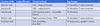
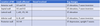
What is the difference between iron deficiency anemia and anemia of chronic disease?

A

In iron-deficiency anemia, the TIBC is higher than 400–450 mcg/dL because stores are low. In anemia of chronic disease, the TIBC is usually below normal because the iron stores are elevated

A total iron-binding capacity (TIBC) test measures the blood’s ability to attach itself to iron and transport it around the body. A transferrin test is similar. If you have iron deficiency (a lack of iron in your blood), your iron level will be low but your TIBC will be high.

17
Q

What is sideroblastic anaemia?

Causes?

A

•Rare

Definition: Anaemia due to defective heam synthesis within the mitochondria.

Causes:

  • Lead poisoning
  • Vitamin B6 deficiency
  • Congenital: X-linked recessive enzyme deficiency
  • Chronic Alcoholism
18
Q

Signs in normocytic anaemia

think of 3As and 2Hs

A
  • A – Acute blood loss
  • A – Anaemia of Chronic Disease
  • A – Aplastic Anaemia
  • H – Haemolytic Anaemia
  • H – Hypothyroidism (can also be macrocytic)
19
Q

What happens in normocytic anaemia?

A
  • Normocytic anaemias: Either increased destruction of RBCs or reduced production of RBCs.
  • Reticular count is used to distinguish the two
  • Reduced production – reticular count is low (failure of bone marrow )
  • Increased destruction – Bone marrow is working fine – compensation from haemolysis so increased reticulocytes
20
Q

What is hereditary spherocytosis?

A
  • Inherited defect in the RBC membrane proteins leading to a haemolytic anaemia
  • Usually autosomal dominant
  • Extra features:
  • Neonatal jaundice, splenomegaly, gallstones
  • Ix: Increased reticulocytes
21
Q

What is sickle cell disease?

A
  • Autosomal recessive mutation in the Hb beta chain at position 6 (glutamic acid to valine)
  • 2 alleles code for the beta chain
  • Sickle cell disease: 2 abnormal alleles
  • Sickle cell trait: 1 abnormal allels
  • Sickle cell crisis: Acute manifestation of sickle cell disease
22
Q

Patho of sickle cell?

A
  • HBS: Crystallises and is insoluble at low PaO2
  • This causes sickling of the RBC
  • Extra features: jaundice, failure to thrive, frontal bossing
23
Q

What is G6PD deficiency?

A
  • Inherited X-linked deficiency resulting in a haemolytic anaemia.
  • Results in a reduced half-life of RBCs (<120days)
  • G6PD is required by cells to prevent them from damage by oxidation.
  • Red cells are under constant stress by oxidants as oxyhaemoglobin is converted to deoxyhaemoglobin

G6PD deficiency is an inherited condition. It is when the body doesn’t have enough of an enzyme called G6PD (glucose-6-phosphate dehydrogenase). This enzyme helps red blood cells work correctly. A lack of this enzyme can cause hemolytic anemia. This is when the red blood cells break down faster than they are made.

24
Q

What is autoimmune haemolytic anaemia?

A
  • Antibody mediated destruction of RBCs
  • Divided into cold or warm depending on what temperature the antibodies bind to RBCs the best.
  • Warm: IgG binds to RBCs; Cold: IgM binds to RBCs.
  • Associated with Autoimmune conditions, infections and drugs
  • Causes extravascular haemolysis (warm) and extra/intravascular haemolysis (cold)
  • Warm: SLE, CLL, Drugs e.g. penicillin
  • Cold: M.pneumonia, EBV
  • Warm vs cold? Coombs test (normally direct)
25
What is the b12 deficiency?
* B12 is absorbed in the ileum bound to intrinsic factor (which made by parietal cells in the stomach) * Pernicious anaemia is an autoimmune reaction against intrinsic factor and parietal cells causing a B12 deficiency. Most common cause in the western world.
26
Risk factors of b21 deficiency?
* Increased age * Vegan Diet (11% on a vegan diet) * Gastric surgery * Malabsorption * Drugs e.g. metformin
27
Folate deficiency RF? Drugs?
* Reduced levels of folic acid * RF: * Increasing age * Malabsorption * Pregnancy * Alcohol * Drugs: e.g. methotrexate, trimethoprim * Folate is absorbed in the jejunum
28
What is Heart Failure?
Characterised by the heart's inability to effectively fill and/or eject (pump) blood, caused by an underlying pathology commonly coronary artery disease or previous MI.
29
Heart Failure can be described as:
- Heart failure with reduced ejection fraction – HFrEF – where there is a pump dysfunction - Heart failure with preserved ejection fraction – HFpEF – due to a filling dysfunction
30
What is the types of heart failure?
biventricular heart failure, left sided heart failure, right sided heart failure and cor pulmonale
31
What is left sided heart failure?
uChange in function of the left ventricle resulting in decreased cardiac output, pulmonary congestion and decreased peripheral perfusion uCan be either with a reduced ejection fraction or preserved ejection fraction
32
What are the risk factors for left heart failure?
- Coronary artery disease → decreases blood supply to cardiomyocytes → decreases contractile strength - Hypertension, aortic stenosis → ↑afterload → ↓stroke volume - Arrhythmias decrease ventricular filling and lead to ineffective contractions - Mitral or aortic regurgitation - Age, Obesity, Diabetes, Smoking, Ethansol, Illicit drug
33
Pathology of left sided failure?
- Underlying disease results in a decrease in the cardiac output results in backup of blood into left atrium, pushed back into pulmonary circulation → ↑pulmonary capillary pressure → pulmonary oedema → ↑gas exchange - RAAS activation due to ↓cardiac output. Angiotensin II production → systemic effects, increasing blood pressure, cardiac hypertrophy, decreased diuresis
34
Consequences and complications for left heart failure?
Effects of compensation: ↑afterload, ↑LV workload, LV remodelling Pulmonary oedema Pulmonary haemorrhage (congested capillaries bursting) Cardiorenal syndrome Death
35
Signs and symptoms for left sided heart failure
uLeft sided heart failure can present as uExertional dyspnoea and can be on rest uOrthopnoea uParoxysmal nocturnal dyspnoea (PND) uCough, restlessness uBibasal crackles on auscultation of lungs – pulmonary oedema uS3/S4 sound
36
What is righ sided heart failure and what causes it?
- Change in function of the right ventricle leading to decreased delivery of blood to the pulmonary circulation and elevated venous pressure - ↑venous pressure leads to systolic volume overload - ↑Right ventricular workload → RV hypertrophy → ↓pumping ability **Caused** by left sided heart failure and the associated pulmonary oedema (most common cause). **Other causes** include RV infarct, bacterial endocarditis, pulmonary valve stenosis, cardiomyopathy.
37
Signs and symptoms of right sided heart failure?
- Peripheral pitting oedema - Fatigue/exercise intolerance – due to poor gas exchange - Hepatojugular reflux - Ascites - Hepatosplenomegaly - S3/S4 sounds
38
Complications of right sided heart failure?
- Failure of left side of heart - Tricuspid regurgitation - Congestive hepatopathy – liver damage due to backup of blood - Cardiac cachexia – nausea, vomiting, anorexia, abdo pain - Eventual death
39
What is Cor Pulmonale?
**Pulmonary Heart Disease** - Right heart failure secondary to pulmonary arterial hypertension - Right ventricular hypertrophy/dysfunction caused by pulmonary hypertension - Presents with signs and symptoms of right sided heart failure
40
41
What are the Diagnostic findings in Heart Failure?
- Elevated BNP/NT-pro-BNP – raised due to stretching of ventricular wall - Chest X-Ray - In Left Heart Failure & biventricular failure – cardiomegaly, pulmonary vascular congestion, Kerley B lines - Echocardiogram - ECG abnormalities – arrhythmias, tall QRS
42
Management for heart failure
- Beta blockers have an evidence base to reduce mortality. (e.g. bisoprolol) - ACE inhibitors (e.g. ramipril) and ARBs (e.g. candesartan) are also used - In fluid overload a loop diuretic can be prescribed e.g. furosemide
43
What is New York Heart Association – Classes of Heart Failure
44
What is compensated and decompensated HF?
•If the dilated ventricle (as seen in systolic left sided HF) is able to maintain cardiac output by this means, the patient is said to be in **compensated heart failure**. However, ventricular dilation comes at the expense of increased wall tension and amplifies the oxygen requirements of an already-compromised myocardium. With time, the failing muscle is no longer able to propel sufficient blood to meet the needs of the body, and the patient develops **decompensated heart failure**.
45
What is cardiomyopathy?
Cardiomyopathy refers to diseases of the heart muscle. These diseases have many causes, signs and symptoms as well as treatments. In most cases, cardiomyopathy causes the heart muscle to become enlarged, thick or rigid.
46
Types of cardiomyopathies
* Hypertrophic Cardiomyopathy * Dilated Cardiomyopathy * Restrictive Cardiomyopathy * Arrhythmogenic Right Ventricular Cardiomyopathy * Takatsubo Cardiomyopathy
47
What is Primary and Secondary Cardiomyopathy?
* When cardiomyopathy develops as a way to compensate for some other underlying disease like hypertension or valve disease its called secondary cardiomyopathy. * If it develops all by itself its called primary cardiomyopathy.
48
What is Hypertrophic Cardiomyopathy?
* Basics: When the walls of the heart become thick, heavy and hypercontractile, essentially the muscle grows a lot larger – and new sarcomeres are added in parallel * Usually LV is affected + muscle growth is asymmetrical meaning that the IV septum grows larger in comparison to the free wall and this causes 2 things: - Take up more room so less blood in the ventricle - More stiffness and less compliant and hence it cant stretch as much – cant fill as much leading to what type of HF?
49
Hypertrophic Cardiomyopathy ## Footnote * In some patient the LV outflow is blocked by the outgrowth of IV septum during \_\_\_\_\_\_\_– Increased blood velocity through smaller aortic valve and this pulls the _____ leaflet of the _____ valve towards the septum (VENTURI EFFECT) causing further obstruction * This is also referred to as _________ \_\_\_\_\_\_\_ \_\_\_\_\_\_\_\_\_ * Causes a ________ \_\_\_\_\_\_\_\_\_ Murmur (like AV valve stenosis) * Bifid Pulse from the mitral valve * S\_ Sound can be heard of atrial depolarization – pushing blood into a non-compliant ventricle during diastole
* In some patient the LV outflow is blocked by the outgrowth of IV septum during **systole** – Increased blood velocity through smaller aortic valve and this pulls the **anterior** leaflet of the **mitral** valve towards the septum (VENTURI EFFECT) causing further obstruction * This is also referred to as **Hypertrophic Obstructive Cardiomyopathy** * Causes a **Cresendo-Decrecendo** Murmur (like AV valve stenosis) * Bifid Pulse from the mitral valve * S**4** Sound can be heard of atrial depolarization – pushing blood into a non-compliant ventricle during diastole
50
Causes of Hypertrophic Cardiomyopathy
* Genetic: Autosomal Dominant: Missense mutation in the Beta Myosin Gene * Freidriches Ataxia: Autosomal Recessive * On Histology: Myocyte Disarray aka Myocytes organization is haywire
51
What is Dilated Cardiomyopathy (MC type)
* Causes all **4 chambers to dilate** * New sarcomeres are added in series causes the muscles to become thin and there is less muscle present for contraction causing weak contractions * This causes reduced stroke volume and leads to **Systolic Heart Failure** * As chambers get larger they tend to stretch out the valves that separate the atrium and ventricles * This causes valves to not close all the way leading to regurgitation (Mitral and tricuspid: Holosystolic murmur) * Arrhythmia complication: stretching of conduction system
52
Dilated Cardiomyopathy : Causes
\> Primary : idiopathic: the most common cause \> inherited: either a familial genetic predisposition to DCM or a specific syndrome e.g. Duchenne muscular dystrophy, or haemochromatosis, sarcoidosis \> Myocarditis: e.g. Coxsackie B, HIV, diphtheria, Chagas disease(protozoan) \> Alcohol is toxigenic \> Drugs like chemotherapy: Daunorubicin and cocaine \> Wet beriberi decreases B1 \> Peripartum cardiomyopathy where DCM can develop in the 3rd trimester of pregnancy or in weeks following pregnancy probably because of of pregnancy associated hypertension.
53
What is Restrictive Cardiomyopathy
* Is where the heart muscle is restricted meaning it becomes stiffer and less compliant * The muscles and size of ventricles stay about the same size or are maybe only slightly enlarged * When blood fills into restricted ventricles, the ventricles aren't allowed to expand.. because they cant stretch .. Less filling ... less pumped out causes diastolic HF
54
Diagnosis of Restrictive Cardiomyopathy
using ECG: smaller QRS complexes, low amplitude signals, Echo
55
Restrictive Cardiomyopathy Causes
Causes include amyloidosis, sarcoidosis, hemochromatosis, endocardial fibro elastosis (children), and Loeffler syndrome (endomyocardial fibrosis with an eosinophilic infiltrate and eosinophilia).
56
57
What is an Abdominal Aortic Aneurysms (AAA)? Causes?
■ An aneurysm is a weakening/thinning of the arterial wall leading to a localized bulging. Definition of AAA: A permanent, localised dilatation in the wall of the abdominal aorta. **Cause**: Primarily from the loss of intima with loss of elastic fibres from the media
58
Risk factors of AAA
o Arterial disease - atherosclerosis - hypertension - diabetes - smokers oConnective tissue disorders - Marfan’s syndrome, Ehler-Danlos syndrome - Extracellular matrix becomes disrupted with changes in the balance of collagen and elastic fibres o Age – over 65 years o Male sex – 3 to 4 times more prevalent o Family history – especially in male first degree relatives
59
Presentation for AAA:
1) Asymptomatic pulsatile abdominal mass (normally diagnosed incidentally) 2) Detected during USS Routine Screening in men over age 65 3) Emergency (rupture) – Sudden onset, severe chest/abdominal pain that radiates to the back – Collapse – Hypotension with tachycardia – Profound anaemia
60
Ix of AAA
– History and examination - examination may identify a expansile, pulsating mass. – Ultrasound - To look at the diameter of the abdominal aorta. – (GOLD STANDARD) CT Angiography - Used preoperatively to check for renal artery involvement and for planning surgery.
61
Management for AAA
o \<5.5 cm on ultrasound - Continued monitoring with ultrasounds o \>5.5 cm on ultrasound - Elective surgery to prevent rupture. o Emergency - Oxygen, 2 large-bore cannulas, fluid management (BP at 100mmHg to prevent dislodging a clot), surgical aneurysm repair **Prognosis:** 80% mortality for ruptured AAA
62
What are the surgical management options for AAA
■ Clamping below the renal artery, then sewing a graft into the aneurysm sac ■ Endovascular Aneurysm Repair (EVAR) - less invasive endoscopic procedure whereby stents are inserted through the femoral artery
63
What is an aortic dissection?
Definition: Blood escapes through the innermost tunica intima layer of the aorta wall, prising apart the media and creating a new lumen
64
Presentation for aortic dissection
- Acute severe tearing or ripping chest pain, radiating to the back - Weak or absent pulses - Systolic BP variation of \>20mmHg between the arms - Hypertension - Aortic regurgitation
65
Ix for Aortic Dissection
Chest X-ray - Widened mediastinum CT Angiograph of Chest-Abdo-Pelvis (For STABLE patients) - False lumen
66
What are the classifications for aortic dissection?
DEBAKEY STANDFORD
67
What is the Debakey classification for aortic dissection?
■ DEBAKEY Classification: * Type 1 - originates in ascending aorta, propagates to at least the aortic arch and possibly beyond it distally * Type 2 - originates in and is confined to the ascending aorta * Type 3 - originates in descending aorta, rarely extends proximally but will extend distally
68
WHat is the standford classification for Aortic Dissection?
* Type A - Involves the ascending aorta (+/- the descending aorta). 2/3rds of cases. * Type B - Involves only the descending aorta. 1/3rd of cases.
69
Management for aortic dissection
To prevent it becoming a AAA. * Type A - Surgery – open surgery or endovascular stent graft repair + control systolic at 100-120mmHg while waiting for intervention * Type B – If no complications, conservative management of bed rest + IV labetalol to reduce BP
70
What is a DVT?
Deep Vein Thrombosis ■ Deep venous thrombosis (DVT) affects the deep veins in the lower leg and the thigh, most commonly popliteal vein, femoral vein and iliac veins
71
Risk factoes fro DVT?
* Obesity * Age * Smoking * female sex * family history * previous DVT and co-occurring conditions (pregnancy or pelvic tumour (causing venous stasis) * Malignancy * pelvic/lower limb orthopaedic surgery * thromboembolic disease (e.g. hypercoagulability)
72
What is Virchows traid of clotting predisposing patient to thrombus formation:
1. Venous stasis: injury or inflammation, air travel 2. Endothelial damage: surgery, malignancy, trauma 3. Raised clotting factors: malignancy, congenital disease, drugs, thrombogenic state
73
Presentation of DVT
o sudden-onset of localised pain o calf tenderness o Asymmetric swelling, redness and tight skin o superficial veins may be engorged o may be a low-grade pyrexia o Some patients are asymptomatic and collapse from a pulmonary embolism o variable oedema in and below the calf and Homan’s sign my be positive
74
Ix for DVT
o Wells’ Score to calculate risk of DVT o D-dimer testing - If it is negative- DVT is highly unlikely (High sensitivity) - If it is positive- it DOES NOT CONFIRM DVT (may be raised in patients with liver disease, rheumatoid disease, inflammation, cancer, trauma, pregnancy, recent surgery etc) o proximal duplex ultrasound o whole-leg ultrasound
75
Management for DVT
§ Low-molecular-weight heparin (LMWH) is initially used § Subsequently converted to warfarin therapy - Warfarin is continued for 3–6 months after a first DVT but is needed lifelong after a second.
76
Carotid Stenosis (Carotid Artery Disease) What is it?
* Carotid Stenosis occurs when plaque (atherosclerosis) build up and narrow the carotid artery, reducing blood flow to the brain. The blockage increases your risk of stroke. * Occlusion or stenosis of the internal carotids is very serious and can lead to a CVA * Atherosclerosis occurs most commonly at the carotid artery bifurcation
77
What are the three ways carotid stenosis increase risk of stroke?
1. Plaque deposits grows larger and larger, severely narrowing/completely occlude the artery à reduced blood flow to brain 2. Plaque deposits can roughen and deform artery wall à causes blood clots to form and block blood flow to brain 3. Plaque deposits rupture and tiny clots break away from repair, travel downstream to clog smaller arteries à blocks blood flow to brain
78
RF for Cartoid Stenosis
Older age PMHx of HTN PMHx of CVD Smoking Diabetes
79
Presentation for carotid stenosis
o Asymptomatic o Cervical bruit o Focal neurological deficit lasting \>24hrs (i.e. stroke) o Focal neurological deficit lasting \<24hrs (i.e. TIA)
80
Ix for carotid stenosis
* BP, glucose, cholesterol, cross-match for blood * Duplex USS – for degree of stenosis * CT Angiography - defines arterial anatomy * MRI Angiography - identifies cerebral infarcts
81
Management for carotid stenosis
Conservative and medical management for asymptomatic patients with less than 70% stenosis: - stop smoking - take aspirin and statin - control hypertension
82
What is a TIA
Transient Ischaemic Attack ■ Definition: A transient episode of neurologic dysfunction caused by focal brain, spinal cord, or retinal ischaemia, without acute infarction. ■ aka ”mini-stroke” – TIAs have a sudden onset and may last from a few mins to 24hrs; most patients have complete resolution of symptoms and signs within 1hr
83
Pathology for TIA
TIAs result when blood flow to the brain is temporarily interrupted and then restored.
84
RF for TIA
Carotid Stenosis - Atrial fibrillation - Valvular disease
85
What are the key diagnostic factors for TIA:
■ Sudden onset ■ Brief duration of symptoms (minutes to 24hrs max) ■ Patient/witness reports of focal neurological deficit - Dysphasia - Unilateral weakness or paralysis - Ataxia, vertigo or loss of balance - Sudden transient loss of vision in one eye
86
Assessment of TIA:
ABCD2 Prognostic Score: Clinical prediction rule used to determine the risk for stroke in the days following a transient ischemic attack
87
Ix for TIA
* Blood glucose * FBC and Platelet count * Prothrombin time, INR * Fasting lipid profile * CT Scan * (GOLD STANDARD) MRI of Head
88
Management for TIA
89
What is Atherosclerosis?
* Arteriosclerosis= Atherosclerosis + Arteriolosclerosis * Multifactorial inflammatory disease of the tunica intima * Buildup of cholesterol plaques in the intima * Cholesterol +WBC * M\>F
90
What are the common sites for arteriosclerosis?
: Abdo CramP: Abdominal artery\> Coronary\> Popliteal\> Carotid\> Circle of Willis
91
What are the modifiable and nonmodifiable atherosclerosis?
_Modifiable_ * Smoking * DM * HTN * Dyslipidemia * Alcohol _Nonmodifiable_ * FH * Age: M\>45, F\>55 * Postmenopausal females
92
Basic principkes of atherosclerosis
* NO\> vasodilation via activation of eNOS -\> stimulation of guanylate cyclase and cGMP production-\> decreasing intracellular Ca2+ decreasing myocyte excitation and contraction. * LDL: transports cholesterol and Ca2+ INTO cell * HDL: transports cholesterol and Ca2+ OUT of cell * In ACS: Adenosine is released by ischaemic myocardium through A1 receptors-\>angina-\> PAIN
93
Atherosclerosis Stage 4
* Fatty streak is **thrombogenic**-\> platelets adhesion -\> PDGF, PGF, TGF-B secretion -\> SMC proliferation and migration of SMC from tunica media into tunica intima * SMC + macrophages secrete ECM ( elastin, collagen, proteoglycan)-\> form a wall around the fatty streak to prevent clotting-\> fibrous cap * LDL-\> calcium depostion * Endothelial cell injury prevents HDL from removing the calcium-\> **stiff fibrous cap**
94
Atherosclerosis Stage 1
* Endothelium becomes dysfunctional * Cause: Chronic stress due to smoking, high BP, hyperlipidemia * Chronic stress causes damage to the glycocalyx barrier which monitors the shear forces in the blood vessels * Permanent hyperglycaemia can destroy the glycocalyx. Regeneration takes 12 hrs. * Glycocalyx dysfunction-\> leaky vessel
95
Complications of atherosclerosis
**•Obstruction:** - 40% luminal obstruction: maximal flow during exercise maintained - \>50% -\> coronary ischemia - \> 70/75% lumen occluded prior to onset of symptoms -\> Downstream cellular injury/death * **Coronary arteries**-\> Angina+ ACS * **Internal carotid+ Middle cerebral-**\> Stroke + cerebral artery * **Mesenteric arteries**-\> acute/chronic mesenteric ischemia * **Popliteal artery**-\> peripheral ischemia ( gangrene + claudication) * **Renal artery**-\> Hypertension ( activation of RAAS) * **Weakening of vessel wall**-\> Abdominal aortic aneurysm below L2 ( no vaso vasorum) -\> hemorrhaging * **Thromboembolism, Cholesterol emboli**-\> livedo reticularis, AKI, gangrene
96
Atherosclerosis Stage 2
* Endothelial damage-\> LDL enters * Excess LDL-\> increases permeability of cells to enter the intimal layer-\> monocytes enter * Monocytes enter through diapedesis * LDL oxidizes and inactivates NO! * NO loses its vasoprotective effect **•-\> ACCUMULATION OF MACROPHAGES + oxLDL**
97
Atherosclerosis Stage 3
* oxLDL releases cholesterol * Monocytes engulf cholesterol-\> die off and form foam cells * Accumulation of foam cells-\> fatty streak
98
Atherosclerosis Stage 6
* BURST: * Foam cells undergo necrosis-\> release of MMPs * IFN-γ induces macrophage MMP expression-\> weakening of cap * IFN-γ inhibits VSMC proliferation and collagen synthesis-\> weakening of cap * Plaque rupture-\> exposure of underlying-\> thrombus formation
99
Atherosclerosis Stage 5
* Atheroma is formed * Grows * \*\*The thinner the fibrous cap, the higher risk of thrombus\*\*
100
Investigations for athersclerosis
•Bloods: - Lipid profile: TC, LDL, HDL, triglycerides - Fasting glucose - FBC - Creatinine - Myocardial damage markers: troponins, CK-MB ( ACS suspected) - Homocysteine - HbA1c - BNP - Thyroid fxn * ECG-\> ACS, underlying hypertrophy * Stress testing unless contraindicated * Echo-\> Valvular heart disease, HF * CT-\> extent of calcification * Coronary angiography
101
Treatment for Atherosclerosis
**_Lifestyle:_** * Smoking cessation * Weight loss **_Underlying comorbidity managed- HTN, DM_** •DM and renal disease treatment goal of BP\< 130/80 mmHg **_Medical:_** * Low dose aspirin, clopidogrel * Statin therapy
102
What is CAD?
Coronary Artery Disease ## Footnote * Disease due to imbalance between myocardial oxygen demand and supply from coronary arteries * Reduced O2 supply to the heart is defined as myocardial ischemia-\> reduced ability of heart to contract * If prolonged ischemia -\> myocardial infarction
103
What causes CAD?
* Atherosclerosis\*\* * Coronary artery embolus: FAT BAT. Classic triad of fat emboli? * Vasculitis * Vasospasm * Aortic stenosis
104
Presentations for CAD
Stable angina, Prinzmetal angina, ACS, sudden cardiac death
105
What is Stable Angina
* Myocardial ischemia due to a plaque occluding \>75% of the coronary artery lumen. * Relieved by rest, nitroglycerin/GTN spray
106
Clinical Features of stable angina
* Deep/poorly localized pain * Squeezing/ crushing/suffocating retrosternal pain * Radiates to the arm, jaw, neck * SOB, nausea, vomiting, diaphoresis, fatigue, dizziness
107
Investigations of stable angina?
•ECG normal, troponins normal, cardiac stress test +
108
A plaque can cause near-total occlusion of the CA but individuals may not develop an infarction- TRUE/FALSE
TRUE- this is called a collateral circulation
109
what is the criteria for stable angina?
1. Substernal chest discomfort 2. Provoked by exertion, stress 3. Relieved by GTN, rest * If all three met-\> typical angina * If 2-\> atypical angina * If 0,1-\> non-cardiac chest pain
110
What is prinzmetal angina?
* vasospasm of a large coronary artery * Transmural ischemia, rest pain, more prolonged than classic angina * ECG: ST elevation * Troponins normal * Women \<50
111
What triggers prinzmetal angina?
* Smoking * Electrolyte disturbance * Cocaine * Cold stimulation
112
Other angina variants:
Angina equivalent syndrome, syndrome X, silent ischemia, nocturnal
113
What is ACS
Acute Coronary Syndrome = plaque disrupted Involves: Unstable angina, NSTEMI, STEMI
114
Clinical features of ACS
* New- onset angina * Acute retrosternal chest pain * Dull/ squeezing/ crushing/ tightness * Radiation to neck, arm, shoulder, epigastrium * Dyspnea * Pallor * Nausea, vomiting * Diaphoresis * Dizziness, lightheadedness, syncope * Silent MI?
115
What are the signs for cardiac shock?
* Tachy/bradycardia * Hypotension * Raised JVP * Murmur due to arrhythmias * Cold extremities
116
ACSyndrome is classed by:
Defined as 2 out of 3: 1. Unstable, ischaemic chest pain 2. Ischaemic ECG changes 3. Raised cardiac biomarkers
117
Comparisons Unstable angina, NSTEMI, STEMI
119
What is the thinking you do if a patient has chest pain?
120
121
What is digoxin used for?
Digoxin is a type of medicine called a cardiac glycoside. It's used to control some heart problems, such as irregular heartbeats (arrhythmias) including atrial fibrillation. It can also help to manage the symptoms of heart failure, usually with other medicines. Digoxin is only available on prescription
122
123
124
125
127
129
What is the blood supply to: SAN: AVN: Bundle of His: LBB: RBB:
* SAN: Usually RCA * AVN: Usually RCA * Bundle of His: RCA + branches of LAD * LBB: LAD. May have collaterals form RCA/LCX - Anterior fascicle: LAD - Posterior fascicle: RCA and may have septal branches of the LAD. •RBB: septal branches of LAD. May have collaterals form RCA/LCX
131
When does S1,2,3,4 occur in the Cardiac cycle
135
It shouldw be remembered that a new left bundle branch block (LBBB) may point towards ___ \_\_\_\_\_\_ \_\_\_\_\_
It shouldw be remembered that a new left bundle branch block (LBBB) may point towards Acute Coronary Syndrome
137
What are: Preload Afterload End diastolic volume (EDV) Ejection systolic volume (ESV)
**Preload-** Volume of blood in ventricles at end of diastole (end diastolic pressure) **Afterload**- Resistance left venricle must overcome to circulate blood **End diastolic volume (EDV)-** amount of blood in the ventricle before contraction **Ejection systolic volume (ESV)-** amount of blood remaining in the heart after ejection
138
Normal heart sounds S1 S2
**•S1- Closure of the AV valves** * At end of phase 1/ beginning of phase 2 * ‘lub’ **•S2-Closure of SL valves** * At end of phase 3/ beginning of phase 4 * ‘dub’
139
Abnorma heart sounds S3 S4
**S3** * AKA ventricular gallop * Occurs just after S2 when the mitral valve opens, allowing for filling of left ventricle * In rapid ventricular filling * Sound is ONLY produced by a **large amount of blood (volume overload) striking overly compliant left ventricle** * Often a sign of systolic heart failure Eg: dilated cardiomyopathy **S4** * AKA atrial gallop * Occurs just before S1 when the atria contract to force blood into left ventricle * Sound is produced ONLY when **left ventricle is not complaint and atrial contraction forces against the wall** * Can be a sign of diastolic heart failure * Eg restrictive and hypertrophic cardiomyopathy •
141
Atrial systole accounts for most of the ventricular filling. True or False?
False
142
What do you expect at the beginning of ventricular contraction? (valves)
AV valves close
143
What does stenosis mean in relation to valves?
* Stenosis = failure of the valve to open completely * Usually chronic and thus well-tolerated due to ventricular hypertrophy or atrial hypertrophy * Causes pressure overload * Diastolic HF: * As the heart fails to fill properly during diastole due to the decreased space in the ventricle
144
What does regurgitation mean in relation to valves
* Insufficiency or regurgitation = failure of a valve to close completely, thus causing reversed flow * Can be acute or chronic * Causes volume overload * Systolic HF: because the ventricles dilate, the walls thin out and there is no proper contraction
145
What is a murmur?
abnormal flow through diseased vavles typically produces abnormal heart sounds ## Footnote * Severe lesions can be palpated as thrills * Depending on the valve involved, murmurs are best heard at different locations on the chest wall
146
•How to describe a murmur?
You look at 1) Timing- systolic or diastolic? 2) Shape? 3) Location? Radiation? 4) Pitch 5) Intensity-Grading?
147
What does the timing of murmurs mean?
* Refers to whether the murmur is systolic or diastolic * Systole occurs between S1 and S2 * Diastole occurs between S2 and S1
148
* A murmur is heard when the diseased valve interrupts the flow of blood during systole or diastole à therefore: * AV regurgitation or semilunar stenosis --\> * AV stenosis or semilunar regurgitation --\>
* A murmur is heard when the diseased valve interrupts the flow of blood during systole or diastole à therefore: * AV regurgitation or semilunar stenosis --\> **systolic murmur** * AV stenosis or semilunar regurgitation --\> **diastolic murmur**
149
Normally: during systole AV closed SL open Problems causing systolic murmurs
Av doesnt close = AV reg SL doesnt open= SL stenosis
150
Normally: during diastole AV open SL close Problems causing diastolic murmurs:
AV doesnt open= AV stenosis SL doesnt close = SL regurg
151
Classification of **systolic** and diastolic murmurs * Mid-systolic murmur (AKA ejection murmur) * Holosystolic murmur * Late systolic murmur
**Mid-systolic murmur (AKA ejection murmur)** * Begins just after S1 heart sound and stops before S2 heart sound * Therefore, S1 and S2 are distinctly audible **Holosystolic murmur** * Begins with immediately after S1 heart sounds and extends up to S2 * S1 and S2 are difficult or impossible to hear **Late systolic murmur** •Starts after S1 and may or may not extend up to S2
152
Classification of systolic and **diastolic** murmurs * Early diastolic * Mid-diastolic * Late diastolic (AKA presystolic)
**Early diastolic** * Starts at the same time as S2 and ends before and ends before S1 * S1 will be distinctly audible and S2 may be difficult to hear **Mid-diastolic** * Starts after S2 and ends before S1 * Both S2 and S1 are distinctly audible **Late diastolic (AKA presystolic)** •Starts after S2 and extend up until S1
153
WHat does it mean by shape of murmurs
Shape refers to the change in intensity of the murmur over time: * Crescendo = progressively gets louder (increases in intensity) * Decrescendo = progressively gets quieter (decreases in intensity) * Crescendo-decrescendo = progressively get louder and then quieter - Has a “diamond shape” •Uniform = does not change in intensity
154
What are the location of murmurs
Location refers to where the murmur is usually heard best * Aortic valve – right sternal edge, 2nd intercostal space * Pulmonary valve – left sternal edge, 2nd intercostal space * Tricuspid valve – left sternal edge, 4th/5th intercostal space * Mitral valve – midclavicular line, 5th intercostal space
155
What does it mean by pitch in relation to valves?
Murmur will be high-pitched if there is a large pressure gradient across the pathological lesion •E.g. aortic stenosis à because there is usually a large pressure gradient between the left ventricular and aorta Murmur will be low-pitched because there is usually a small pressure gradient •E.g. mitral stenosis à because there is a lower pressure gradient between the left atrium and left ventricle during diastole
156
What can go wrong with the valves?
There are three main types: 1. Damage to collagen of the leaflets 2. Nodular calcification e.g. aortic valve stenosis 3. Fibrotic thickening (key feature in rheumatic heart disease)
157
What causes valvular problems?
What can cause problems? 1. Inflammation 2. Congenital HD 3. Underlying HD 4. Infections
158
Investigations for valvular heart disease?
•TTE (trans-thoracic echocardiogram) - Performed in all patients •TOE (trans-oesophageal echocardiogram) - Performed in 20% of patients * Stress echocardiogram = performed when the patient is exercising or by giving drugs to increase the heart rate, e.g. dobutamine * Catheterisation = angiogram is performed to assess the structure and function or coronary arteries
159
What is aortic stenosis? Causes?
* Narrowing of the aortic valve * Causes obstruction of outflow of blood from LV through aorta * Can cause LV hypertrophy -\>LV HeartFailure-\> diastolic HF Causes * Mechanical wear and tear * Bicuspid valve * Rheumatic HD * Willams syndrome * Pagets disease (+hypercalcemia)
160
Symptoms and consequences of **aortic stenosis**
symptoms * Syncope * Angina pectoris * Dyspnoea Consequences 1. LV hypertrophy 2. Increased oxygen demand of the heart 3. Diastolic HF (doesn’t fill properly) 4. Reduced SV-\> Compensatory TachyC-\> Impaired coronary filling 5. Eventually RHF
161
What is Aortic Regurgitation? Causes?
What happens? * Incomplete closure of aortic valve * Causes backflow of blood from aorta to LV * Can cause LV dialation -\>LV HF-\> systolic HF Causes * Bacteria infection (infective endocarditis) * Chronic Inflammation * Aortic root dilation
162
Symtoms and Consequences of **Aortic regurgitation**
Symptoms * Do not develop until LVF * Dyspnea on exertion * Orthopnea * Paroxysmal nocturnal dyspnea * Angina pectoris Consequences Acute -During diastole, blood flows back-\> Ventricular pressure increases-\> pressure transmits backwards into pulmonary circulation à pulmonary oedema and dyspnoea Chronic -Overtime, increased left ventricular end-diastolic volume à left ventricular dilation à LVF urgit
163
Whats is mitral stenosis? Causes?
What happens? * Narrowing of mitral valve * obstruction of blood flow into the left ventricle * Can cause LA hypertrophy Causes * Rheumatic fever * Calcification * Carcinoid * Congenital
164
Symptoms and Consequences of Mitrals stenosis?
Symptoms * Dyspnea on exertion * Orthopnea * Paroxysmal nocturnal dyspnea * Pulmonary congestion * Later-\> symptoms of RHF Consequences 1. Incr volume and pressure in LA 2. Dilation of LA 3. Backflow of blood into pulmonary circulation 4. Pulmonary congestion, edema 5. Increased pulmonary pressure 6. Eventually RHF as it finds it harder to pump
165
What happens in mitral regurgitation? Causes?
What happens? * incomplete closure of the mitral valve * Leakage of blood from the left ventricle into the left atrium * Can cause LA dialation -\>LV HF-\> systolic HF Causes? * Mitral valve prolapse * Heart attack * LV heart failure * Rheumatic fever
166
Symptoms and Consequences of mitral regurg
Symptoms? * Palpitation * Dyspnoea * orthopnoea * Fatigue and lethargy * Pulmonary congestion * Later-\> symptoms of RHF Consequences Acute 1. No dilation of LA-\> LA pressure increases-\> incr in pulmonary pressure-\> pulmonary edema 2. Backflow of blood into pulmonary circulation Chronic 1. Eccentric hypertrophy 2. left atrial dilation as it is accommodated
167
PULMONARY REGURG What happens? Causes?
**What happens?** * Incomplete closure of the pulmonary valve * backflow from the pulmonary artery to the right ventricle * Can cause LV hypertrophy -\>LV HF-\> diastolic HF **Causes?** Occurs secondary to pulmonary HTN Infective endocarditis Rheumatic endocarditis
168
What happens when there is high blood pressure
169
PULMONARY REGURG Symptoms Consequences
**Symptoms** * Due to backed up blood * Distended neck veins * Swelling in ankles and feet * Hepatosplenomegaly **Consequences** 1. Blood flow back into RV 2. Increased volume of blood 3. Eccentric hypertrophy-\> RV dilation 4. Systolic RHF
170
TRICUSPID REGURGITATION What happens? Causes?
**What happens?** * Incomplete closure of the tricuspid valve * Leakage of blood from the right ventricle into the right atrium * Can cause RA dilation -\>right sided HF due to the increase of volume and pressure **Causes?** * Pulmonary HTN * Heart attack * Congenital (ebstein abnormality) * Rheumatic fever
171
TRICUSPID REGURGITATION Symptoms Consequences
**Symptoms?** * Dyspnoea * Right-sided heart failure: * Distended neck veins * Swelling of ankles and feet * Hepatosplenomegaly * Ascites **Consequences** * Extra blood that flowed back into the atrium, flows back into the ventricle during diastole à increase in right ventricular pre-load * In order to deal with the extra work, the right ventricle becomes larger à eccentric ventricular hypertrophy * Eventually, the right ventricle might not be able to keep up à right sided-heart failure
172
TRICUSPID STENOSIS What happens? Causes?
**What happens?** * Narrowing of tricuspid valve * obstruction of blood flow into the right ventricle * Can cause RA hypertrophy **Causes** * Rheumatic fever
173
TRICUSPID STENOSIS Symptoms Consequences
**Symptoms?** * dysphagia * Right sided heart failure * Distended neck veins * Swelling of ankles and feet * Hepatosplenomegaly **Consequences** 1. Incr volume and pressure in RA 2. hypertrophy of RA 3. Backflow of blood into systemic circulation (vena cava) 4. Right sided HF
174
Pulmonary stenosis What happens? Causes?
**What happens?** * Narrowing of the pulmonary valve * Causes obstruction of outflow of blood from RV through pulmonary trunk * Can cause RV hypertrophy -\>RV HF-\> diastolic HF **Causes?** * Mechanical wear and tear * Rheumatic fever * Often congenital * Tetralogy of Fallot
175
Pulmonary Stenosis Symptoms and Consequences
**Symptoms** * Due to backed up blood Distended neck veins Swelling in ankles and feet Hepatosplenomegaly * Because blood cannot get to the lungs Cyanosis SOB and fatigue à especially during exercise **Consequences** 1. High pulmonary pressure 2. RV hypertrophy 3. Diastolic HF (doesn’t fill properly) 4. Blood gets backed up 5. Right HF
176
How do you calculate the mean arterial Pressure (MAP)
Diastolic Pressure+[1/3×(Systolic Pressure-Diastolic Pressure)] usually 93 mmHg MAP=Cardiac Output × Systemic Vascular Resistance Cardiac Output=Stroke Volume × Heart Rate
177
Regulatory Mechanisms for blood pressure Short term Long term
**Short term:** * Nervous System (baroreceptors) * Chemical (chemoreceptors) **Long term:** •Hormonal (RAAS)
179