CVS Diseases Flashcards

1
Q

What is Atherosclerosis?

A
  • Arteriosclerosis= Atherosclerosis + Arteriolosclerosis
  • Multifactorial inflammatory disease of the tunica intima
  • Buildup of cholesterol plaques in the intima
  • Cholesterol +WBC
  • M>F
2
Q

What are the common sites for arteriosclerosis?

A

: Abdo CramP:

Abdominal artery> Coronary> Popliteal> Carotid> Circle of Willis

3
Q

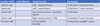
What are the modifiable and nonmodifiable atherosclerosis?

A

Modifiable

  • Smoking
  • DM
  • HTN
  • Dyslipidemia
  • Alcohol

Nonmodifiable

  • FH
  • Age: M>45, F>55
  • Postmenopausal females
4
Q

Basic principkes of atherosclerosis

A
  • NO> vasodilation via activation of eNOS -> stimulation of guanylate cyclase and cGMP production-> decreasing intracellular Ca2+ decreasing myocyte excitation and contraction.
  • LDL: transports cholesterol and Ca2+ INTO cell
  • HDL: transports cholesterol and Ca2+ OUT of cell
  • In ACS: Adenosine is released by ischaemic myocardium through A1 receptors->angina-> PAIN
5
Q

Atherosclerosis

Stage 1

A
  • Endothelium becomes dysfunctional
  • Cause: Chronic stress due to smoking, high BP, hyperlipidemia
  • Chronic stress causes damage to the glycocalyx barrier which monitors the shear forces in the blood vessels
  • Permanent hyperglycaemia can destroy the glycocalyx. Regeneration takes 12 hrs.
  • Glycocalyx dysfunction-> leaky vessel
6
Q

Atherosclerosis

Stage 2

A
  • Endothelial damage-> LDL enters
  • Excess LDL-> increases permeability of cells to enter the intimal layer-> monocytes enter
  • Monocytes enter through diapedesis
  • LDL oxidizes and inactivates NO!
  • NO loses its vasoprotective effect

•-> ACCUMULATION OF MACROPHAGES + oxLDL

7
Q

Atherosclerosis

Stage 3

A
  • oxLDL releases cholesterol
  • Monocytes engulf cholesterol-> die off and form foam cells
  • Accumulation of foam cells-> fatty streak
8
Q

Atherosclerosis

Stage 4

A
  • Fatty streak is thrombogenic-> platelets adhesion -> PDGF, PGF, TGF-B secretion -> SMC proliferation and migration of SMC from tunica media into tunica intima
  • SMC + macrophages secrete ECM ( elastin, collagen, proteoglycan)-> form a wall around the fatty streak to prevent clotting-> fibrous cap
  • LDL-> calcium depostion
  • Endothelial cell injury prevents HDL from removing the calcium-> stiff fibrous cap
9
Q

Atherosclerosis

Stage 5

A
  • Atheroma is formed
  • Grows
  • **The thinner the fibrous cap, the higher risk of thrombus**
10
Q

Atherosclerosis

Stage 6

A
  • BURST:
  • Foam cells undergo necrosis-> release of MMPs
  • IFN-γ induces macrophage MMP expression-> weakening of cap
  • IFN-γ inhibits VSMC proliferation and collagen synthesis-> weakening of cap
  • Plaque rupture-> exposure of underlying-> thrombus formation
11
Q

Complications of atherosclerosis

A

•Obstruction:

  • 40% luminal obstruction: maximal flow during exercise maintained
  • >50% -> coronary ischemia
  • > 70/75% lumen occluded prior to onset of symptoms -> Downstream cellular injury/death
  • Coronary arteries-> Angina+ ACS
  • Internal carotid+ Middle cerebral-> Stroke + cerebral artery
  • Mesenteric arteries-> acute/chronic mesenteric ischemia
  • Popliteal artery-> peripheral ischemia ( gangrene + claudication)
  • Renal artery-> Hypertension ( activation of RAAS)
  • Weakening of vessel wall-> Abdominal aortic aneurysm below L2 ( no vaso vasorum) -> hemorrhaging
  • Thromboembolism, Cholesterol emboli-> livedo reticularis, AKI, gangrene
12
Q

Investigations for athersclerosis

A

•Bloods:

  • Lipid profile: TC, LDL, HDL, triglycerides
  • Fasting glucose
  • FBC
  • Creatinine
  • Myocardial damage markers: troponins, CK-MB ( ACS suspected)
  • Homocysteine
  • HbA1c
  • BNP
  • Thyroid fxn
  • ECG-> ACS, underlying hypertrophy
  • Stress testing unless contraindicated
  • Echo-> Valvular heart disease, HF
  • CT-> extent of calcification
  • Coronary angiography
13
Q

Treatment for Atherosclerosis

A

Lifestyle:

  • Smoking cessation
  • Weight loss

Underlying comorbidity managed- HTN, DM

•DM and renal disease treatment goal of BP< 130/80 mmHg

Medical:

  • Low dose aspirin, clopidogrel
  • Statin therapy
14
Q

What is CAD?

A

Coronary Artery Disease

  • Disease due to imbalance between myocardial oxygen demand and supply from coronary arteries
  • Reduced O2 supply to the heart is defined as myocardial ischemia-> reduced ability of heart to contract
  • If prolonged ischemia -> myocardial infarction
15
Q

What causes CAD?

A
  • Atherosclerosis**
  • Coronary artery embolus: FAT BAT. Classic triad of fat emboli?
  • Vasculitis
  • Vasospasm
  • Aortic stenosis
16
Q

Presentations for CAD

A

Stable angina, Prinzmetal angina, ACS, sudden cardiac death

17
Q

What is Stable Angina

A
  • Myocardial ischemia due to a plaque occluding >75% of the coronary artery lumen.
  • Relieved by rest, nitroglycerin/GTN spray
18
Q

Clinical Features of stable angina

A
  • Deep/poorly localized pain
  • Squeezing/ crushing/suffocating retrosternal pain
  • Radiates to the arm, jaw, neck
  • SOB, nausea, vomiting, diaphoresis, fatigue, dizziness
19
Q

Investigations of stable angina?

A

•ECG normal, troponins normal, cardiac stress test +

20
Q

A plaque can cause near-total occlusion of the CA but individuals may not develop an infarction-

TRUE/FALSE

A

TRUE- this is called a collateral circulation

21
Q

what is the criteria for stable angina?

A
  1. Substernal chest discomfort
  2. Provoked by exertion, stress
  3. Relieved by GTN, rest
  • If all three met-> typical angina
  • If 2-> atypical angina
  • If 0,1-> non-cardiac chest pain
22
Q

What is prinzmetal angina?

A
  • vasospasm of a large coronary artery
  • Transmural ischemia, rest pain, more prolonged than classic angina
  • ECG: ST elevation
  • Troponins normal
  • Women <50
23
Q

What triggers prinzmetal angina?

A
  • Smoking
  • Electrolyte disturbance
  • Cocaine
  • Cold stimulation
24
Q

Other angina variants:

A

Angina equivalent syndrome, syndrome X, silent ischemia, nocturnal

25
What is ACS
Acute Coronary Syndrome = plaque disrupted Involves: Unstable angina, NSTEMI, STEMI
26
Clinical features of ACS
* New- onset angina * Acute retrosternal chest pain * Dull/ squeezing/ crushing/ tightness * Radiation to neck, arm, shoulder, epigastrium * Dyspnea * Pallor * Nausea, vomiting * Diaphoresis * Dizziness, lightheadedness, syncope * Silent MI?
27
What are the signs for cardiac shock?
* Tachy/bradycardia * Hypotension * Raised JVP * Murmur due to arrhythmias * Cold extremities
28
ACSyndrome is classed by:
Defined as 2 out of 3: 1. Unstable, ischaemic chest pain 2. Ischaemic ECG changes 3. Raised cardiac biomarkers
29
Comparisons Unstable angina, NSTEMI, STEMI
30
What is digoxin used for?
Digoxin is a type of medicine called a cardiac glycoside. It's used to control some heart problems, such as irregular heartbeats (arrhythmias) including atrial fibrillation. It can also help to manage the symptoms of heart failure, usually with other medicines. Digoxin is only available on prescription
31
What is the thinking you do if a patient has chest pain?
32
33
What is the blood supply to: SAN: AVN: Bundle of His: LBB: RBB:
* SAN: Usually RCA * AVN: Usually RCA * Bundle of His: RCA + branches of LAD * LBB: LAD. May have collaterals form RCA/LCX - Anterior fascicle: LAD - Posterior fascicle: RCA and may have septal branches of the LAD. •RBB: septal branches of LAD. May have collaterals form RCA/LCX
34
35
36
37
38
It shouldw be remembered that a new left bundle branch block (LBBB) may point towards ___ \_\_\_\_\_\_ \_\_\_\_\_
It shouldw be remembered that a new left bundle branch block (LBBB) may point towards Acute Coronary Syndrome
39
40
What are: Preload Afterload End diastolic volume (EDV) Ejection systolic volume (ESV)
**Preload-** Volume of blood in ventricles at end of diastole (end diastolic pressure) **Afterload**- Resistance left venricle must overcome to circulate blood **End diastolic volume (EDV)-** amount of blood in the ventricle before contraction **Ejection systolic volume (ESV)-** amount of blood remaining in the heart after ejection
41
Normal heart sounds S1 S2
**•S1- Closure of the AV valves** * At end of phase 1/ beginning of phase 2 * ‘lub’ **•S2-Closure of SL valves** * At end of phase 3/ beginning of phase 4 * ‘dub’
42
Abnorma heart sounds S3 S4
**S3** * AKA ventricular gallop * Occurs just after S2 when the mitral valve opens, allowing for filling of left ventricle * In rapid ventricular filling * Sound is ONLY produced by a **large amount of blood (volume overload) striking overly compliant left ventricle** * Often a sign of systolic heart failure Eg: dilated cardiomyopathy **S4** * AKA atrial gallop * Occurs just before S1 when the atria contract to force blood into left ventricle * Sound is produced ONLY when **left ventricle is not complaint and atrial contraction forces against the wall** * Can be a sign of diastolic heart failure * Eg restrictive and hypertrophic cardiomyopathy •
43
When does S1,2,3,4 occur in the Cardiac cycle
44
Atrial systole accounts for most of the ventricular filling. True or False?
False
45
What do you expect at the beginning of ventricular contraction? (valves)
AV valves close
46
What does stenosis mean in relation to valves?
* Stenosis = failure of the valve to open completely * Usually chronic and thus well-tolerated due to ventricular hypertrophy or atrial hypertrophy * Causes pressure overload * Diastolic HF: * As the heart fails to fill properly during diastole due to the decreased space in the ventricle
47
What does regurgitation mean in relation to valves
* Insufficiency or regurgitation = failure of a valve to close completely, thus causing reversed flow * Can be acute or chronic * Causes volume overload * Systolic HF: because the ventricles dilate, the walls thin out and there is no proper contraction
48
What is a murmur?
abnormal flow through diseased vavles typically produces abnormal heart sounds ## Footnote * Severe lesions can be palpated as thrills * Depending on the valve involved, murmurs are best heard at different locations on the chest wall
49
•How to describe a murmur?
You look at 1) Timing- systolic or diastolic? 2) Shape? 3) Location? Radiation? 4) Pitch 5) Intensity-Grading?
50
What does the timing of murmurs mean?
* Refers to whether the murmur is systolic or diastolic * Systole occurs between S1 and S2 * Diastole occurs between S2 and S1
51
* A murmur is heard when the diseased valve interrupts the flow of blood during systole or diastole à therefore: * AV regurgitation or semilunar stenosis --\> * AV stenosis or semilunar regurgitation --\>
* A murmur is heard when the diseased valve interrupts the flow of blood during systole or diastole à therefore: * AV regurgitation or semilunar stenosis --\> **systolic murmur** * AV stenosis or semilunar regurgitation --\> **diastolic murmur**
52
Normally: during systole AV closed SL open Problems causing systolic murmurs
Av doesnt close = AV reg SL doesnt open= SL stenosis
53
Normally: during diastole AV open SL close Problems causing diastolic murmurs:
AV doesnt open= AV stenosis SL doesnt close = SL regurg
54
Classification of **systolic** and diastolic murmurs * Mid-systolic murmur (AKA ejection murmur) * Holosystolic murmur * Late systolic murmur
**Mid-systolic murmur (AKA ejection murmur)** * Begins just after S1 heart sound and stops before S2 heart sound * Therefore, S1 and S2 are distinctly audible **Holosystolic murmur** * Begins with immediately after S1 heart sounds and extends up to S2 * S1 and S2 are difficult or impossible to hear **Late systolic murmur** •Starts after S1 and may or may not extend up to S2
55
Classification of systolic and **diastolic** murmurs * Early diastolic * Mid-diastolic * Late diastolic (AKA presystolic)
**Early diastolic** * Starts at the same time as S2 and ends before and ends before S1 * S1 will be distinctly audible and S2 may be difficult to hear **Mid-diastolic** * Starts after S2 and ends before S1 * Both S2 and S1 are distinctly audible **Late diastolic (AKA presystolic)** •Starts after S2 and extend up until S1
56
WHat does it mean by shape of murmurs
Shape refers to the change in intensity of the murmur over time: * Crescendo = progressively gets louder (increases in intensity) * Decrescendo = progressively gets quieter (decreases in intensity) * Crescendo-decrescendo = progressively get louder and then quieter - Has a “diamond shape” •Uniform = does not change in intensity
57
What are the location of murmurs
Location refers to where the murmur is usually heard best * Aortic valve – right sternal edge, 2nd intercostal space * Pulmonary valve – left sternal edge, 2nd intercostal space * Tricuspid valve – left sternal edge, 4th/5th intercostal space * Mitral valve – midclavicular line, 5th intercostal space
58
What does it mean by pitch in relation to valves?
Murmur will be high-pitched if there is a large pressure gradient across the pathological lesion •E.g. aortic stenosis à because there is usually a large pressure gradient between the left ventricular and aorta Murmur will be low-pitched because there is usually a small pressure gradient •E.g. mitral stenosis à because there is a lower pressure gradient between the left atrium and left ventricle during diastole
59
What can go wrong with the valves?
There are three main types: 1. Damage to collagen of the leaflets 2. Nodular calcification e.g. aortic valve stenosis 3. Fibrotic thickening (key feature in rheumatic heart disease)
60
What causes valvular problems?
What can cause problems? 1. Inflammation 2. Congenital HD 3. Underlying HD 4. Infections
61
Investigations for valvular heart disease?
•TTE (trans-thoracic echocardiogram) - Performed in all patients •TOE (trans-oesophageal echocardiogram) - Performed in 20% of patients * Stress echocardiogram = performed when the patient is exercising or by giving drugs to increase the heart rate, e.g. dobutamine * Catheterisation = angiogram is performed to assess the structure and function or coronary arteries
62
What is aortic stenosis? Causes?
* Narrowing of the aortic valve * Causes obstruction of outflow of blood from LV through aorta * Can cause LV hypertrophy -\>LV HeartFailure-\> diastolic HF Causes * Mechanical wear and tear * Bicuspid valve * Rheumatic HD * Willams syndrome * Pagets disease (+hypercalcemia)
63
Symptoms and consequences of **aortic stenosis**
symptoms * Syncope * Angina pectoris * Dyspnoea Consequences 1. LV hypertrophy 2. Increased oxygen demand of the heart 3. Diastolic HF (doesn’t fill properly) 4. Reduced SV-\> Compensatory TachyC-\> Impaired coronary filling 5. Eventually RHF
64
What is Aortic Regurgitation? Causes?
What happens? * Incomplete closure of aortic valve * Causes backflow of blood from aorta to LV * Can cause LV dialation -\>LV HF-\> systolic HF Causes * Bacteria infection (infective endocarditis) * Chronic Inflammation * Aortic root dilation
65
Symtoms and Consequences of **Aortic regurgitation**
Symptoms * Do not develop until LVF * Dyspnea on exertion * Orthopnea * Paroxysmal nocturnal dyspnea * Angina pectoris Consequences Acute -During diastole, blood flows back-\> Ventricular pressure increases-\> pressure transmits backwards into pulmonary circulation à pulmonary oedema and dyspnoea Chronic -Overtime, increased left ventricular end-diastolic volume à left ventricular dilation à LVF urgit
66
Whats is mitral stenosis? Causes?
What happens? * Narrowing of mitral valve * obstruction of blood flow into the left ventricle * Can cause LA hypertrophy Causes * Rheumatic fever * Calcification * Carcinoid * Congenital
67
Symptoms and Consequences of Mitrals stenosis?
Symptoms * Dyspnea on exertion * Orthopnea * Paroxysmal nocturnal dyspnea * Pulmonary congestion * Later-\> symptoms of RHF Consequences 1. Incr volume and pressure in LA 2. Dilation of LA 3. Backflow of blood into pulmonary circulation 4. Pulmonary congestion, edema 5. Increased pulmonary pressure 6. Eventually RHF as it finds it harder to pump
68
What happens in mitral regurgitation? Causes?
What happens? * incomplete closure of the mitral valve * Leakage of blood from the left ventricle into the left atrium * Can cause LA dialation -\>LV HF-\> systolic HF Causes? * Mitral valve prolapse * Heart attack * LV heart failure * Rheumatic fever
69
Symptoms and Consequences of mitral regurg
Symptoms? * Palpitation * Dyspnoea * orthopnoea * Fatigue and lethargy * Pulmonary congestion * Later-\> symptoms of RHF Consequences Acute 1. No dilation of LA-\> LA pressure increases-\> incr in pulmonary pressure-\> pulmonary edema 2. Backflow of blood into pulmonary circulation Chronic 1. Eccentric hypertrophy 2. left atrial dilation as it is accommodated
70
PULMONARY REGURG What happens? Causes?
**What happens?** * Incomplete closure of the pulmonary valve * backflow from the pulmonary artery to the right ventricle * Can cause LV hypertrophy -\>LV HF-\> diastolic HF **Causes?** Occurs secondary to pulmonary HTN Infective endocarditis Rheumatic endocarditis
71
PULMONARY REGURG Symptoms Consequences
**Symptoms** * Due to backed up blood * Distended neck veins * Swelling in ankles and feet * Hepatosplenomegaly **Consequences** 1. Blood flow back into RV 2. Increased volume of blood 3. Eccentric hypertrophy-\> RV dilation 4. Systolic RHF
72
TRICUSPID REGURGITATION What happens? Causes?
**What happens?** * Incomplete closure of the tricuspid valve * Leakage of blood from the right ventricle into the right atrium * Can cause RA dilation -\>right sided HF due to the increase of volume and pressure **Causes?** * Pulmonary HTN * Heart attack * Congenital (ebstein abnormality) * Rheumatic fever
73
TRICUSPID REGURGITATION Symptoms Consequences
**Symptoms?** * Dyspnoea * Right-sided heart failure: * Distended neck veins * Swelling of ankles and feet * Hepatosplenomegaly * Ascites **Consequences** * Extra blood that flowed back into the atrium, flows back into the ventricle during diastole à increase in right ventricular pre-load * In order to deal with the extra work, the right ventricle becomes larger à eccentric ventricular hypertrophy * Eventually, the right ventricle might not be able to keep up à right sided-heart failure
74
TRICUSPID STENOSIS What happens? Causes?
**What happens?** * Narrowing of tricuspid valve * obstruction of blood flow into the right ventricle * Can cause RA hypertrophy **Causes** * Rheumatic fever
75
TRICUSPID STENOSIS Symptoms Consequences
**Symptoms?** * dysphagia * Right sided heart failure * Distended neck veins * Swelling of ankles and feet * Hepatosplenomegaly **Consequences** 1. Incr volume and pressure in RA 2. hypertrophy of RA 3. Backflow of blood into systemic circulation (vena cava) 4. Right sided HF
76
Pulmonary stenosis What happens? Causes?
**What happens?** * Narrowing of the pulmonary valve * Causes obstruction of outflow of blood from RV through pulmonary trunk * Can cause RV hypertrophy -\>RV HF-\> diastolic HF **Causes?** * Mechanical wear and tear * Rheumatic fever * Often congenital * Tetralogy of Fallot
77
Pulmonary Stenosis Symptoms and Consequences
**Symptoms** * Due to backed up blood Distended neck veins Swelling in ankles and feet Hepatosplenomegaly * Because blood cannot get to the lungs Cyanosis SOB and fatigue à especially during exercise **Consequences** 1. High pulmonary pressure 2. RV hypertrophy 3. Diastolic HF (doesn’t fill properly) 4. Blood gets backed up 5. Right HF
78
How do you calculate the mean arterial Pressure (MAP)
Diastolic Pressure+[1/3×(Systolic Pressure-Diastolic Pressure)] usually 93 mmHg MAP=Cardiac Output × Systemic Vascular Resistance Cardiac Output=Stroke Volume × Heart Rate
79
Regulatory Mechanisms for blood pressure Short term Long term
**Short term:** * Nervous System (baroreceptors) * Chemical (chemoreceptors) **Long term:** •Hormonal (RAAS)
80
What happens when there is high blood pressure
81