MSK HARC booklet Flashcards

1
Q

WHat is another name for shoulder joint?

A

glenohumeral joint

2
Q
A
3
Q

. The articular surfaces are covered by ______ ______

A

hyaline cartilage

4
Q

The stability of the shoulder joint is dependent on: (3)

A
  • Glenoid labrum – a fibrocartilaginous rim that deepens the glenoid cavity
  • Rotator cuff muscles
  • Ligaments
5
Q
A
6
Q

You should appreciate that the tendons of rotator muscles blend with the glenohumeral joint capsule to form a_________ _______that surrounds the anterior, posterior, and superior aspects of the joint.

A

a musculotendinous colla

7
Q
A
8
Q
A
9
Q

determine the structures that make up the boundaries of the axilla.

Medial:

Lateral:

Anterior:

Posterior:

A

Medial: Upper ribs and intercostal spaces covered by the superior part of serratus anterior

Lateral: Intertubercular groove of the humerus

Anterior: Pectoralis major overlying pectoralis minor and subclavius

Posterior: Subscapularis above; teres major and latissimus dorsi below

10
Q

Name the structures that can be found within the axilla.

A

Axillary artery (and its tributaries); axillary vein (and its tributaries); cords of the brachial plexus; proximal parts of the coracobrachialis and biceps brachii; lymph nodes; axillary process of the breast.

11
Q
A
12
Q

What are functions of the ligament before?

  • Glenohumeral ligament: .
  • Coracohumeral ligament:
  • Transverse humeral ligament:
  • Coracoacromial ligament:
A
  • Glenohumeral ligament: three weak bands of fibrous tissue that run from the glenoid cavity to the lesser tubercle of the humerus extending inferiorly to the anatomical neck.
  • Coracohumeral ligament: stretches between the base of the coracoid process of the scapula to the greater tubercle of the humerus.
  • Transverse humeral ligament: bridges the gap between the two tubercles of the humerus. Holds the long head of the biceps in the intertubercular groove.
  • Coracoacromial ligament: an accessory ligament that extends between the coracoid process and acromion of the scapula
13
Q
A
14
Q

The shoulder joint is the most commonly dislocated large joint. In which direction does it normally dislocate?

A

Anterior (95%). Most often due to excessive extension and lateral rotation of the arm, or a blow to a fully abducted arm.

15
Q

Which nerve is most at risk of damage due to an inferior dislocation at the shoulder joint?

A

The axillary nerve runs close to the joint and surgical neck of the humerus. It can be damaged due to direct compression of the nerve inferiorly as it leaves the axilla by passing through the quadrangular space. This would result in paralysis of the deltoid muscle and loss of sensation over the upper lateral side of the arm (regimental badge area). Injury to the axillary nerve during shoulder dislocation is as high as 40%.

16
Q

Wear of the rotator cuff is usually related to what?

A

age

17
Q

Inflammation and tearing of the rotator cuff is associated with _______ ______ ____

A

Inflammation and tearing of the rotator cuff is associated with excessive repetitive use

18
Q

Which muscle of the rotator cuff is most frequently injured?

A

The supraspinatus. It passes beneath the acromion and acromioclavicular ligament and is therefore most exposed to friction against them during abduction of the shoulder.

19
Q

Under normal circumstances the amount of friction between the supraspinatus tendon and the acromion is reduced to a minimum by the large s_________ _____, which extends laterally beneath the deltoid.

A

Under normal circumstances the amount of friction between the supraspinatus tendon and the acromion is reduced to a minimum by the large subacromial bursa, which extends laterally beneath the deltoid.

20
Q

Degenerative changes in the bursa are followed by degenerative changes in the underlying supraspinatus tendon

What is this called?

A

subacromial bursitis, supraspinatus tendinitis, or pericapsulitis

21
Q

What is ‘painful arc syndrome’?

A

It is the presence of a spasm of pain in the middle range of abduction, which is characteristic of supraspinatus tendinitis. When the glenohumeral joint is adducted, initially no pain is felt, but as the arm is abducted to 50-130 degrees pain occurs as the diseased area impinges on the acromion.
22
Q
A
23
Q

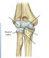
Which of the articulations are primarily involved with the hinge-like flexion and extension of the forearm on the arm?

A

The articulation between the trochlea of the humerus and the trochlea notch of the ulna, and the articulation between the capitulum of the humerus and the head of the radius.

24
Q

Which of the articulations is involved with pronation and supination of the forearm?

A

The articulation between the head of the radius and the radial notch of the ulna.

25
Q

The elbow joint is supported during movement by the ______ membrane of the joint capsule, which overlies the synovial membrane and encloses the joint.

The ________ membrane is thickened to form ligaments, which support the joint during specific movements:

A

The elbow joint is supported during movement by the fibrous membrane of the joint capsule, which overlies the synovial membrane and encloses the joint.

The fibrous membrane is thickened to form ligaments, which support the joint during specific movements:

26
Q

• Radial and ulnar collateral ligaments:

how are they formed?

what movements does it allow?

A

o Formed by medial and lateral thickening of the fibrous membrane.

o Support the flexion and extension movements of the elbow joint.

27
Q

• Annular ligament of the radius:

how is formed ?

functions?

A

o Formed by the lateral free margin of the fibrous membrane that passes around and cuffs the head of the radius.

o Blends with the radial collateral ligament.

o Holds the radial head in place at the proximal radioulnar jo

28
Q
A
29
Q
A
30
Q
  • The _________ artery is the continuation of the axillary artery at the inferior border of the teres major. It runs medially down the arm giving off several branches, including the profunda brachii artery, before dividing into the radial artery and ulnar artery at the cubital fossa.
  • The ______ artery passes along the lateral aspect of the forearm. Its branches in the forearm include the radial recurrent artery and the superficial palmar branch.
  • The _____ artery is larger than the radial artery and passes along the medial aspect of the forearm. Its branches in the forearm include the ulnar recurrent artery and the common interosseous artery.
A
  • The brachial artery is the continuation of the axillary artery at the inferior border of the teres major. It runs medially down the arm giving off several branches, including the profunda brachii artery, before dividing into the radial artery and ulnar artery at the cubital fossa.
  • The radial artery passes along the lateral aspect of the forearm. Its branches in the forearm include the radial recurrent artery and the superficial palmar branch.
  • The ulnar artery is larger than the radial artery and passes along the medial aspect of the forearm. Its branches in the forearm include the ulnar recurrent artery and the common interosseous artery.
31
Q

Elbow dislocations are common. In which direction does the elbow most commonly dislocate?

A

Posterior. Usually following a fall on an outstretched hand. The distal end of the humerus is driven through the weak anterior part of the fibrous layer of the joint capsule and the radius and ulna dislocate posteriorly

32
Q

Which nerve may be damaged by a posterior dislocation of the elbow?

A

The ulnar nerve, as it passes posterior to the medial epicondyle.

33
Q

Dislocations of the elbow occur most frequently in children. Why is this?

A

The parts of the bone that stabilise the joint are incompletely developed. The elbow has six ossification centres around the articular surfaces of the joint. Due to the timing of the closure of these centres, various points are weak across the child’s growth

  • The bony fit support provided by the olecranon is not fully formed until the age of 9 meaning that the elbow joint has an inbuilt weakness until that point, which leaves the child more prone to posterior dislocations at the elbow.
  • The final ossification centre is at the lateral epicondyle and does not close until the age of 11.
34
Q

The wrist joint is a synovial joint between the:

A
  • Concave surface of distal end of the radius and the articular disc overlying the distal end of the ulna
  • Convex-oval shaped surface formed by the proximal carpal bones - scaphoid, lunate, and triquetral
35
Q
A
36
Q
A
37
Q

. A combination of all 4 movements is described as circumduction.

What are these movements

A

flexion, extension, abduction, and adduction

38
Q

These muscles are responsible for producing movement at the wrist joint.

A
39
Q

: Collectively the carpal bones form an arch. What is the name of the membranous band that spans from medial to lateral across the carpal arch?

A

Transverse carpal ligament/ flexor retinaculum.

40
Q

: What is the name of the passage between the membranous band above and the carpal bones below?

A

Carpal tunnel.

41
Q

What is carpal tunnel syndrome?

A

An entrapment mononeuropathy caused by compression of the median nerve as it passes through the tunnel between the flexor retinaculum and carpal arch. Typically caused by an injury that results in inflammation of the synovial tendon sheaths, decreasing the space available for the structures running through the carpal tunnel compressing them and compromising their functions.

42
Q

Carpal tunnel syndrome causes?

A

Mostly idiopathic. It is associated with obesity, hypothyroidism, arthritis, trauma, and repetitive strain injury.

43
Q

Carpal tunnel syndrome

how does it present?

A

Numbness, pain and tingling in the lateral palm, thumb, index, middle and lateral half of the ring finger. Weakened or inactive thenar muscles – may see muscle wasting.

44
Q

There are ___ synovial carpometacarpal joints between the base of the metacarpals and the related distal row of carpal bones.

A

There are five synovial carpometacarpal joints between the base of the metacarpals and the related distal row of carpal bones.

45
Q

The saddle joint, between the ____ _______ and the _____ permits a wide range of movement of the thumb. This is not a feature of the other digits, whose movements are restricted to limited gliding movements.

A

The saddle joint, between the first metacarpal and the trapezium permits a wide range of movement of the thumb. This is not a feature of the other digits, whose movements are restricted to limited gliding movements.

46
Q
A
47
Q
A
48
Q

What is the collective name for the abductor pollicis brevis, flexor pollicis brevis, and opponens pollicis?

A

Thenar muscles

49
Q

What is the collective name for the equivalent muscles of the little finger?

A

Hypothenar muscles: abductor digiti minimi, flexor digiti minimi, opponens digiti minimi.

50
Q

The metacarpophalangeal joints are between the____ _____ __ __ _______and the ______ ______ of the digits.

A

The metacarpophalangeal joints are between the distal heads of the metacarpals and the proximal phalanges of the digits.

51
Q

The proximal and distal interphalangeal joints are _____ joints that occur between the proximal and middle phalanges, and between the middle and distal phalanges, respectively. The thumb only has ___ phalanges and therefore possesses a single interphalangeal joint.

A

The proximal and distal interphalangeal joints are hinge joints that occur between the proximal and middle phalanges, and between the middle and distal phalanges, respectively. The thumb only has 2 phalanges and therefore possesses a single interphalangeal joint.

52
Q

What movements occur at the interphalangeal joints and which muscles are responsible for those movements?

A

Flexion and extension.

53
Q

What is the difference between intrinsic and extrinsic muscles of the hand? Look back through station C and try to classify the muscles previously mentioned.

A

Intrinsic muscles originate within the area they act upon i.e. located entirely in the hand. For example, adductor pollicis. Extrinsic muscles originate outside of their area of action i.e. originate in the forearm but insert in the hand. For example, flexor digitorum superficialis

54
Q

In terms of innervation and action, what is unusual about the adductor pollicis muscle?

A

It is innervated by the ulnar nerve but acts on the thumb whose muscles usually are innervated by median nerve.

55
Q

what is Tenosynovitis

A

is the inflammation of the tendons and their synovial sheaths resulting in the swelling of the digits and compromised movement. It is caused by injuries to the fingers, such as cuts and bites that can result in infection. The infection could potentially spread depending on the level of treatment received.

56
Q

How could an injury in the little finger or thumb result in infection spread through the wrist into the forearm?

A

In the thumb and little finger, the tendon sheath usually extends into the radial and ulnar bursae, respectively (see diagram). This means that if infection is not treated, it can spread into the palm and then into the distal forearm due to the continuous nature of the sheath. This is unlikely with the 2nd, 3rd , and 4th digits due to them possessing their own sheaths

57
Q

The primary function of the lower limb is to support the weight of the body and to provide a stable foundation in standing, walking and running.

What structures play crucial role in this?

A

The pelvic girdle and hip joints play a crucial role in achieving this

58
Q

: What type of cartilage coats the femoral head and the acetabulum?

A

Hyaline cartilage – translucent covering which is found on many articular surfaces. Hyaline is said to mean ‘glass like’.

59
Q

The sartorius muscle crosses the hip joint and the knee joint, what action does it have on the latter?

A

Knee flexion, it is the only muscle that is capable of flexing the hip and the knee. This is particularly useful if you are trying to look at the bottom of your shoe!

60
Q

The rectus femoris is part of quadriceps femoris and also crosses the hip and knee joints, what action does it have on the latter?

A

Knee extension, so it flexes the hip and extends the knee alongside the other three muscles which make up the quadriceps femoris. The rectus femoris inserts distally into the quadriceps tendon on the anterior surface which causes extension of the knee. Sartorius passes onto the posteroinferior surface of the knee medially inserting into the pes anserinus, as it moves from the anterior to posterior aspects of the joint this makes it a knee flexor not extensor

61
Q

Why is it important that the two anastomoses are not purely made up of branches from femoral artery?

A

Femoral artery obstruction or even external iliac artery obstruction would cut off blood supply to the femoral head leading to avascular necrosis causing weakness in the joint and increasing joint fracture risk. By having a connection to the internal iliac artery through the superior and/or inferior gluteal artery, this gives the hip joint better arterial coverage in the event of femoral artery obstruction.

62
Q

Which vessels do you think are most likely to be compromised during hip dislocation and result in avascular necrosis (AVN) of the femoral head?

A

The most likely arteries to be compromised and cause AVN are the medial circumflex femoral artery and the artery to head of femur (depends on how developed this is). As the femoral head has limited independent blood supply, any disruption via compression of a vessel or trauma to the femoral neck can cause significant AVN.

63
Q

Why are the elderly more prone to hip fractures?

A

Increased chance of osteoporosis which makes the bones break more easily coupled with being more of a fall risk. Both are linked to natural degeneration of the body and clinical issues associated with the ageing process.

64
Q

When a hip is fractured, particularly at the neck of femur, it may disrupt this blood supply and result in ______ ______ of the femoral head.

A

When a hip is fractured, particularly at the neck of femur, it may disrupt this blood supply and result in avascular necrosis of the femoral head.

65
Q

Why is the femoral head at such risk of AVN in a neck of femur fracture? Refer back to the diagram below

A

Only the acetabular branch of the obturator artery (if it is present) goes directly to the femoral head. The majority of its blood supply has to come from arteries distally and they use the neck of femur as a conduit to gain access to the femoral head. If this area is fractured, the arteries will most likely be damaged and therefore arterial supply is lost.

66
Q

Using the pelvis model with ligaments displayed and the diagram below, In what direction are hip dislocations most likely to occur and why?

A

Hip dislocations tend to occur posteriorly (85%) as a result of when the individual suffers high impact trauma to the pelvis i.e., car accidents. Lack of ligamentous support posteriorly, particularly when with flexed hips seen in a seated position

67
Q

: If the femoral head is has suffered significant avascular necrosis, what would be the best course of treatment? Use the bony femurs with implants to help work this out.

A

Total hip replacement (arthroplasty) – replacement of the femoral head and neck with an implant and this would articulate with a replacement acetabulum

68
Q
A
69
Q

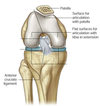
The knee is the largest and most complex synovial joint in the body. It consists of two articulations within a single capsule; the_______ joint and the _________ joint

A

The knee is the largest and most complex synovial joint in the body. It consists of two articulations within a single capsule; the tibiofemoral joint and the patellofemoral joint

70
Q

Can you recall the function of the menisci?

A

The menisci have several functions. Primarily, they serve to amplify the contact surface between the tibia and femur. This helps to evenly distribute the forces experienced and improves stability. They act as mild shock absorbers, attenuating some of the pressure that would be exerted onto the tibia. They’re also very important for proprioception.

71
Q

Which meniscus disc is attached to an extracapsular ligament?

A

The medial meniscus is attached to the medial collateral ligament which clinically means that damage to one often results in the other.

72
Q

The patella (P) is a ________ bone; it is formed inside the common quadriceps tendon (QT). A triangular shape, it has an apex, lying ________. The apex is connected to the _____ _______ by the patellar tendon (PT), a continuation of the common quadriceps tendon. The posterior surface of the patella articulates with the _____ of the femur. The upper, lateral and medial margins give attachment to the different parts of the ________ ______ muscle the large muscle (consisting of rectus femoris and the three vasti muscles).

A

The patella (P) is a sesamoid bone; it is formed inside the common quadriceps tendon (QT). A triangular shape, it has an apex, lying inferiorly. The apex is connected to the tibial tuberosity by the patellar tendon (PT), a continuation of the common quadriceps tendon. The posterior surface of the patella articulates with the condyles of the femur. The upper, lateral and medial margins give attachment to the different parts of the quadriceps femoris muscle the large muscle (consisting of rectus femoris and the three vasti muscles).

73
Q

The lateral femoral condyle projects more anteriorly than the medial femoral condyle to help prevent patellar dislocation/subluxation. Why do you need this preventative arrangement and are there any others around the knee joint?

A

It is designed to resist the lateral pull of the quadriceps femoris, the lateral aspect in modern humans is particularly well developed so causes a lateral drag which is not resisted could result in patellar dislocation/subluxation. The more horizontally arranged fibres of the lower VM help limit the patellar drag

74
Q

The patellar position is crucial to knee stability; can you think of any group of individuals who would be at higher risk of patellar dislocation?

A

Professional athletes: particularly those who train their quadriceps extensively. The vastus medialis lower fibres are harder to train therefore are inherently weaker.

75
Q

Which ligament would be damaged in the incident demonstrated on the image on the right?

A

High level impact on the lateral knee surface is an example of valgus force being applied to the knee joint. This would result in damage to the MCL as it is stretched by the force being applied laterally.

76
Q

What effect would impact on the medial knee surface have?

A

The LCL would be damaged. Essentially the opposite of what you have seen in the previous question as varus forces are generated in this example. In both, while there would be superficial soft tissue damage on the impact surface, the actual ligamentous damage would be contralateral.

77
Q

What are the Extracapsular Ligaments and Intracapsular Ligaments

A

Extracapsular Ligaments: medial collateral (MCL) and lateral collateral (LCL)

Intracapsular Ligaments: The cruciate ligaments (ACL and PCL)

78
Q

ACL attaches to the ________ ____, they limit excessive tibial displacement in the antero-posterior plane

A

ACL attaches to the anterior tibia, they limit excessive tibial displacement in the antero-posterior plane

79
Q

The PCL is resisting what movement of the femur?

A

Anterior femoral translation – the sliding forward of the femur against the tibia. The ACL resists posterior femoral translation – the sliding backwards of the femur against the tibia. You can discuss the function of cruciate ligaments as relating to the tibia or femur so be careful when reading questions!

80
Q

What is the most common cause of an ACL tear/sprain?

A

Non-contact injury resulting from excessive medial rotation of the tibia and excessive lateral rotation of the femuron the standing leg. Common in football players, particularly when they are trying to turn to run after the ball.

81
Q

An individual who suffers from an ACL tear often damages other areas in the knee joint, can you think of what these might be?

A

Medial Meniscus tear and damage to medial collateral ligament due to excessive force now being placed through the medial aspect of the joint – known as the unhappy triad of injury presentation.

82
Q
A
83
Q

What three muscles make up the hamstrings?

A

The 3 hamstrings are biceps femoris, semimembranosus and semitendinosus.

84
Q

What four muscles make up the quadriceps femoris?

A

The quadriceps femoris consists of rectus femoris and the 3 vasti muscles, medius, lateralis and intermedius.

85
Q

Which three muscles in the posterior compartment of the leg are weak knee flexors?

A

Any muscle which crosses the knee joint at its posterior aspect is capable of producing knee flexion. This includes popliteus, both heads of gastrocnemius and the plantaris, which are all muscles of the posterior compartment of the leg

86
Q

what is the true ankle joint?

A

talocrural joint- a deep-socketed synovial hinge joint

87
Q

The talocrural joint is formed between the______ ends of the tibia (T) and fibula (F), and the upper part of the body of the talus (Ta). While the leg bones remain reasonably fixed, the talus moves on the _________ axis to form the hinge.

A

The talocrural joint is formed between the inferior ends of the tibia (T) and fibula (F), and the upper part of the body of the talus (Ta). While the leg bones remain reasonably fixed, the talus moves on the transverse axis to form the hinge.

88
Q
A
89
Q

Largest bone on foot?

what does it articulate with

A

calcaneum

articulates above with the talus and in front with the cuboid.

90
Q

The _____ articulates above at the ankle joint with the tibia and fibula, below with the calcaneum and on front with the navicular bone

A

The talus articulates above at the ankle joint with the tibia and fibula, below with the calcaneum and on front with the navicular bone

91
Q

The tuberosity of the _________ bone can be seen and felt on the medial border of the foot one inch in front and below the medial malleolus.

A

The tuberosity of the navicular bone can be seen and felt on the medial border of the foot one inch in front and below the medial malleolus.

92
Q

The ______ bone is on the lateral aspect of the foot.

A

The cuboid bone is on the lateral aspect of the foot.

93
Q

The three small, wedge-shaped cuneiform bones articulate proximally with the navicular bone and distally with the first three metatarsal bones. Their wedge shape contributes greatly to the _______ and _________ of the transverse arch of the foot

A

The three small, wedge-shaped cuneiform bones articulate proximally with the navicular bone and distally with the first three metatarsal bones. Their wedge shape contributes greatly to the formation and maintenance of the transverse arch of the foot

94
Q

The _____ metatarsal bone is large and strong and plays an important role in supporting the weight of the body.

A

The first metatarsal bone is large and strong and plays an important role in supporting the weight of the body.

95
Q
A
96
Q

The foot has two important functions

What are they?

A

: to support the weight of the body and to serve as a lever to propel the body forward in walking and running demonstrated in the gait cycle diagram above

97
Q

The foot has three such arches, which are

A

the medial longitudinal, lateral longitudinal and transverse arches

98
Q

Which bones form the medial longitudinal arch of the foot?

A

The calcaneus, the talus, the navicular bone, the three cuneiform bones and the first three metatarsal bones

99
Q

Which bones form the lateral longitudinal arch of the foot?

A

The calcaneus, the cuboid, and the 4th and 5th metatarsal bones

100
Q

Which bones form the transverse arch of the foot?

A

The three cuneiform bones, cuboid, and bases of the five metatarsal bones.

101
Q
A