HARC - Urogenital Flashcards

1
Q

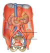
Anatomy of urinary system

A
2
Q

Anatomy of the kidneys in situ

A
3
Q

Anatomy of the kidney

A
4
Q

Anatomy of the bladder

A
5
Q

Anatomy of the pelvis

A
6
Q

Anatomy of the pelvis

A
7
Q

Anatomy of the male genitalia

A
8
Q

Anatomy of the male genitalia

A
9
Q

Anatomy of the testes

A
10
Q

Anatomy of female genitalia (simplified)

A
11
Q

Anatomy of the uterus and vagina

A
12
Q

Anatomy of the uterus and vagina

A
13
Q
A
14
Q

RAAS

A
15
Q

Bladder – urine and fluid balance

A
16
Q

Female – genitalia and hormones

A
17
Q

Female – genitalia

A
18
Q
A
19
Q
A
20
Q
A
21
Q
A
22
Q

: From where does each of these vessels originate?

Ureter

Renal Vein

Renal Artery

A

The ureter is the continuation of the renal pelvis,

the artery is a direct branch of the abdominal aorta

the renal vein is the venous drainage from the kidney.

23
Q

In what order are they arranged at the hilum?

Ureter, Renal artery and vein

(from Posterior to Anterior)

A

Ureter, Renal Vein, Renal Artery

24
Q

To where does the venous blood from the suprarenal (adrenal) glands drain?

A

Right-inferior vena cava; left= left renal vein.

25
Q
A
26
Q
A
27
Q
A
28
Q

How are the kidneys anchored to the posterior abdominal wall?

A

They are retroperitoneal and occupy a space between the peritoneum and posterior abdominal wall. They are also supported by their surrounding perirenal fat and fascia. Sometimes this gradually breaks down and the kidney drops (nephroptosis) which is potentially damaging.

29
Q

How would you describe the transit of the ureters from the abdomen to the pelvis?

What is the potential consequence of this?

A

After descending in a straight line on top of the psoas major and just lateral to the transverse processes of the lumbar vertebrae, they must turn and cross the pelvic brim. As they do this, they cross the common iliac artery.

This is one of three major points of constriction for the ureters and ureteric calculi (stones) commonly present here. The other two are at the renal hilum and the vesicoureteric junction

30
Q

How do you think a horseshoe kidney develops?

A

Fusion of the inferior poles of the two kidneys during development, prior to ascent of the metanephros

31
Q

At what level would you find a horseshoe kidney, and why?

A

Approximately L3. It usually cannot ascend further than the inferior mesenteric artery (L3). This is variable as the IMA is not always at L3 (can be a little higher).

32
Q

What affect does horshoe kidney have on kidney function?

A

Traditionally no effect on urine production. However, can have issues due to excessive blood supply and/or tangles.

33
Q

Features and surfaces of the bladder:

A

BOLD are surfaces

34
Q
A
35
Q

What ligament anchors the bladder to the anterior abdominal wall? Hint: think of embryological remnants.

A

The median umbilical ligament is a remnant of the embryonic urachus (primitive waste disposal) which exited fetus via umbilical cord. Hence, ligament passes toward umbilicus.

36
Q

Which sphincter is located here?

A

Internal urethral sphincter

37
Q
A
38
Q

Which sphincter is located here?

A

Internal urethral sphincter more specifically sphincter urethrovaginalis and compressor urethrae in the female

39
Q

Pelvic Floor

A
40
Q

Pelvic Floor

A
41
Q

Name all the muscles

A
42
Q
A
43
Q

Why is the perineal body such an important structure in the pelvic floor?

A

It is the central tendon of the pelvic floor. Damage to a tendon heals very slowly and this could lead to an inherent weakness in the pelvic floor. This is of particular risk during childbirth.

44
Q

Why is the position of the pudendal canal important during childbirth?

A

It contains the pudendal nerve. A pudendal nerve block is routinely administered transvaginally due to the close proximity of the canal to the lateral vaginal wall

It may also be the case that compression of the pudendal canal causes temporary paralysis of the pudendal nerve, affecting continence. This is possible because the canal lies along the ischiopubic ramus and the fetus may compress it during delivery.

45
Q

Sensory innervation:

Name the purple nerves

A
46
Q

Note the curvature and orientation of the uterus. How might you describe this position?

A

The most common arrangement is anteverted (at the cervix) and anteflexed (at the uterine body) but this may not always be the case and some prosections may differ.

47
Q

Which reproductive structure(s) does the broad ligament overlie?

A

The broad ligament can be divided into 3 segments: the mesometrium is the largest part, covering the uterus, the mesosalpinx covers the uterine tubes in the superior margin of the broad ligaments and the mesovarium suspends the ovaries from its posterior margin

48
Q
A

Uterine/Fallopian Tube

Ovary

Fundus

Body/uterus

Cervis

Vagina

49
Q

What is the difference between the internal and external os of the cervix?

A

The central canal of the cervix runs through its entire length and is mostly narrow. It has two dilations (openings): the internal os, opening into the uterus and the external os, opening into the vaginal canal.

50
Q

How does the uterus relate to the bladder and rectum

A

The rectum is most posterior, separated from the uterus by the recto-uterine pouch (of Douglas). The bladder is most anterior, separated from the uterus by the Vesico-uterine pouch (Dunne’s pouch) and is situated just posterior to the pubic symphysis. During pregnancy the uterus expands and compresses the bladder into the pubis, causing a decreased capacity and potentially urinary problems.

51
Q

How does the vagina relate to the urethra and rectum?

A

Vaginal canal is situated between the urethra (anterior) and rectum (posterior) in the mid-sagittal plane, with their external openings consistent with this.

52
Q

How does the broad ligament relate to the uterine tubes and ureter?

A

Uterine artery crosses the ureter at the base of the broad ligament. While the uterine tubes are enclosed in the superior margins of the broad ligament.

53
Q
A
54
Q
A
55
Q
A

Ductus deferens

Bulbourethral glands

Epididymis

Seminal Vesicle

Spermatic cord

Prostatic urethra

56
Q
A

Bladder

Prostrate

57
Q

What is the prostate for? Which can be palpated during a digital rectal examination (DRE)?

A

Main function is to secrete prostatic fluid, which makes up ~30% of ejaculate and makes it an alkaline solution to counteract the acidic environment of the vagina and prolong life of spermatozoa. Posterior lobe can be palpate in DRE.

58
Q

Bulbourethral glands Q2: Where are they and what do they produce?

A

They are located in the deep perineal pouch, just posterolateral to the membranous urethra. They contribute to lubrication of the urethra and pre-ejaculatory emissions.

59
Q
  • Seminal vesicles Q3: Where are they and what do they produce?
A
  • Located at the posterior surface of the bladder, just anterior to the rectum.
  • They contribute the majority of semen produced and contain a high fructose content to supply spermatozoa with energy.
  • Fluid is highly alkaline again to neutralize vaginal pH
60
Q

How do the seminal vesicles relate to the ductus deferens?

A

Start off as blind-ended sacs developing from the ductus deferens and are found immediately lateral to each ductus deferens and joins the duct to form the ejaculatory duct.

61
Q

What is the cremasteric reflex?

A

A reflex elicited by the gentle stimulation of the skin of the scrotum or anterior thigh and is controlled by the sensory part of the femoral branch of the genitofemoral nerve and the ilioinguinal nerve (which travel into the spinal cord to elicit the reflex). Motor component is controlled by the genital branch of the genitofemoral nerve (innervation to cremaster muscle) See my lecture for detail too.

62
Q

Which nerves are in the spermatic cord?

A

Sympathetic nerves and genital branch of genitofemoral – not the ilioinguinal nerve which runs on the superior surface of the spermatic cord.

63
Q

What layers of the abdominal wall do the testes pass through during their descent?

What do these layers contribute towards the spermatic coverings?

Where are the deep and superficial inguinal rings located?

A

All layers of abdominal wall. Some ambiguity regarding transversus abdominis. See lecture slide for layers and mnemonic :)

The deep inguinal ring is located at a midpoint between the ASIS and the pubic symphysis while the superficial is just superior to the pubic tubercle. See lecture for landmarks.

64
Q

Which male reproductive structure would be found in the deep perineal pouch?

A

Bulbourethral glands. Unlike female equivalent (greater vestibular glands) which are in the superficial perineal pouch.

65
Q

Name these?

A

Corpora cavernosa

Corpus spongiosum

Bulbourethral glands

66
Q

Now identify the following muscles:

A

Ischiocavernosus

Bulbospongiosus

Superficial transverse perineal muscles

67
Q
A

Glans clitoris (with corpora cavernosa*)

Clitoral hood

Urethra

Vestibule

Labia minora

68
Q

Now identify the following muscles:

 Ischiocavernosus  Bulbospongiosus  Superficial transverse perineal muscles

A
69
Q
A
70
Q
A
71
Q
A
72
Q
A
73
Q
A
74
Q
A