HARC - Cardiovascular Flashcards
1
Q
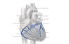
Anatomy of Heart

A

2
Q

A

3
Q

A

4
Q

A

5
Q

A

6
Q

A

7
Q

A

8
Q

A

9
Q
Name the black labels

A
Depolarization
Action potential
Repolarization
Refractory period
10
Q
Which action potential graph is this? (nodal cells or cardio myocytes)

A
Nodal Cells
11
Q
Which action potential graph is this? (nodal cells or cardio myocytes)

A
cardio myocytes
12
Q
Innervation of the heart

A

13
Q

A

14
Q

A

15
Q

A

16
Q

A

17
Q
The Circulatory System

A

18
Q
The Circulatory System

A

19
Q
The Circulatory System

A

20
Q

A

21
Q

A

22
Q
A

23
Q
Muscle Contraction

A

24
Q
Muscle Contraction

A

25
Muscle Contraction


26
Muscle Contraction

27
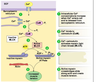
Muscle Contraction


28


29


30


31


32


33
What is the ligamentum arteriosum a remnant of?
It is a remnant of the ductus arteriosus which connects the pulmonary trunk to the aorta to divert blood away from the lungs in the fetus
34


35
What shape is the pectinate muscle?
Pectin = comb.
36


37


38
What is the function of the chordae tendineae?
They prevent prolapse of the valves after they close by becoming tense and pull on the cusps holding them in a closed position.
39


40
Which chamber of the heart does not send its venous drainage to the coronary sinus?
Why?
Right ventricle because the coronary sinus drains directly into this chamber. Anterior cardiac veins overlying the right ventricle drain into the right atrium instead of going to the coronary sinus.
Some veins called venae cordis minimae are very small and drain directly into the chambers of the heart and are found in large numbers in the right atrium and right ventricle. The small cardiac vein may drain to the coronary sinus, right atrium, middle cardiac vein or be absent.
41


42


43
What is mediastinum?
The mediastinum is a space that lies between, and separates, the cavities of the two lungs.
44
What does mediastinum consist of?
It contains various structures and allows other structures to pass through the thorax either to the abdomen, heart or head and neck.
45
Name the different parts of the mediastinum (Labels A - E)

A= Anterior mediastinum
B= Middle mediastinum
C= Posterior mediastinum
D= inferior mediastinum
E= Superior mediastinum

46
A transverse plane is indicated by the yellow dotted line. It passes through the sternal angle at what vertebral level?

The sternal angle is found at the second rib (costal cartilages) but at the vertebral level T4/T5
47
The middle part of the mediastinum contains\_\_\_\_\_\_\_\_
the pericardium, the heart and the roots of the great vessels
48
The pericardium is a _________ \_\_\_\_\_\_\_\_\_\_that encloses the heart and the roots of the great vessels - ascending aorta, pulmonary trunk and superior vena cava.
The pericardium is a **fibroserous membrane** that encloses the heart and the roots of the great vessels - ascending aorta, pulmonary trunk and superior vena cava.
49
How is the visceral pericardium also known?
Epicardium – forms the outer layer of the heart
50
. If bleeding occurred into the intact pericardial sac, how would the heart function be impaired?
_Cardiac tamponade_
and the build up of blood/fluid would impair the contraction power of the heart walls thereby decreasing the ability of the heart to pump blood. The reason being that the fibrous pericardium cannot expand as easily with the increasing volume, thereby pushing on the heart muscle instead.
51
The phrenic nerve runs ______ to the hilum of the lung
The vagus runs ________ to the hilum of the lung
anterior
posterior
52
Identify the phrenic and vagus nerves on both the left and right side of the thorax


53
Q6. Which branch of the vagus nerve curves underneath the arch of the aorta?
Left recurrent laryngeal nerve which goes on to supply the muscles of the larynx except for cricothyroid.
54
The recurrent laryngeal nerves are branches from\_\_\_\_\_\_\_
he Vagus (Cranial nerve X) that supply motor innervation to the muscles of the larynx
55
Why are recurrent laryngeal nerves called recurrent?
They are called “recurrent” because when they branch off from the vagus, they re-ascend the neck (hence recurrent) to supply the larynx - they also have differing anatomical pathways because one branch gets caught around the arch of the aorta.
56


57
What is the conduction system influenced by?
the sympathetic (fight, flight freeze response) or parasympathetic (rest and digest) components of the autonomic nervous system (ANS).
58
. Which node is known as the ‘pacemaker’ of the heart due to the propagation of impulses here?
SA node
59
Which papillary muscle does the moderator band supply?
Anterior papillary muscle
60
Does a moderator band also exist on the left hand side of the heart?
Yes, however its hidden by lots of left ventricular tissue
61


62


63
1. Atrial depolarisation (P wave) – beginning of atrial contraction
2. Ventricles are depolarised (ST segment) – semilunar valves open
3. Ventricular depolarisation (QRS complex) – AV valves close
4. Impulse travels from SA node to AV node (PR segment) – atria relax (diastole)
5. Ventricular repolarisation (T wave) – beginning of ventricular relaxation
Put it in correlation to A-E
A = 1
B = 4
C = 3
D = 2
E = 5
64


65
Identify the locations of where the stethoscope will be placed to hear the four valve sounds on the skeleton. The following are NOT in the correct order, use the diagram to help:
Mitral valve

Mitral valve = 5th intercostal space

66
Identify the locations of where the stethoscope will be placed to hear the four valve sounds on the skeleton. The following are NOT in the correct order, use the diagram to help:
Aortic

Aortic = medial end of right second intercostal space

67
Identify the locations of where the stethoscope will be placed to hear the four valve sounds on the skeleton. The following are NOT in the correct order, use the diagram to help:
Tricuspid

Tricuspid = left of lower part of sternum near 5th intercostal space

68
Identify the locations of where the stethoscope will be placed to hear the four valve sounds on the skeleton. The following are NOT in the correct order, use the diagram to help:
Pulmonary

Pulmonary = medial end of left second intercostal space

69
. The average adult pulse rate at rest is
60 - 80 beats per minute
70
What can cause your average heart rate to be higher?
exercise, injury, illness and emotion may produce much faster rates
71
What causes a pulse to be generated?
Most pulses are named after the artery where it can be found. Below are examples of where pulses can be felt in 10 major locations on each side of the body. The accessible radial artery in the wrist is one of the most routinely palpated. You can palpate a pulse by putting a gentle pressure over the artery with the pads of the index and middle fingers.
72
The femoral vessels and nerves lie in an area known as......
the femoral triangle in the upper 1/3 of the anterior thigh and is continuous below with the adductor canal.
73
The borders of the femoral triangle are:
Lateral border =
Medial border =
Superior border =
Lateral border = sartorius
Medial border = adductor longus
Superior border = inguinal ligament
74
Match these to the correct boxes:
Great saphenous vein
Femoral nerve
Deep femoral artery
Adductor Longus muscle
Sartorius muscle
Inguinal Ligament
Femoral Vein


75
Match these with the correct arrows:
Biceps brachii tendon
Cubital fossa
Brachial artery
Median nerve


76
Head fibula
Medial head of gastrocnemius muscle
Small saphenous vein
Lateral head of gastrocmemius
Semimembranosus muscle
Common fibular nerve
Biceps femoris muscle and tendon
Tibial nerve
Popliteal artery
Popliteal artery
Penetrates deep fascia
Semitendinosus tendon


77
How do you perform an Allen's test?
The Allen test is performed by having the patient clench their fist several times while the operator occludes the radial and ulnar artery at the wrist. The patient then extends their fingers, palm up, which should show a "blanched" hand.

78
Name all 10 pulse points
Superficial temporal
Facial
Common carotid
Axillary
Brachial
Radial
Ulnar
Femoral
Popliteal
Dorsalis pedis
Posterior tibial
79
Locate the **Superficial temporal** pulse points on yourselves
Imagine a line joining the top of the ear (pinna) to the side of the eye (orbit). 2cm above the middle of the line is the superficial temporal pulse. The image shows an alternative closer to the ear. It is a branch of the **external carotid artery**

80
Locate the **Facial** pulse points on yourselves
Where the artery crosses the mandible (jaw) near to the submandibular gland (salivary gland under jaw). It is a branch of the **external carotid artery**

81
Locate the **Common carotid** pulse points on yourselves
This pulse is located lateral to the thyroid cartilage to gently press the common carotid against the transverse process of C3. Do not push hard and do not palpate both sides simultaneously. It is a branch of the aortic arch (left side) and brachiocephalic artery (right side). The terminal branch is the external carotid artery and can be palpated as seen below in picture C2.

82
Locate the **Axillary** pulse points on yourselves
– palpation of the axillary artery in the armpit
D

83
Locate the Brachial pulse points on yourselves
– continuation of axillary artery into the upper limb. It is most commonly palpated in the antecubital fossa medial to the tendon of the biceps
E

84
Locate the **Radial** pulse points on yourselves
– pulse can be felt by placing 2 finger pads under the base of the thumb and pushing lightly against the radius. Radial pulse can also be felt in the anatomical snuffbox
F

85
Locate the **Ulnar** pulse points on yourselves
taken on the opposite side of the wrist at the same level as the radial artery
G

86
Locate the **Femoral** pulse points on yourselves
– Located at the midpoint of the inguinal ligament (halfway between pubis and ASIS)

87
Locate the **Popliteal** pulse points on yourselves
– located in the popliteal fossa (behind the knee) and press deep against the popliteal surface of the femur. It is a branch of the femoral artery
I

88
Locate the **Dorsalis pedis** pulse points on yourselves
– Between the tendons of extensors hallucis and digitorum longus (top of the foot)
J

89
Locate the **Posterior tibial** pulse points on yourselves
Posterior to the medial malleolus on the medial side of the ankle
K

90
There are two groups of veins – What are they?
deep and superficial
91


92
What are:
Preload
Afterload
End diastolic volume (EDV)
Ejection systolic volume (ESV)
****
Preload- Volume of blood in ventricles at end of diastole (end diastolic pressure)
Afterload- Resistance left venricle must overcome to circulate blood
End diastolic volume (EDV)- amount of blood in the ventricle before contraction
Ejection systolic volume (ESV)- amount of blood remaining in the heart after ejection
93


94
What happens when you have low blood pressure?

95
Peripheral Chemoreceptors

96
Staging of Hypertension

97
What is primary and secondary hypertension
**primary hypertension**
elevated blood pressure without an identifiable cause; also known as essential or idiopathic hypertension
**secondary hypertension**
elevated blood pressure with an underlying cause or pathology such as from kidney disease or endocrine disease
98
What are the causes of primary hypertension
none
99
Risk Factors for Primary hypertension

100
Causes of secondary hypertension?
* Renal Disease (most common)
* Endocrine Disease
* Vascular Disease
* Drugs

101
