Embryology Flashcards
MSK:
• Mesenchyme is the same as_____ ______ _______
embryonic connective tissue
MSK:
What is mesenchyme?
a loosely organized, mainly mesodermal embryonic tissue which develops into connective and skeletal tissues, including blood and lymph.
From the mesoderm
MSK:
The mesenchymal cells migrate and differentiate into what?
into many different types of primitive cell lines
MSK:
Examples of 3 different types of primitive cell lines?
- Fibroblasts (adult connective tissue forming cells)
- Chondroblasts (cartilage forming cells)
- Osteoblasts (bone forming cells)
MSK:
What does the mesenchyme in the paraxial mesoderm do?
it will transform into osteoblasts that will form the bony elements of the vertebral column
MSK:
What does the mesenchyme in the somatopleuric mesoderm do?
will transform into osteoblasts that will form the pelvic/pectoral girdles and also the bones of the upper and lower limbs
MSK:
What is the process of bone formation known as?
Ossification
MSK:
Whw does bone start to develop?
develops during the intra uterine life through 2 types of ossification
- Membranous type (intermembranous)
- Intra cartilgainous type (endochondral)
MSK:
What is intermembranous ossifiction?
(mesenchymal tissue will directly transform into bone) eg: the flat bones of the skull
MSK:
What is endochondral ossifiction?
(mesenchymal tissue first gives rise to a hyaline cartilage model of the bone, then the osteoblasts will convert that model into bone) eg: long and irregular bones
MSK:
What is the entire muscular system of the body developed from?
mesoderm
• The entire muscular system of the body develops from the mesoderm (except from the muscles of the_____, which develop from the _______ of the optic cups)
iris
ectoderm
The cardiac muscles of the heart develop from the _________ ______ surrounding the primitive heart tube
splanchnic mesoderm
The smooth muscles of the GI tract develop from the _______ _______ surrounding the gut tube
splanchnic mesoderm
MSK:
How many pharyngeal arches are there in the human development?
5
MSK:
Each pharyngeal arch has its own:
Cartilage skeleton,
muscular component,
sensory nerve supply,
motor nerve supply
MSK:
Muscles of mastication are dervied from which pharyngeal arch?
Which nerve is the muscle innervated by?
derived from the 1st pharyngeal arch and are therefore innervated by the Trigeminal nerve (V)
MSK:
Muscles of facial expression are dervied from which pharyngeal arch?
Which nerve is the muscle innervated by?
derived from the 2nd pharyngeal arch and therefore are innervated by the Facial nerve (VII)
Uro:
What are the 3 stages in the development of the kidney:
- Pronephros
- Mesonephros
- Metanephros
Uro:
Embryology of the kidney


Uro:
Embryology of Kidney


Uro:
Embryology of Kidney


Uro:
When doe the pronephros develope?
It develops during the 4th week of uterine life in the cervical region of the intermediate mesoderm
Uro:
What does pronephros contain?
The pronephros contains lots of segmental vesicles and has the pronephric duct that grows caudally toward the cloaca
- The pronephric duct is in the embryo and thus cannot filter materials outside the embryo.
- Therefore it is said that the pronephros kidney is non-functional in humans thus it degenerates.
Uro:
When is mesonephros developed?
- The Mesonephros kidney follows on from the development pronephros at around the end of the 4th week of uterine life
- It also comes from intermediate mesoderm and is located in the thoraco-lumbar area where the pronephric duct that was developed in the pronephros stage continue to elongated caudally and become the mesonephric tubules.
Uro:
Does the mesonephros degenerate?
Most of this tubules eventually degenerates but leave behind the Mesonephric duct or Wolffian duct which extends towards the cloaca
Uro:
The mesonephros is the functioning kidney during the __ trimester and it produces urine during weeks____
1st
6-10
Uro:
When does Metanephros develop?
This kidney begins to develop in the 5th week of the embryonic period and
appears in the sacral region of the intermediate mesoderm
Uro:
What happens to metanephros during the fifth week?
the mesonephric duct from the mesonephros kidney develops an outgrowth called the ureteric duct. This duct is close to the attachment of the cloaca.
The ureteric duct eventually forms the ureter, renal pelvis, major calyces, minor calyces and collecting tubules
- The ureteric bud interacts with a portion of undifferentiated intermediate mesoderm called the metanephric mesenchyme.
- This interaction induces differentiation and thus the formation of the renal tubules, glomerulus, loops of Henle, and the distal/proximal convoluted tubules
- If there is an abnormal interaction between the ureteric bud and the metanephric mesenchyme, several congenital malformation of the kidney may result.
Uro:
Embryology of the Kidney
Part A


Uro:
Embryology of the Kidney
Part B

Uro:
Embryology of the Kidney
Part C and D


Uro:
Embryology of the Kidney
Part E


Uro:
What is Sexual differentiation?
is the process of the development of the differences between males and females from an undifferentiated zygote.
Uro:
What carries the essential gene that determines testicular formation?
Y chromosome
Uro:
What induces differentiation of cells derived from the genital ridges into testes?
A gene in the sex-determining region of the short arm of the Y, known as SRY leads to the formation of testis determining factor (TDF), which binds to DNA and induces differentiation of cells derived from the genital ridges into testes
Uro:
When are the gonads distinguishable by?
weeks 6-8 of gestation.
Uro:


Uro:
Outline the embryology of testes
- Between the 3rd month and the end of pregnancy, the testes become transferred from the lumbar area into the future scrotum.
- This transfer is due to a combination of growth processes and hormonal influences.
- The gubernaculum testis arises in the course of the 7th week from the lower gubernaculum, after the mesonephros has atrophied. Cranially it has its origin at the testis and inserts in the region of the genital swelling (future scrotum).
- At the same time, at the inguinal canal along the lower gubernaculum, an evagination of the peritoneum arises, the vaginal process, on which the testes will slide through the inguinal canal.
Uro:
Embryology of the ovaries
Mesonephric duct (Wolffian duct) degenerates, small remnants may remain as epoophoron and paroophoron (in the mesentery of the ovary) and Gartner’s cyst’s (near vagina)
- Paramesonephric duct (Müllerian duct) grows and forms the oviduct (uterine horn) and the end opens into the peritoneal cavity and terminates in fimbria (finger-like extensions extending from the ovary)
- Away from the ovary, the two paramesonephric ducts fuse in the midline to form the uterus
- Cortical sex cords form after the primary sex cords degenerate and the mesothelium forms secondary cords
- The surrounding connective tissue differentiates to form follicle cells.
Uro:
Embryology of the uterus and vagina
The entire vagina is formed from the paramesonephric (Müllerian) duct and does not have a contribution from the urogenital endoderm
- The initially paired ducts fuse in the midline forming the single body of the uterus
- The ducts remain separate laterally where they form the uterine tubes (Fallopian tubes, uterine horns)
- The ducts peripheral attachment site to the urogenital sinus wall is described as the Müllerian tubercle
- The fused ducts also generate the vagina, under the influence of BMP4
- Oestrogen will also later alter the vaginal epithelium
What are the three germ layers?
Ectoderm, mesoderm and endoderm
Mesoderm splits into……
somatic mesoderm (next to endoderm) and splanchnic mesoderm (next to ectoderm)
three main body cavities will form in the space between…..
the two mesoderm’s
The three main body cavities are:
the pleural cavity,
pericardial cavity
the peritoneum
________ and _______ mesoderm become the two pleural layers (visceral and parietal respectively)
Splanchnic
somatic
Resp:
In what week does the laryngotracheal groove form?
week 4 of development
Resp:
What happens in week 4 of development?
- the laryngotracheal groove forms
- This gives rise to the trachea, bronchi and the tracheobronchial tree
- It then develops into the laryngotracheal diverticulum which by the end of week week 4 forms a globular respiratory bud
Resp:
• The respiratory bud then divides into 2 outpouchings called ____ ______ __
Primary bronchial buds
These buds continue to grow and secondary and tertiary bronchial buds develop
Resp:
What happens to the bronchial buds by the end of week 5?
By week 5, the bronchial buds have enlarged and their connections to the trachea form the main bronchi
Resp:
What happens after week 5? (lungs)
the lungs begin maturation
This continues up until the age of 8
Resp;
Lung maturation
Pseudoglandular stage is between how many weeks?
Resp;
Lung maturation
Canalicular stage is between how many weeks?
Canalicular stage 16-26 weeks
Resp;
Lung maturation
Terminal sac stage is between how many weeks?
Terminal sac stage 26 weeks to birth
Resp:
Lung maturation
Alveolar stage is between how many weeks?
Alveolar stage 32 weeks – 8 years old
Resp:
What happens in the Pseudoglandular stage?
- By 16 weeks, all the major elements of the lungs have formed…
- EXCEPT those involved in gas exchange
- Therefore, respiration isn’t possible and a fetus born at this stage is unable to survive
Resp:
What happens in the Canalicular stage?
- Cranial segments of lungs develop faster than caudal
- Lumen of bronchi and terminal bronchioles are enlarging
- Lung tissue becomes vascularised
- Respiration is possible towards the middle/end of this stage
- It is possible because of the primordial alveolar sacs & vascularisation
Resp:
What happens in the Terminal sac stage?
- More terminal sacs develop (they have a very thin epithelium)
- Capillaries bulge into developing alveoli and adequate gas exchange can occur if the foetus is born after 26 weeks
- Lung surfactant starts to be produced
- Capillary networks proliferate rapidly around the alveoli
Resp:
What happens in the Alveolar stage?
- Epithelial lining becomes a thin, squamous epithelial layer (these are type 1 pneumocytes)
- By the late fetal stage, the lungs are capable of respiration because the alveolar capillary membrane is sufficiently thin enough to allow gas exchange
Resp:
What are the key difference between the lungs before and after birth:
- Before birth, the lungs are dependent on the placenta for gas exchange
- After birth, the lungs must become capable to carry out gas exchange themselves (ie: produce surfactant, good circulation)
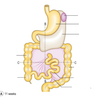
The primitive gut forms during what week?
4th week
The flat embryonic discs folds to…….
forma tubular structure
Forma tubular structure is divided into 3 distinct regions. What are they?
Foregut
Midgut
Hindgut
Gastro:
What does the foregut give rise to?
esophagus
stomach
liver
gallbladder
bile ducts
pancreas
proximal duodenum.
Gastro:
What does the midgut give rise to?
distal duodenum
jejunum
ileum
cecum
appendix
ascending colon
proximal 2/3 of transverse colon.
Gastro:
The hingut becomes?
distal 1/3 of the transverse colon
descending colon
sigmoid colon
the upper anal canal.
Gastro:
GI embryology = mesoderm, ectoderm or endoderm
ectoderm
Gastro:
Connective tissue, smooth muscle and serosa all arise from the __________ ___________(NOT THE ECTODERM!)
splanchnopleuric mesoderm
Gastro:


Gastro:
★ Primitive stomach rotates ________
★ Uneven growth (the p_______ side grows more rapidly causes the J shape of the stomach and the formation of the lesser and greater curvatures)
★ When it rotates, it pulls the _______ ________ over the top of itself – this forms the lesser sac
★ Meanwhile, the _______ is growing in the dorsal mesogastrium so therefore, it is pulled by the stomach rotation into the ____ ________ _____
★ Thus _____ ______ligament between the stomach and the spleen
★ Between the stomach and the liver = l_____ ______ with the free border carrying the hepatic artery, the bile duct and the ____ ______ ___
★ Primitive stomach rotates 90 degrees
★ Uneven growth (the posterior side grows more rapidly causes the J shape of the stomach and the formation of the lesser and greater curvatures)
★ When it rotates, it pulls the dorsal mesogastrium over the top of itself – this forms the lesser sac
★ Meanwhile, the spleen is growing in the dorsal mesogastrium so therefore, it is pulled by the stomach rotation into the left hypochondrial region
★ Thus gastro-splenic ligament between the stomach and the spleen
★ Between the stomach and the liver = lesser omentum with the free border carrying the hepatic artery, the bile duct and the hepatic portal vein
Gastro:
STOMACH


Gastro:
STOMACH


Gastro:
DUODENUM


Gastro:
- DUODENUM
★The duodenum is derived from the _____ and _____ (half and half separated by the entry of the bile duct)
★ It is a solid structure at first, and so must undergo _________ (a process to restore the hollow lumen of the duodenum)
★ Failure of recanalization = _________ _______
★ Duodenum rotates to the _____ (stomach rotates to the left)
★ Duodenum has a ______ ____ _____(FG&MG)
★The duodenum is derived from the foregut and midgut (half and half separated by the entry of the bile duct)
★ It is a solid structure at first, and so must undergo recanalization (a process to restore the hollow lumen of the duodenum)
★ Failure of recanalization = Duodenal stenosis
★ Duodenum rotates to the right (stomach rotates to the left)
★ Duodenum has a dual blood supply (FG&MG)
Gastro:
Pancreas


Gastro:
PANCREAS
★ The pancreas develops from a ____ and_____ bud
★ The dorsal bud = in the dorsal _______
★ The ventral bud = in the ventral _______
★ The buds _____ and come together in the same plane, they then fuse together
★ The duct of the larger dorsal bud forms the _____ ______ _______
★ The pancreas develops from a dorsal and ventral bud
★ The dorsal bud = in the dorsal mesentery
★ The ventral bud = in the ventral mesentery
★ The buds twist and come together in the same plane, they then fuse together
★ The duct of the larger dorsal bud forms the main pancreatic duct
Gastro:
LIVER/BILIARY TREE


Gastro:
LIVER/BILIARY TREE
★The liver forms from the h_______ d_______
★ It enlarges to fill the s_______ t_________ (the space it will occupy)
★ The liver is large in size (see diagram!) – by___weeks, it is forming blood cells
★ The gall bladder duct is solid at first and must undergo r________
★ Failure to recanalize =_______ _______ ______
★The liver forms from the hepatic diverticulum
★ It enlarges to fill the septum transversium (the space it will occupy)
★ The liver is large in size (see diagram!) – by 10 weeks, it is forming blood cells
★ The gall bladder duct is solid at first and must undergo recanalization
★ Failure to recanalize = Extrahepatic biliary atresia
Gastro:
MIDGUT


Gastro:
MIDGUT


Gastro:
MIDGUT
★ At first, the midgut has a wide communication with the y____ ____
★ This narrows to form the_______ _______duct
★ The midgut rapidly elongates and can no longer fit in the abdominal cavity
★ It therefore herniates out of the abdomen and into the ______ _______
★ It undergoes __ ____ _________rotation on departure, and ____ ____ ________rotation on return to the abdomen
★ At first, the midgut has a wide communication with the yolk sac.
★ This narrows to form the vitello-intestinal duct
★ The midgut rapidly elongates and can no longer fit in the abdominal cavity
★ It therefore herniates out of the abdomen and into the umbilical cord
★ It undergoes 90 degrees anticlockwise rotation on departure, and 180 degrees clockwise rotation on return to the abdomen
Gastro:
HINDGUT

Gastro:
HINDGUT
★ The endodermal lining also forms the epithelial lining of the______r and u______
★ The _____ ______ opens
★ ________ ______ divides the cloaca anteriorly (urogenital sinus) and posteriorly (rectum)
★ Mucosa of the upper ½ of the anal canal is from hindgut _______
★ Mucosa of the lower ½ of the anal canal is from the ________ (the proctodaeum)
★ Pectinate line demarcates upper from lower half
★ It fills completely and ___________ occurs in wk9
★ The endodermal lining also forms the epithelial lining of the bladder and urethra
★ The primitive cloaca opens
★ Mesenchymal tissue divides the cloaca anteriorly (urogenital sinus) and posteriorly (rectum)
★ Mucosa of the upper ½ of the anal canal is from hindgut endoderm
★ Mucosa of the lower ½ of the anal canal is from the ectoderm (the proctodaeum)
★ Pectinate line demarcates upper from lower half
★ It fills completely and recanalization occurs in wk9
what is the first organ to develop within the embryo?
the heart
Cvs:
When does the heart start to function?
It starts to function at the beginning of the fourth week (when the nutritional and oxygen requirements of the growing embryo can no longer be met by diffusion from the placenta)
Prenatally, the lungs are not active in gas exchange and therefore the pulmonary vessels are _________
vasoconstricted
Primordial heart and vascular system starts to develop in weeks…..
3 and 4 of pregnancy
Cvs:
oxygenated or deoxygenated
Normal veins =
Fetal veins =
Normal arteries =
Fetal arteries =
Normal veins = Deoxygenated blood
Fetal veins = Oxygenated blood
Normal arteries = Oxygenated blood
Fetal arteries = Deoxygenated blood
CVS:
Step 1 - Fusing


CVS:
Step 1 - Fusing
what happens?
� Start with 2 endocardial tubes (Endothelial strands)
� Lateral folding of these tubes fuses the 2 into 1 endocardial tube
� The pericardial cavity is present around the 1 endocardial tube
CVS:
Step 2 - Folding


CVS:
Step 2 - Folding
� ____ _______ and develops dilations and constrictions
� _____ ______ and the ventricle grow much faster than the rest of the heart and therefore has to fold onto itself creating a U-shape bulboventricular loop
� It then keeps expanding to form a _ - shaped loop
� Tube elongates and develops dilations and constrictions
� Bulbis cordis and the ventricle grow much faster than the rest of the heart and therefore has to fold onto itself creating a U-shape bulboventricular loop
� It then keeps expanding to form a S - shaped loop
CVS:
Embryology – Blood flow at this stage


CVS:
Step 3 - Partitioning
What the happens here
3 things now divide 1 common tube into 4 chambers (LA,LV,RA,RV)
3 Steps:
- Part the AV canal
- Part the atria
- Part the ventricles
CVS:
Outilines what happens in Step 3
- The endocardial cushions form AV valves
- Membranous Septum Primum grows from the roof of the atria
- Little holes allow blood to move from atria to atria via Foramen Primum
- Little holes become one big hole between atria for continuous blood flow via Foramen Secundum
- Septum Secundum grows downwards and overlaps Foramen Secundum to form a valve like hole between the atria (Aka Foramen Ovale)
- Crescenteric muscle forms a median ridge in the floor of the ventricles
- Myoblasts proliferate and grow upwards to form an interventricular septum
CVS:


CVS:
SHUNTS


CVS:
What day does cardiac cells start to beat?
Day 22/23
CVS:
What is the purpose of a yolk sac
provides enough nutrients to the embryo
CVS:
Where does the fetus get its oxygen from before birth?
Placenta
CVS:
Identify A- F

A = placenta
B = Umbilical vein
C = Ductus venosus
D = Foramen Ovale
E = Ductus arteriosus
F = Umbilical arteries

CVS:
What is the purpose of each of these structures in the fetus?
Placenta
Umbilical vein
Ductus venosus
Foramen Ovale
Ductus arteriosus
Umbilical arteries
Left umbilical vein brings oxygenated blood to the fetus
Ductus venosus shunts blood away from the liver to the heart via the IVC
Foramen ovale shunts blood from the right to the left to avoid the pulmonary circulation as it is not needed
Ductus arteriosus shunts any blood from the pulmonary trunk to the arch of the aorta
Umbilical arteries carry deoxygenated blood back to the placenta to be oxygenated again
CVS:
What are these embryological structures called in the adult? (there are 5 structures in total)
Left umbilical vein =
Ductus venosus =
Foramen ovale =
Ductus arteriosus =
Umbilical arteries =
Left umbilical vein = round ligament of Teres (in falciform ligament)
Ductus venosus = ligamentum venosum
Foramen ovale = fossa ovalis
Ductus arteriosus = ligamentum arteriosum
Umbilical arteries = medial umbilical ligaments
CVS:
Which one is fetal haemoglobin and which one is maternal haemoglobin?

A = fetal haemoglobin
B = maternal haemoglobin

CVS:
Why is there a difference in affinity between the adult haemoglobin and fetal haemoglobin (diagram B)?

Maternal blood in the placenta is at a lower pO2 and needs to release oxygen as well as take up CO2 from the fetal blood. The fetus has to be able to uptake oxygen more efficiently and so needs to have a higher affinity in order to do so. If a fetus were to have adult haemoglobin, the respiratory gases would bind equally between the two circulations and the fetus would become hypoxic.
CVS:
What is patent Forament Ovale?
A patent foramen ovale is a type of atrial septal defect that occurs when the atrial walls between the right and left atria fail to fuse and close the connection between them. This can lead to blood bypassing the right ventricle and escaping into the left atrium without having picked up oxygen in the lungs
CVS:
which one is before birth and which one is after?


Neuro:
Where is the nervous system derived from?
Ectoderm
At the beginning of week 3, a visible line/streak of cells forms on the epiblast layer?
What is this called?
Primitive streak
This streak deepens to form a groove and a pit forms at the cranial end of the primitive streak. This is called?
primitive node
CVS:


CVS:
• The ectoderm cells tunnel from the primitive node _________ and cranially forming a hollow tube known as the ________
The ectoderm cells tunnel from the primitive node longitudinally and cranially forming a hollow tube known as the notochord
CVS:
The notochord becomes the nucleus pulposus of the_________ _____

The notochord becomes the nucleus pulposus of the intervertebral disc
CVS:
Growth factors from the axial mesoderm act on the ectoderm, causing ________ cells to differentiate into____________ ______ (this is the neural plate)
Growth factors from the axial mesoderm act on the ectoderm, causing ectoderm cells to differentiate into neuro-ectoblast cells (this is the neural plate)
CVS:
The cranial end of the neural plate will become the ______ and the caudal end will become the ______ ____
The cranial end of the neural plate will become the brain, and the caudal end will become the spinal cord
CVS:


CVS:
- As the neural plate forms, it sinks in the midline to form the _____ ______
- There are folds either side of the neural groove that by day __ meet in the middle, this forms the neural tube
- The neural tube cells will make up all of the matter in the ____ _____
- As the neural plate forms, it sinks in the midline to form the neural groove
- There are folds either side of the neural groove that by day 25 meet in the middle, this forms the neural tube
- The neural tube cells will make up all of the matter in the spinal cord
CVS:


CVS:
- Neural folds merge in the______ of the embryonic disk first and spread cranially and caudally
- (An error here will cause ____ _____)
- This whole process is called _________

- Neural folds merge in the middle of the embryonic disk first and spread cranially and caudally
- (An error here will cause spina bifida)
- This whole process is called neurulation
CVS:
In the neural tube…
- _______ portion (AKA alar plate) will form the ________ area of the spinal cord
- ______ portion (AKA basal plate) will form the _____ area of the spinal cord
- Dorsal portion (AKA alar plate) will form the sensory area of the spinal cord
- Ventral portion (AKA basal plate) will form the motor area of the spinal cord
CVS:


CVS:
- During neurulation, some cells ‘break off’ from the ____ aspect of the neural tube (these are neural crest cells)
- _____ ______ _____ will become the sensory neurones of the dorsal root ganglia
- Neural crest cells extend axons both peripherally and centrally (explaining why we find pseudo-unipolar neurones in the dorsal ganglia)
- On the other hand, motor axons just extend from neurones in the ____ _____
- During neurulation, some cells ‘break off’ from the dorsal aspect of the neural tube (these are neural crest cells)
- Neural crest cells will become the sensory neurones of the dorsal root ganglia
- Neural crest cells extend axons both peripherally and centrally (explaining why we find pseudo-unipolar neurones in the dorsal ganglia)
- On the other hand, motor axons just extend from neurones in the basal plate
CVS:
Embryology of the nervous system- Spinal cord
- As the mesoderm develops, it becomes _______
- Axons extend peripherally from the ____ _____ (motor) and neural crest cells (sensory) and grow into the adjacent mesoderm
- This forms a _____
- Each pair of somites will turn into a_____ or ______
- The segmentation of the neural tube explains the segmentation of the spinal cord and why different muscle groups are associated with certain spinal levels
- As the mesoderm develops, it becomes segmented
- Axons extend peripherally from the alar plate (motor) and neural crest cells (sensory) and grow into the adjacent mesoderm
- This forms a ‘somite’
- Each pair of somites will turn into a myotome or dermatome
- The segmentation of the neural tube explains the segmentation of the spinal cord and why different muscle groups are associated with certain spinal levels
CVS:
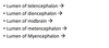
At the cranial end of the neural tube, it becomes pinched to form 3 distinct primary brain vesicles/regions
What are they?
- Forebrain (Prosencephalon)
- Midbrain (Mesencephalon)
- Hindbrain (Rombencephalon)
CVS:
- The wall of the BRAIN vesicles will become the ____ ______
- The lumen of the vesicles will become the _______ and _____ _____
- The wall of the BRAIN vesicles will become the brain tissue
- The lumen of the vesicles will become the ventricles and spinal canal
CVS:
During what week does the foerbrain and hindbrain subdivide?
Week 5
CVS:
The forebrain subdivides into:
- Telencephalon (à Cerebral cortices)
- Diencephalon (à Basal ganglia/hypothalamus)
CVS:
The hindbrain subdivides into:
- Metencephalon (à Pons and cerebellum)
- Myencephalon (à Medulla Oblongata)
CVS:
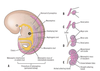
The primary brain vesicles


CVS:
forebrain and hindbrain both subdivide


CVS:

CVS:
- By 26 weeks, the lateral sulcus is evident
- By 30 weeks, the cerebellum is distinct
- By 26 weeks, the lateral sulcus is evident
- By 30 weeks, the cerebellum is distinct
