HARC - MSK Flashcards
Anatomy of the musculoskeletal system


Anatomy of the musculoskeletal system


Anatomy of the musculoskeletal system


Anatomy of the musculoskeletal system


Anatomy of the musculoskeletal system


Basic Principles of MSK


Basic Principles of MSK


The skull


What is pterion?


Spinal cord


Intervertebral discs


ARM ANTERIOR:
What is this superficial muscle and what its function?

Biceps brachii – FLEXES ARM/SUPINATES FOREARM

ARM ANTERIOR:
What is the deeper muscles and what are their functions?

Brachialis – FLEXES THE FOREARM
Coracobrachialis – WEAKLY ADDUCTS/FLEXES ARM

ARM ANTERIOR:
What nerve innervates these muscles?
Biceps Brachii
Brachialis
Coracobrachialis
Musculocutaneous nerve
ARM POSTERIOR:
What muscle is this? And what is its function?
And what is it inervated by?

Triceps brachii – EXTENDS FOREARM

FOREARM ANTERIOR:
What is the superficial muscles shown on the picture?
What is its functions?
What nerve innervates them?

Left to right…
Pronator Teres – PRONATE FOREARM
Flexor carpi radialis – FLEX AND ABDUCT WRIST
Palmaris Longus – FLEXES WRIST
Flexor carpi ulnaris – FLEX AND ADDUCT WRIST
MEDIAN NERVE

FOREARM ANTERIOR:
What is the deep muscles shown on the picture?
What is its functions?
What nerve innervates them?

At the bottom…
Pronator quadratus – PRONATES THE FOREARM
Left to right…
Flexor pollicis longus – FLEXES THE THUMB
Flexor digitorum profundus – FLEXES DISTAL PHALANGES
MEDIAN NERVE

FOREARM POSTERIOR:
What is the superficial muscles shown on the picture?
What is its functions?
What nerve innervates them?

Left to right…
B Brachioradialis (missing) – FLEXES FOREARM
U Extensor carpi ulnaris – EXTENDS/ADDUCTS WRIST
M Extensor digit minimi – EXTENDS WRIST & LITTLE FINGER
D Extensor digitorum – EXTENDS MEDIAL 4 DIGITS
A Anconeus – EXTENDS AND STABILISES THE ELBOW
R Extensor carpi radialis brevis – EXTENDS/ABDUCTS WRIST
R Extensor carpi radialis longus – EXTENDS/ABDUCTS WRIST
RADIAL NERVE

FOREARM POSTERIOR:
What is the deep muscles shown on the picture?
What is its functions?
What nerve innervates them?

Left to right…
Left to right…
I Extensor indices – EXTENDS THE INDEX FINGER
E Extensor pollicis longus – EXTENDS THE THUMB
E Extensor pollicis brevis– EXTENDS THE THUMB
A Abductor pollicis longus – ABDUCTS THE THUMB
S Supinator - SUPINATES FOREARM
POSTERIOR INTEROSSEUS NERVE

Shoulder anterior


Shoulder anterior


Shoulder anterior


Shoulder posterior


Shoulder posterior

Shoulder Posterior


The Rotator cuff muscles


The Rotator cuff muscles


The Rotator cuff muscles


The Thorax


The Thorax


GLUTEAL REGION:
What is the superficial muscles shown on the picture?
What is its functions?
What nerve innervates them?

Left to right…
Ma Gluteus Maximus – EXTENDS/MEDIALLY ROTATES THE THIGH
Me Gluteus Medius – ABDUCTS/MEDIALLY ROTATES THE THIGH
Mi Gluteus Minimus – ABDUCTS/MEDIALLY ROTATES THE THIGH (Tensor Fascia Latae – FLEXES & ABDUCTS THIGH)
Ma – Inferior gluteal nerve
Me/Mi/T – Superior gluteal nerve
GLUTEAL REGION:
What is the deep muscles shown on the picture?
What is its functions?
What nerve innervates them?

Left to right…
O Obturator Internus – ABDUCTS FLEXED THIGH & ABDUCTS/LATERALLY ROTATES EXTENDED HIP
GS Gemellus Superior - LATERALLY ROTATES EXTENDED THIGH
GI Gemellus Inferior – LATERALLY ROTATES EXTENDED THIGH
P Piriformis – LATERALLY ROTATES THE THIGH
Q Quadratus Femoris – ADDUCTS/LATERALLY ROTATES THE THIGH
P – Piriformis nerve
GS&O – Obturator nerve
GI&Q – Quadratus nerve
THIGH ANTERIOR:
What are muscles shown on the picture?
What is its functions?
What nerve innervates them?
L F M S
Left to right…
L Vastus lateralis – EXTENDS LEG
F Rectus Femoris – EXTENDS LEG & FLEXES THIGH
M Vastus Medialis – EXTENDS LEG
S Sartorius – FLEXES/ABDUCTS/LAT ROTATES THIGH. FLEXES LEG
FEMORAL NERVE
THIGH ANTERIOR:
What are muscles shown on the picture?
What is its functions?
What nerve innervates them?

I Iliacus – FLEXES THIGH
P Psoas Major – FLEXES THIGH
FEMORAL NERVE
What are the Quad muscles
Vastus Lateralis
Rectus Femoris
Vastus Medialis
What are the iliopsoas muscles?
iliacus
psoas major
THIGH MEDIAL:
What are the muscles shown on the picture?
What is its functions?
What nerve innervates them?

Left to right…
G Gracilis – ADDUCTS THIGH AND FLEXES LEG
L Adductor Longus – ADDUCTS & MEDIALLY ROTATES THIGH
B Adductor Brevis – ADDUCTS & MEDIALLY ROTATES THIGH
M Adductor Mangus – ADDUCTS & MEDIALLY ROTATES THIGH
P Pectineus – ADDUCTS/MEDIALLY ROTATES THIGH (Obturator Externus – LATERALLY ROTATES HIP)
P – Femoral nerve
Adductors/G/O – Obturator nerve
THIGH POSTERIOR:
What are the muscles shown on the picture?
What is its functions?
What nerve innervates them?

Left to right…
M Semimembranosus – FLEXES/MEDIALLY ROTATES LEG
T Semitendanosus – FLEXES/MEDIALLY ROTATES LEG
B Biceps Femoris – EXTENDS THIGH/FLEXES LEG
SCIATIC NERVE
LOWER LEG ANTERIOR:
What are the muscles shown on the picture?
What is its functions?
What nerve innervates them?

Left to right…
Fibularis/Peroneus tertius – DORSIFLEXES & EVERTS FOOT
Extensor digitorum longus – DORSIFLEXES FOOT & EXTENDS TOES
Extensor Hallucis longus – DORSIFLEXES FOOT & EXTENDS BIG TOE
Tibialis anterior - DORSIFLEXES & INVERTS FOOT
THE DEEP BRANCH OF THE COMMON PERONEAL NERVE
LOWER LEG LATERAL:
What are the muscles shown on the picture?
What is its functions?
What nerve innervates them?

Left to right…
Fibularis/Peroneus Longus – PLANTARFLEXES & EVERTS FOOT Fibularis/Peroneus Brevis – PLANTARFLEXES & EVERTS FOOT
THE SUPERFICIAL BRANCH OF THE COMMON PERONEAL NERVE
LOWER LEG POSTERIOR:
What are the superficial muscles shown on the picture?
What is its functions?
What nerve innervates them?

Top to bottom…
Plantaris – PLANTARFLEX FOOT AND FLEX KNEE
Gastrocnemius – PLANTARFLEX FOOT AND FLEX KNEE
Soleus – PLANTARFLEX FOOT
THE TIBIAL BRANCH OF THE SCIATIC NERVE
Bone
Remodeling Cycle


These hormones would take action if the paitent was hypOcalcemic


Vitamin D: Step 1


Vitamin D: Step 2


Vitamin D: Step 3


Vitamin D Summary:


What is the pectoral girdle composed of?
Scapula and Clavicle
Is the pectoral girdle part of the appendicular or axial skeleton?
appendicular


What joints are involved in the pectoral girdle?
What type of joints are they?
What are their function?
Sternoclavicular - synovial plane joint. Increases freedom of movement for scapula. Mostly slight protraction/retraction and elevation/depression
Acromioclavicular - Synovial plane joint. Gliding movement.
Glenohumeral - Synovial ball and socket. full range of circumduction plus protraction/retraction and medial/lateral rotation.
What joints are involved in the elbow?
What type of joints are they?
What are their function?
Radiohumeral - Synovial ball and socket. Mainly pronation/supination. It goes through the flexion/extension passively.
Ulnohumeral - Synovial hinge. Flexion/extension.
Proximal radioulnar - Synovial pivot joint. Permits pronation/supination
What are the five characteristics of a synovial joint?
- Capsule,
- Supporting ligaments,
- Joint cavity,
- Articular cartilage
- Synovial
Can you classify the bones into axial or appendicular?
The axial skeleton makes up our central axis and consists of the following bones: skull, vertebrae, ribs and sternum. The appendicular skeleton consists of the limbs and girdles. The girdles are the attachment points for the limbs
Biceps brachii
Brachialis
Coracobrachialis
Triceps brachii
To which compartments do these muscles belong?
What is their nerve supply?
BBC → Anterior compartment of upper arm – Musculocutaneous nerve
Triceps brachi → Posterior compartment of upper arm – Radial nerve


What is the primary function of the rotator cuff muscles?
Rotator cuff forms protective sheath for shoulder joint behind, on top and in front. It lacks inferiorly hence shoulder dislocations occurs through this pathway
Shoulder


Elbow

radial annular ligament
radial collateral ligament
Ulnar collateral ligament

Elbow ligaments


Elbow Ligaments


What is the importance of the radial annular ligament (RAL)?
What 2 movements of the forearm occur here?
: Head of radius held against ulna by annular ligament. Allows head of radius to rotate within ligament for supination and pronation
Name all the carpal bones?
scaphoid.
lunate.
triquetral.
pisiform.
hamate
capitate.
trapezium.
trapezoid.
.
Which carpal bone is the largest?
Capitate
- Which carpal bone has a hook?
Hamate
Which carpal bone is the smallest?
Pisiform
Which carpal bone is a sesamoid bone?
Pisiform
Which carpal bone is under the thumb?
Trapezium
Which carpal bones articulate with the radius?
Scaphoid and lunate
Which carpal bones articulate with the ulna?
Trick question – the ulna does not articulate with the carpal bones. There is a triangular fibrocartilage disc instead
Bones of the hand are called?
the metacarpals and phalanges
How many phalanges are there in your body?
56
What is the significance of the blood supply of the scaphoid?
Where can you palpate the scaphoid? What else is in this region?
Palpable in anatomical snuffbox. Shape and blood supply are important. Has a narrow waist which is prone to fracture on outstretched hand. Since proximal portion articulates with radius, blood vessels can only enter the proximal portion through the distal waist and ascend upwards. If the waist is fractured, the proximal portion may undergo avascular necrosis.

scaphoid.
lunate.
triquetral.
pisiform.
trapezium.
trapezoid.
capitate.
hamate


how many muscle groups are in the anterior compartment of the forearm?
Three – Superficial, Intermediate and Deep flexors
how many muscle groups are in the posterior compartment?
One
Which nerve is associated with the ‘funny bone?’
Compression of the ulnar nerve at the elbow during its superficial course behind the medial epicondyle of the humerus (you can see the groove it takes).


Can you name the contents of the carpal tunnel?
Tendons of digitorum supericialis,
flexor digitorum profundus and flexor pollicis longus
median nerve.
What structure forms the roof of this tunnel?
To which bones is this structure attached?
The flexor retinaculum running from trapezium and scaphoid laterally to pisiform and hamate medially.


Innervation


A patient complains of loss of feeling in the lateral three and a half digits on her left hand. She also has muscle wasting of the thumb and complains of difficulty fastening buttons
What condition is this and which nerve is affected?
Carpal tunnel syndrome.
The median nerve, which provides sensory innervation to the lateral 3 ½ digits is being compressed within the carpal tunnel. Atrophy of the thenar eminence is also caused by this and results in difficulty in opposition of the thumb
If you see a patient with hypothenar wasting =
think of ulnar nerve lesion
If you see a patient with thenar wasting =
think median nerve lesion
If you see patient with both thenar and hypothenar =
think brachial plexus lesion and/or T1 compression (such as Pancoast’s tumour in lung cancer)
Can you articulate C1 and C2 together?
yes


What type of joint is made between skull and C1 and between C1 and C2, and what type of movement does it produce?
A synovial pivot joint between the odontoid process/dens (C2) and the anterior arch of C1. It permits lateral rotation (shaking your head “no”). Skull and C1 is nodding movement – saying “yes”
Which features allow the vertebral arteries a passageway through the neck?
Transverse foramina (foramina transversarium). Small holes in the transverse process allow the vertebral arteries to pass between C6 and C1, before entering the skull through the foramen magnum
How bones are in the skull?
19
Name these

Frontal
Parietal
Occipital
temporal
Sphenoid
Zygomatic
Ethmoid
Mandibular
Maxilla
Lacrimal
Nasal
Volmer


: What is the function of the IV discs, and which type of joint are they part of?
It forms a symphysis (secondary cartilaginous joint) between the two vertebrae. Serving to separate them and allow passage of the spinal nerves. They also provide an effective mechanism of distributing and dampening hydraulic pressure under compressive loading.


Coracoid process
Acromion
How does the clavicle articulate with the scapula?
The acromioclavicular joint is a synovial plane joint formed between the acromion process and the lateral margin of the clavicle.


: Between which of the intercostal layers does the neurovascular bundle of the costal groove run?
Between the innermost and internal intercostal muscle layers.
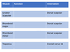
Functions of the following muscles
Serratus anterior
Pectoralis major
Pectoralis minor
Serratus anterior - Protraction and stabilisation of scapula, essential for flexion of the arm at the shoulder. Resists scapular winging.
Pectoralis major - Adduction, medial rotation and flexion of the shoulder. Accessory muscle of respiration.
Pectoralis minor - Depression and rotation of scapula. Assists in elevation of ribs.
The long thoracic nerve (“of Bell”) innervates serratus anterior and is prone to damage during surgery and lymph node removal for breast cancer – what would be the resulting effect to serratus anterior?
Winging of the scapula due to the course of the nerve over the medial wall of the axilla.
Functions of the following muscles?
Rectus abdominis
External oblique
Internal oblique
Transversalis abdominis
Rectus abdominis (with linea alba and semilunaris) - Flexion of lumbar vertebral column.
External oblique - Unilateral contraction=rotation of trunk. Bilateral for compression and stabilisation.
Internal oblique - Unilateral contraction = rotation of trunk. Bilateral for compression and stabilisation.
Transversalis abdominis - Abdominal compression and stability.


What are attachments of the capsule of the hip joint?
Anterior: acetabular margin to the intertrochanteric line. Posterior: acetabular margin to about half-way down the neck of the femur. Inferior: extends somewhat down medial border of the femur to provide some slack on abduction of the lower limb .


What and where are the 2 anastomoses that supply head of femur and hip joint?
Cruciate and trochanteric posterior to hip joint and neck of femur. Receive blood from gluteal and obturator arteries (from internal iliac) and from circumflex branches of profunda femoris.
Why might avascular necrosis of the femoral head occur if femoral neck is fractured?
Same reason as scaphoid necrosis – blood supply supplies the head of femur and joint from distal to proximal so a fracture of the neck would rupture the blood supply.


What is the primary function of gluteus medius and gluteus minimus?
They act together to abduct the thigh and in conjunction with the fascia lata of the thigh, which help you stand on one leg. They prevent the pelvis from dropping on the other side thereby allowing a smooth and balanced gait by balancing the pelvis.
If they were paralysed, how would this be presented in the patient’s gait?
If gluteus medius and minimus are paralysed on one side, this is would result in a waddling gait called Trendelbergs sign.
What is the innervation for these 3 muscles? And where do these nerves originate from?
gluteus medius and gluteus minimus and gluteus maximus
Superior gluteal nerve supplies the gluteus medius and gluteus minimus. Inferior gluteal nerve supplies the gluteus maximus


Rectus femoris
Vastus lateralis
Vastus medialis
Vastus intermedius
What group are these muscles in?
Quadriceps Femoris
Name the muscles in the anterior compartment of the thigh?
Iliopsoas
Sartorius
Rectus femoris
Vastus lateralis
Vastus medialis
Vastus intermedius
What are the muscles in the posterior compartment of the thigh?
Biceps femoris
Semimembranosus
Semitendinosus
What are the muscles of the medial adductor compartment?
Adductor magnus
Adductor longus
Adductor brevis
Gracilis
The sciatic nerve is the biggest nerve of the thigh – how does it get from the pelvis and into the posterior compartment of the thigh?
Greater sciatic foramen


What type of joint is formed between these proximal parts of these two bones?
The proximal tibiofibular joint is a primary cartilaginous joint. The distal tibiofibular joint is a syndesmosis.


What are the 7 tarsal bones?
Talus, Calcaneus, Navicular, Cuboid, 3 Cuneiforms (medial, middle, lateral)
How many tarsals, metatarsals and phalanges are in the toe?
7 t
5 m
14 p
The common fibular nerve wraps around the neck of the fibula as it enters the lateral compartment of the leg – if the nerve is damaged by say a fracture to the neck of the fibula, what would be the resulting effect?
: Foot drop


Can you articulate the head of the talus with the inferior surface of the tibia? What shape is this called?
A mortice joint – stable when the joint is flat but unstable when moved in a plantarflexion direction – like when you wear heels and the ankle joint becomes unstable and wobbly.
Ligaments of the Ankle Joint
Lateral ligaments:
Calcaneofibular: runs from the calcaneus to the fibula
Posterior talofibular: runs from the talus to the posterior part of the fibula
Anterior talofibular: runs from talus to anterior part of the fibula
Ligaments of the Ankle Joint
Medial Ligaments:
Deltoid (medial): runs from medial malleolus and spreads out in a triangle shape
What is the function of the cruciate ligaments?
To prevent anterior and posterior displacement.
What is the function of the collateral ligaments?
to prevent lateral or medial displacement.


How might you test whether a patient has a damaged ACL? Damaged PCL? Damaged collateral ligaments?
Anterior draw test most common method. Most ACL injuries are sustained through nonimpact injury as a result of excessive translation. Posterior draw test for PCL.
What is the function of a meniscus?
: Their most important function is to increase the area of contact between the femoral condyle and tibia. It also acts as a reservoir for synovial fluid, which is released at the point of articulation under compressive loading.
There are three muscle compartments that are found in the leg
What are they?
Posterior compartment: superficial and deep parts
Lateral compartment: fibularis longus and fibularis brevis
Anterior compartment: tibialis anterior, extensor hallucis longus and extensor digitorum longus
Gastrocnemius
Soleus
Popliteus
Tibialis posterior
Flexor hallucis longus
Flexor digitorum longus
fibularis longus
fibularis brevis
: tibialis anterior,
extensor hallucis longus
extensor digitorum longus

