GI histology Flashcards
What are the main layers of the GI tract?
- Mucosa 2. Submucosa 3. Muscularis Propria 4. Adventitia
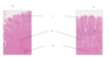
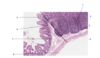
Name the following components on the histological image of the oesphagus below:

- muscularis mucosa
- stratified squamous epithelium
- mucosa
- submucosa
- muscularis
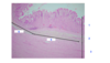
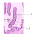
Name the following components on the histological image of the oesphagus shown below:
- stratified squamous epithelium
- lamina propria
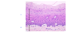
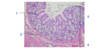
Name the following components on the histological slide of the stomach below:
- mucosa
- submucosa
- muscularis
- not exist- fun
- fundus
- pyloris
Name the following images on the histological slide shown below:
- columnar epithelium
- glands
- muscularis mucosa
- fundus
- pylorus
Name each of the following components:

- Fundus glands
- Pylorus glands
- parietal cells
- mucus cells
- peptic cells
Name the following cells that are present in the image of the gastric glands below:

- parietal cells
- cheif cells
Name the structure that is shown in the duodenum below and state the name of the individual cells that are present:

- Paneth cells- secrete defensive molecules
- Goblet cells- mucin for lubrication and protection
- Neuroendocrine cells- local acting hormones that are involved in regulating motility and secretion
Name the following structures in the jenenum below:

- Simple columnar epithelium
- villus
- lamina propria
- serosa
- mucosa
- muscularis
Name the following features on the jejenum below:

- lamina propria
- crypt
- muscularis mucosa
- serosa
- mucosa
- submucosa
- muscularis
Name the following components of the ileum on the diagram below:

- myenteric plexus
- serosa
- outer longitudinal muscle
- submucosal plexus
- inner circular muscle
Name the following components in the diagram below:
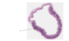
- Peyeers patch
Name the following components in the diagram below:
- Peyers patch
- Mucosa
- Submucosa
- Muscularis
- Lamina Propria
- Muscularis mucosa
- Crypt
- Serosa
Name the following components of the colon:
- colonic glands
- muscularis mucosa
- mucosa
- submucosa
Name the component of the rumen and the structures that are shown within the diagram below:

Its reticulum.
- Primary fold
- Secondary fold
- Stratified squamous epithelium
Name this component of the rumen:

Rumen
Label the following components of the rumen within the diagram below:
- papillae
- stratified squamous epithelium
Name the location of this tissue and name the different layers of it:

Tissue sample taken from the omasum:
- Muscularis mucosa
- Inner layer of the tunica muscularis
- Tunica muscularis
State the location of this tissue:

Abomasum
Name the following components of the diagram below:

- portal vein
- hepatic artery
- bile duct
Name each of the following components of the pancreas on the diagram below:

- Acinar cells
- Secretory cells
- Duct
Name each of the following components of the pancreas on the diagram below:

- Islet of Langerhans
- Capillaries
- Exocrine cells
Name the following components in the diagram below:

- Right uterine horn
- Ovary
- Bladder
- Lateral vesicular ligament
- ureter
- urethra
- Body of the uterus
- Broad ligament
- Not there
- Uterine tube
- fimbriae
- cervix
- vagina
- clittoris
- vestibule of the vagina
- Anus
