FINAL SKIN REVIEW Flashcards
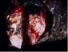
Which of the following conditions you will investigate in a dog presented with this picture?

Staphylococcal pyoderma
food allergy
immune mediated skin conditions
parasitic infections
neoplasm
Dermatitis in bacteriology is often called
pyoderma
Panniculitis
subcutaneous tissue
Cellulitis
dermis and Subcutaneous fat
Most pyoderma/skin infection are due to
Coagulase positive staph
Are skin infections usually the primary issue?
No! Usually problems with the skin are secondary!
Therefore…….
Always investigate underlying causes such as allergy, ectoparasites, Immunosuppressive conditions”
What is the most common gram negative organism that can be involved with a pyoderma skin lesion?
Pseudomonas
A common bacterial isolate from canine pyoderma is
Staphylococcus pseudintermedius
Fungal differentials
- Actinomycosis- gram positive
- Nocardiosis- gram positive
- Mycobacteriosis- gram positive
What is the diagnostic workup for skin conditions?
- Skin scrapings to rule-out parasites such as Demodex
- Fungal culture to rule-out deep fungal infection
- Skin cytology by performing an impression smear of pustules, papules, crusts or draining tract fluid:
- Bacterial culture / susceptibility testing using fresh purulent discharge or a tissue
- Skin biopsy for dermatohistopathology
What is the treatment?
Systemic antibiotics based on culture and susceptibility (up to 8 weeks or longer in refractory cases)
Fluoroquinolones(broad spectrum): (tissue penetration, Activity against Gram positives and negatives, uptake by macrophages increases penetration and concentration)
Clindamycin (consider inducible clindamycin resistance- macrolide restisttant ) Antibacterial topical therapy (Chlorhexidine)

Inducible clindamycin resistance
All staphylococcus isolates which are macrolide(erythromycin) resistant should be considered Clindamycin resistant unless otherwise confirmed by a D-test
How should you submit a superficial lesion?
Superficial lesions- A culturette swab in transport media
What should you submit for an abscess
Abscess –Fine needle aspirates or contents in anaerobic
transport media
What should you submit for a granulomatous lesion?
Granulomatous lesions- Sample for Biopsy and a fresh piece of tissue for culture
What is recommended for a non-resolving lesion?
For any non-resolving lesions a biopsy is always recommended
Can you submit a dry swab for culture?
Do not submit dry swabs for cultures
Anaerobic infections
Foul smelling discharge, necrotic gangrenous tissue and abscess formation, free gas in tissue, black discoloration of exudates,Sulphur granules in discharge
What do you have to do to take an aerobic culture?
Disinfect skin surface with 70% alcohol, allow to dry
Aspirate specimen directly into the syringe.
Remove air from syringe.
Aseptically transfer material into an anaerobic transport media
Greasy pig disease is caused by
Staphylococcus hyicus
Hemophilius parasuis causes
gram negative
causes blue ears in pigs
GLASSER”S DISEASE
Bumble Foot in birds
Staphylococcus aureus
Botryomycosis-
Botryomycosis- Rodents, Human, Horses Chronic pyogranulomatous inflammation
Most common isolate : S. aureus
deep in the tissue
Wound infections, draining tracts, Abscess
What are your top bacterias that you are considering?
Gram Positive anaerobes
Clostridium
Nocardia
Actinomyces
Gram Negative Anaerobes
Fusobacterium
Bacteroides
Dichelobacter

Mycobacteria- negatively stained with Gimesa Stain
Negative (“Not Gram Negative”) stained rods
Dogs and cats presented with Non healing cutaneous lesions and subcutaneous nodules

Dogs and cats presented with Non healing cutaneous lesions and subcutaneous nodules “Mycobacterium”

Dermatophilus congolensis

Dermatophilus congolensis

Trichophyton verrucosum

Trichophyton verrucosum

IN cats:
• Mycobacterial infections(Feline Leprosy)
- Yersinia pestis (lymph node abscess)- Plague
- Cat bite abscess- Pasteurella sp

Actinomyces bovis- lumpy Jaw, pyogranulomatous osteomyelitis
Foot rot- Fusobacterium Necrophorum in cattle, sheep, goat

Mycotoxins (Sporidesmin; Pithomyces chartarum)-Facial eczema due to photosensitization)
fungal toxins that can cause skin lesions due to photosenitization
Mycotic infections of the skin
• Dermatophytosis
- Trychophyton
- Microsporum
- Epidermophyton
Mycotic infections of the skin in Dogs
• Dogs: M.canis, M. gypsium, T. mentagrophytes
Mycotic infections of the skin in Cats
• Cats: M. canis
Mycotic infections of the skin in Equine:
Equine: T. mentagrophytes, T equinum
Mycotic infections of the skin in Cattle:
Cattle: T. verrucosum
Mycotic infections of the skin in Pigs:
Pigs: M. nanum
You can use a Wood’s lamp for?
Microsporum canis
Which medium do you use to test for Dermatophytes?
Dermatophyte test medium- will change the color; but you should not worry about the color change
always also follow up with a Lactophenol cotton blue staining

Persian cat: Pseudomycetoma
Microsporum canis
In cats, Micropsorum canis can go deeper causing these types of lesions.
Which species shows no lesions of Microsporum canis?
Cats
Sometimes, owners will come in and say that they have these lesions, but their cats don’t have any. What will you do? Test the cat! You take a toothbrush and comb the cat and then you send it to the lab.
Where do you collect samples from in a ring worm infection?
MARGINS because is it clearned in the middle
Griseofulvin
Act only against dermatophytes, Need oral administration and the drug reaches the superficial dead epithelium
- high concentration in the stratum corneum
Cryptococcus neoformans
Lungs, CNS, Eyes, Skin)
very common in cats
Blastomyces dermatitidis
Lungs-primary, Skin lesions in disseminated disease
Sporothrix schenckii
cutaneous/lymphatics

Blastomyces dermatitidis- Broad based budding yeast

Cryptococcus neoformans: Capsule

Histoplasma capsulatum:Intracellular small yeasts

Coccidioides immitis- spherule with endosporulation HUGE

Pythiosis and Lagenidiosis
Pythiosis and Lagenidiosis
Horses, dogs and humans
Cutaneous, vascular, ocular, gastrointestinal and a systemic form
Prognosis for most cases is poor
Radical surgery, antifungal drugs, immunotherapy or a combination of these therapies.
After resection, medical therapy using Itraconazole or Terbinafine 10 – 20 percent of dogs respond.
Avoiding stagnant waters

Cutaneous Lagenidiosis

Erysipelothrix rhusiopathiae
Pathognomonic skin lesions observed in swine erysipelas is a local suppurative dermatitis.
True or False
FALSE- those skin lesions are a septicemia that causes vascultis and this those skin lesions.
Strangles, Lymphadenitis
Streptococcus equi subsp equi;
lymphatanitis lesions
Ulcerative lymphangitis (Pigeon
fever)
Corynebacterium pseudotuberculosis- you will see lymphacutaenous lesions in the legs and brisket region
HORSES
Glanders
Burkholderia mallei- horses;
pneunomia, farcy, abscess in the skin
Melioidosis
Burkholderia pseduomallei
abscesses in horses

Pythium insidiosum
oomycosis
Kunkers( coral like necrotic debris)- what comes out of the lesions
Leeches, Bursatti, SWAMP CANCER
What is the best treatment for a mature staphyloccocal abscess?
SURGERY
