Urinary System Flashcards
(72 cards)
Functions of the Urinary System (8)
- Filters Wastes from Blood
- Regulates Ion Levels in Plasma
- Regulates Blood pH
- Conserves Valuable Nutrients
- Regulates Blood Volume
- Regulates RBC Production
- Stores Urine
- Excretes Urine
Organs of the Urinary System
- Kidneys
- Ureters
- Urinary Bladder
- Urethra
Kidney Location
At level of which vertebrae?
Which is higher?
- against dorsal body wall
- retroperitoneal
- at level of T12 to L3
- left kidney higher than right

Two Coverings of the Kidneys
Fibrous Capsule - tough dense CT, surrounds each kidney, giving it shape and a barrier
Adipose Capsule - external to fibrous capsule, surrounds and protects kidney, keeping it in correct location
What is this entire organ?

kidney
What are the three regions of the kidney?
Renal Cortex - outer region; granular appearance
Renal Medulla - inner to cortex; darker
Renal Sinus - large space medial to hilum containing calices, blood vessels, renal pelvis, nerves and fat

Renal Cortex
- outer layer of kidney containing renal corpuscles and parts of renal tubules

Renal Medulla
- inner layer of kidney containing renal pyramids with loops of Henle
What’s this outer layer?

Renal Capsule
- outer dense CT layer
- thin, transparent and lies directly on renal surface
What is this space or notch?

Renal Hilus
- notch in kidney where renal artery and nerve enter and renal vein and ureter exit

Renal Pyramids
- cone-shaped medullary structures containing loops of henle
- 6-18 per medulla
- wide, outer edge is base
- narrow, inner edge near papillae is apex

Renal Columns
- cortical tissue between pyramids
- path of interlobar arteries and veins

Renal Papilla
- pointed end of renal pyramid
- site of collecting duct drainage into calyx

Minor Calyx
- funnel-like structure which collects urine from collecting ducts at papillae

Major Calyx (pl. Calices)
- collects urine from minor calyx and drains it to renal pelvis

Renal Pelvis
- chamber formed by merging of the two major calyces
- connected to ureter
What is the entire structure highlighted by the green line?
What type is it? And what is the other type?

Nephron
- the functional unit of the kidney
- shown here is a Juxtamedullary Nephron, meaning its corpuscle is close to the medulla and its loop of Henle extends deep into the medulla to create an osmotic gradient for creating different concentrations of urine (15% of nephrons are JM)
- the other kind is a Cortical Nephron, its corpuscle is further into the cortex and its loop of Henle just barely dips into the medulla (85% of nephrons)
What are the two components of the nephron?
Renal Corpuscle - glomerulus and Bowman’s capsule
Renal Tubule - PCT, loop of Henle, DCT
What is the entire bracketed structure?

Renal Corpuscle
- site of filtration within nephron
- spherical structure located in cortex
- made up of outer Bowman’s capsule and inner glomerulus
What is it?
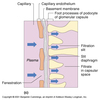
What are its layers and their cells?

Bowman’s (or Glomerular) Capsule
- double walled capsule around glomerulus
- receives filtrate and empties into PCT
Two Layers:
- Visceral Layer (inner) - branching podocytes interdigitate and cling to glomerulus
- Parietal Layer (outer) - simple squamous epithelium
What is this bundle of tubes?

Glomerulus
- a knot of specialized capillaries within a Bowman’s capsule
- capillaries are fenestrated and porous to allow filtration into capsular space
What is the lumen of the Bowman’s capsule called?
Capsular Space
What are the capillaries of the glomerulus called?
Glomerular capillaries
(duh)


