QRS complex
ventricular systole (contraction, depolarization)
What is this and how can you tell?
PVC (spikes between beats)
leukopenia
low WBC count (usually viral infection)
What are the 3 artery groups?
- arterioles- small, control blood pressure
- muscular (distributing) arteries- mostly smooth muscle, vasoconstrict/vasodilate to move blood
- elastic (conducting) arteries- have elastic fibers, when blood from the left ventricle surges in, the artery stretches then recoils to propel blood (ex- aorta, pulmonary, brachiocephalic & carotid arteries)
autorhythmic
“cardiac muscles have the ability to start their own action potentials”
What happens after prothrombinase acts?
a blood clot is formed
What are the 3 special immune system cells and what do they do?
- NK cells (natural killer)- kill body cells that aren’t right (tumors) (Police)
- B cells- lymphocytes that combat extracellular antigens, have the capacity to make a memory of that antigen” function by producing plasma cells which make antibodies (taggers) (Mothership)
- T cells- combat intracellular antigens, these kill infected cells (Mr. T)
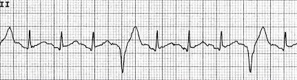
What is this and how can you tell?
Bundle branch block (wide QRS complex)
What are the 5 qualities of heart cells?
-elongated -involuntary -branched cells -intercalated discs -autorhythmic
What is bulk flow movement and how does it happen?
moves large amounts of fluid across capillary walls
- large amounts of fluid enters capillaries on arteriole end
- this generates “blood hydrostatic pressure” (BHP)
- BHP forces fluid out of capillaries (filtration)
- as more fluid leaves, BHP goes down and albumin concentration goes up (albumin is a protein that maintains fluid concentration in the blood)
- high albumin concentration creates “blood colloidal osmotic pressure” (BCOP), drawing fluid back into capillaries (reabsorption) as it enter the venule
EKG problem with multiple extra P waves?
heart block
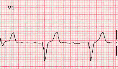
EKG problem with a dipped Q wave?
previous heart attack
What is this and how can you tell?
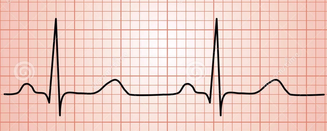
Normal
P wave
atrial systole (contraction, depolarization)
stroke volume
avg. 70mL, amount pumped out by the left ventricle with each contraction
What are the 3 layers of the heart?
- endocardium- innermost layer (includes valves)
- myocardium- muscle layer (left ventricle is the thickest)
- epicardium (visceral pericardium)- membranes on the outside of the heart
What stimulates leukocyte differentiation?
interleukin & colony stimulating factors
Why is blood pressure in veins lower than arteries?
-veins are farther from the heart -veins go from small to large (arteries are opposite)
What is hemoglobin and what role does iron play with it?
protein that carries oxygen inside a RBC, oxygen binds to the iron in the hemoglobin
B cells
lymphocytes that combat extracellular antigens, have the capacity to make a memory of that antigen” function by producing plasma cells which make antibodies (Mothership)
What is this and how can you tell?
Atrial fibrillation (rapid beats, faintness, spikey)
What is a neutrophil?
phagocytic WBC that is first to a wound site
What growth factor is most important to hemopoiesis, why, & where is it made?
erythropoietin- growth factor manufactured by kidney, stimulates stem cells to become RBCs
What is the tunica interna also called?
endothelium


