Structure of glands in the digestive system week 3 Flashcards
(26 cards)
What is the morphology of salivary glands?
What is found btwn lobules?
What are the 2 kinds of ducts?
Compound Tubuloacinar Morphology
- lobular organization with septae
- interlobular ducts (in septae) and excretory ducts
- intralobular ducts
What are 2 physical features of intralobular ducts?
intercalated and striated
What are the 3 main salivary glands?
- parotid
- submandibular
- sublingual

What are histological features of the parotid gland?
only serous acini, prominent striated ducts, often fatty infiltration

What are histological features of the submandibular gland?
mostly serous with some mucus acini. many striated intralobular ducts. excretory: wharton’s duct beneath tongue at frenulum
in pic: note demilunes: mucus acini with surrounding serous cells

What are histological features of the sublingual gland?
mostly mucus acini, some serous cells. no appreciable striated ducts. multiple excretory ducts beneath tongue.
note large ducts in attached pic.

What is the composition of saliva?
Complex fluid of neutral pH composed of mucins and many proteins/glycoproteins and with an ionic composition high in calcium and phosphate, bicarbonate, and potassium, but low in sodium and chloride.
What glands provide mucins in saliva?
What type of acini provide enzymatic and proteinaceous/glycoprotein components of saliva?
- Mucins from the mucus acini in the sublingual and submandubular glands.
- The enzymatic and proteinaceous/glycoprotein components of saliva are derived from serous acini and serous demilunes (esp. lysozyme).
What is contained within striated ducts? What is the importance of them?
Striated ducts have extensive mitochondrial basal infoldings of their basal membrane. They are important in modifying acinar secretion to produce a watery hypotonic secretion by removing sodium and chloride and introducing secreting potassium and bicarbonate.
Which salivary gland produces the most saliva per day?
What are important functions of saliva?
- Saliva is produced at about 1 liter/day with contributions of ~30% (parotid), ~65% (submandibular) and ~5% (sublingual). It has pre-digestive actions (esp. α-amylase [starch], some lipase and protease activity) and provides lubrication for bolus formation during chewing and swallowing. Saliva, including secretions from minor mucosal glands, are important in taste.
- Important antibacterial/viral activity (IgA, lysozyme, lactoferrin).
Salivary secretion is primarily under
a. neural control
b. hormonal control
What is the effect of cholinergic sympathetic innervation on saliva? Beta adrenergic innervation?
Salivary secretion is primarily under neural (not hormonal) control. Cholinergic parasympathetic innervation increases acinar secretion and a watery saliva; Adrenergic (β) innervation alters blood flow but also stimulates acinar secretion to produce a thicker mucoid saliva.
What are the histological features of the exocrine pancreas? (morphology, cell types, secretions, stimulation of secretions)
- Compound Tubuloacinar Morphology
- Serous Acini (secrete lipases, proteases, amylases, nucleotidases)- stimulated by CCK and VIP, inhibited by pancreatic polypeptide
- Centroacinar Cell –pale staining cell at origin of ducts secrete bicarbonate stimulated by secretin
• Both are stimulated by parasympathetic and inhibited by sympathetic (largely through decreased blood flow) innervation

What cell types are present within Islets of Langerhans? What do they each secrete?
What is the most prominent cell type?
- alpha cells (α) – glucagon (15-20 % of Islets)
- beta cells (β) - insulin (60-70% of Islets)
- delta cells (δ) - somatostatin (5-10% of Islets)
- other minor cell types are also present and have an impact on gastrointestinal function along with their counterparts in the gut mucosa.
Attached pic: Arrows point to islets. Note that the panreas is arrannged in lobules. Note septa marking boundaries of ducts.

How is insulin released from beta cells controlled?
How are the 3 cell types within Islets distributed?
What regulatory role do the secretions of alpha, beta, and delta cells have on one another?
- Beta cells are primarily responsive to elevated blood glucose levels, secreting insulin to enhance uptake and utilization of glucose by peripheral tissues and the liver.
- They are under control of parasympathetic (+) and sympathetic (-) innervation as well as circulating metabolites and hormones.
Alpha and delta cells tend to have a more peripheral distribution in the islet with first access to blood flow to the center of the islet, so that the beta cell is under inhibitory modulation of both somatostatin and glucagon from these cells. Somatostatin also inhibits glucagon secretion.

The liver also has exocrine and endocrine features.
What is released a result of endocrine fxn? Exocrine fxn?
• Also a mixed exocrine and endocrine gland
• Endocrine and exocrine functions performed by the same cells (unusual feature of the liver)
Endocrine – plasma proteins and lipoproteins
Exocrine – bile
T or F: Lobular structure provides a blood “filter” with both portal venous input from the intestine and systemic arterial input.
True.

The classical liver lobule is organized as plates of ______ surrounded by ______ that contain blood flowing from branches of the hepatic portal vein and hepatic artery to a central vein in the center of the lobule.
hepatocytes
sinusoids

How do metabolites and oxygen delivered into sinusoids have access to hepatocytes?
What does the wall of sinusoids contain? What is the fxn of this?
Metabolites and oxygen delivered into sinusoids by the portal/hepatic artery at the lobule peripherally have access through the incomplete sinusoidal wall to hepatocytes that line the sinusoids in plates organized in vertical radial arrays. The incomplete wall of each sinusoid consists of fenestrated endothelial cells and interspersed fixed macrophages called Kupffer cells. The latter form a filtration system to extract microbial antigen antibody complexes, heme-breakdown products, and other metabolites in blood passing through the sinusoid.
Where do Spaces of Disse’ lie? What is the purpose?
What are bile canaliculi?
What issues with the bile duct system can cause jaundice?
Molecules can also diffuse into the extrasinusoidal Space of Disse’ which lies between the sinusoidal lining cells and the hepatocyte. Products produced by hepatocytes are also secreted into this space from which they gain access to the circulation.
Small channels formed by folds in the plasma membrane form continuous channels (bile canaliculi) that convey bile to the bile ducts.
Bile is secreted into bile canaliculi which are channels formed between adjacent hepatocytes by adherens type junctions. These channels form an anastamosing network within each hepatic plate that deliver bile to small ducts at the perimeter of the lobule and to the bile duct component of the portal triad. The portal biliary system converges to form the hepatic duct. The common bile duct is formed by union of the hepatic duct with the cystic duct from the gall bladder where bile is stored and concentrated. Obstructions of the biliary system in the major ducts (e.g. gall stones) or by pathology or scarring in the liver parenchyma produce bile stasis and expansion of the canalicular system. This can results in escape of bile pigments into the sinusoidal blood, a cause of jaundice.

What fxn of the liver does the classical lobule define?
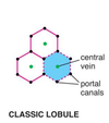
The classical lobule defines the endocrine function of the liver (protein production). It is centered on central veins.

What are the boundaries of portal lobules? What function of the liver do portal lobules define?
The PORTAL LOBULE, highlighting the exocrine function of the liver in bile secretion, is centered on the portal triad (branch of hepatic portal vein, hepatic artery and a bile duct). A portal lobule is a triangular area defined by lines connecting the central veins of three adjacent classical lobules with the portal triad and its bile duct at its center.

What are the boundaries of the liver acinus? What function of the liver do liver acini define?
What are the zones within liver acini?
What does the nutrient/oxygen gradient within liver acini determine?
The LIVER ACINUS defines a functional gradient of hepatocyte metabolism that differs between the perimeter of the classical lobule and the central region. This metabolic gradient is created by the concentration of metabolites adjacent to the portal triads (periportal zones) which are high in oxygen, glucose, and other metabolites, and the oxygen/nutrient poor regions of the sinusoids closer to the central veins.
The acinus is physically defined by a triangle with its base extending between two portal triads to its apex at the central vein. It is subdivided into Zones I, II, III. Zone I, closest to the base has high oxygen and glucose; zone III closest to the central vein is glucose and oxygen poor. This gradient determines the patterns of glycogen deposition and removal as well as exposure of hepatocytes to natural metabolites or toxins. The acinus is an important pathophysiologic concept that influences patterns of cellular degeneration seen in liver pathology, e.g. the preference for centrolobular degeneration in some forms of poisoning.

How can CHF lead to liver necrosis?
Blood supply to liver is compromised due to CHF. Supply of blood going to zone I: still reasonably oxygenated. By the time blood reaches zone III, there is not enough nutrients or oxygen present for cells in this area. Secondary liver failure can develop as a result. Because the liver is so highly vascularized, hepatocytes are more sensitive to lack of blood. Also, still have to deal with toxins

What are Ito (hepatic stellate cells)? What is their fxn? What role may they play in liver fibrosis?
What is the role of Kupffer cells?
Ito or Hepatic stellate cells store vitamin A, remodel ECM, produce growth factors and cytokines, regulate sinusoidal lumen size in response to prostaglandins and thromboxanes. In liver disease these cells become more fibroblast like in character and secrete collagen and contribute to liver fibrosis.
Kupffer Cells – are monocytes derived cells that contribute to the sinusoid wall. Are phagocytic cells that can take up and dispose of senescent red cells and compensate if this function in lost in the spleen.


