muscle charts (spine/LE) COPY COPY Flashcards
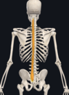
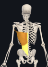
erector spinae
intermediate back layer
- attachments: TLF attached to sacrum, iliac crest, spinous processes L5-T11, supraspinous ligament, angles of ribs > costal angles (iliocostalis), ribs and transverse processes (longissimus), and spinous processes (spinalis)
- spinalis: thoracis, cervicis, capitis
- longissimus: capitis, cervicis, thoracis
- iliocostalis: cervicis, thoracis, lumborum
- action: extension and lateral flexion of vertebral column
- innervation: dorsal rami of spinal nerves

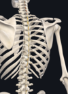
semispinalis
deep, transversospinales group
- attachments: transverse processes > distant superior spinous processes
- semispinalis: capitis, cervicis, thoracis
- action: extension and contralateral rotation of vertebral column
- innervation: dorsal rami of spinal nerves

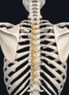
multifidus
deep, transversospinales group
- attachments: sacrum, posterior SI ligament, posterior iliac spine, transverse processes > more superior spinous processes
- multifidus: sacral, lumbar, thoracic, cervical
- action: extension and contralateral rotation of vertebral column
- innervation: dorsal rami of spinal nerves
rotatores longus and brevis
deep, transversospinales group
- attachments: transverse processes > spinous processes of vertebra above (1-2 levels)
- action: extension and contralateral rotation of vertebral column
- innervation: dorsal rami of nerves
interspinales
deep
- attachments: between adjacent spinous processes of adjacent vertebrae
- cervical: 6
- thoracic: 2-3
- lumbar: 4
- action: extension of spine
- innervation: dorsal rami of spinal nerves
intertransversarii
deep
- attachments: between transverse processes of adjacent vertebrae
- action: lateral flexion of spine
- innervation: dorsal rami of spinal nerve

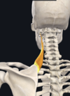
splenius cervicis
superficial
- attachments: spinous process of T3-T6 > posterior tubercles of transverse processes of first 3 cervical vertebrae
- action: extension, lateral flexion, ipsilateral rotation of head and neck
- innervation: dorsal rami of spinal nerves
splenius capitis
superficial
- attachments: ligamentum nuchae and spinous processes of T4-C7 > lateral 1/3 of superior nuchal line
- action: extension, lateral flexion, ipsilateral rotation
- innervation: dorsal rami of spinal nerves

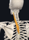
longus colli
- attachments: anterior tubercles and bodies T3-C3 > anterior tubercles and bodies C6-C1
- action: flexion, lateral flexion, ipsilateral rotation
- innervation: ventral rami of spinal nerves (C2-7)

longus capitis
- attachments: anterior tubercles C3-C6 > basilar portion occipital bone
- action: flexion, ipsilateral rotation
- innervation: ventral rami of spinal nerve (C1-3)

latissimus dorsi
- attachments: lower thoracic spinous processes, thoracolumbar fascia, iliac crest > floor of intertubercular sulcus of humerus
- action: adduction, extension, medial rotation of humerus
- innervation: thoracodorsal nerve (C6-8)
levator scapulae
- attachments: transverse processes of C1-4 > superior angle and superomedial border of scapula
- action: elevation and downward rotation of scapula
- innervation: dorsal scapular nerve (C4-5) - sensory C3-4
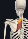
rhomboids
- attachments: spinous processes T2-3, ligamentum nuchae, spinous processes C7-T1 > medial border of scapula
- action: scapular retraction, elevation and upward rotation
- innervation: dorsal scapular nerve (C4-5) - sensory C3-4
trapezius
- attachments: superior nuchal line, external occipital protuberance, ligamentum nuchae, cervical and thoracic spinous processes > clavicle, acromion, and scapular spine
- action: scap elevation, retraction, depression, upward rotation
- innervation: accessory nerve (CN XI) - sensory C3-4

extrinsic superficial muscle layer
axio-appendicular
- trapezius
- latissimus dorsi
- levator scapulae
- rhomboids
CN XI/ventral rami of spinal nn. to dorsum of body
extrinsic intermediate muscles
- serratus posterior superior
- serratus posterior inferior
intercostal nn. at corresponding levels
intrinsic superficial muscles
- splenius capitis
- splenius cervicis
dorsal rami of spinal nn.
intrinsic intermediate layer
erector spinae
- spinalis
- thoracis, cervicis, (semi) capitis
- longissimus
- capitis, cervicis, thoracis
- iliocostalis
- cervicis, thoracis, lumborum
lateral br. of dorsal rami of spinal nn.
intrinsic deep layer
- semi spinalis
- capitis, thoracis, cervicis
- multifidus
- rotatores
- longus, brevis
- interspinales, intertransversarii
- function in proprioception
dorsal rami of spinal nn.
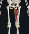
adductor brevis
- attachments: inferior pubic ramus > distal 2/3 pectineal line, medial lip linea aspera
- action: adducts and flexes hip
- innervation: obturator nerve (L2-4)

adductor longus
- attachments: pubic crest > medial lip linea aspera
- action: adduction, medial rotation, flexion of hip
- innervation: obturator nerve (L2-4)
adductor magnus
- attachments: inferior pubic ramus, ischial ramus, ischial tuberosity >
- hamstring: medial supracondylar ridge, adductor tubercle of femur
- adductor: gluteal tuberosity, linea aspera
- action:
- hamstring: extends hip
- adductor: adducts and flexes hip
- innervation:
- hamstring: tibial division of sciatic nerve (L4-S1)
- adductor: obturator nerve (L2-4)

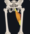
gracilis
- attachments: body of pubis, inferior pubic ramus > medial surface of tibia (lateral to sartorius inseration, distal to condyle, proximal to semitendinosus insertion)
- action:
- flexes and medially rotates knee
- adducts hip
- innervation: obturator nerve (L2-4)
pectineus
- attachments: superior pubic ramus > femur between lesser trochanter and linea aspera (pectineal line)
- action: adducts, flexes hip
- innervation: femoral nerve and obturator nerve (L2-4)

obturator externus
- attachments: ramis of pubis and ischium, external surface obturator membrane
- action: laterally rotates hip
- innervation: obturator nerve (L3-4)

biceps femoris
- attachments:
- long head: ischial tuberosity, sacrotuberous ligament > lateral side of fibular head
- short head: lateral lip of linia aspera, lateral supracondylar ridge > lateral side of fibular head
- action:
- LH: extends hip, flexes and laterally rotates knee
- SH: flexes and laterally rotates knee
- innervation:
- LH: tibial division of sciatic nerve (L5-S3)
- SH: fibular division of sciatic nerve (L5-S2)
semitendinosus
- attachments: ischial tuberosity > proximal, medial tibia
- action: extends hip, flexes and medially rotates knee
- innervation: tibial division of sciatic nerve (L4-S2)

semimembranosus
- attachments: ischial tuberosity > posterior medial tibial condyle
- action: extends hip, flexes and medially rotates knee
- innervation: tibial divison of sciatic nerve (L4-S2)

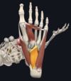
extensor digitorum longus
- attachments: lateral tibial condyle, proximal 3/4 of fibula, CIM > dorsal digital expansions of toes 2-5
- action: dorsiflexes ankle, extends digits 2-5 MP and IP
- innervation: deep fibular (L4-S1)

extensor hallucis longus
- attachments: middle 1/2 of fibular surface, CIM > distal phalangeal base of 1st toe
- action: dorsiflexes ankle, extends great toe MP and IP
- innervation: deep fibular nerve (L4-S1)

fibularis tertius
- attachments: distal fibula, CIM > base of 5th metatarsal
- action: everts foot, dorsiflexes ankle
- innervation: deep fibular nerve (L4-S1)

tibialis anterior
- attachments: lateral condyle, proximal 2/3 of lateral tibial surface, CIM > medial cuneiform, adjacent 1st metatarsal
- action: dorsiflexes ankle, inverts foot
- innervation: deep fibular nerve (L4-S1)

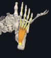
fibularis longus
- attachments: head and proximal 2/3 of fibula > lateral aspects of 1st metatarsal, adjacent medial cuneiform
- action: everts foot, plantar flexes ankle, depresses 1st metatarsal head
- innervation: superficial fibular nerve (L4-S1)

fibularis brevis
- attachments: distal 2/3 of fibula > lateral base of 5th metatarsal
- action: everts foot, plantar flexes ankle
- innervation: superficial fibular nerve (L4-S1)

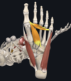
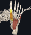
flexor digitorum longus
- attachments: posterior tibia distal to soleal line > plantar surface of distal phalangeal bases
- action: plantar flexes ankle, flexes digits 2-5 MP and IP
- innervation: tibial nerve (L5-S2)

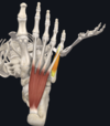
flexor hallucis longus
- attachments: distal 2/3 of posterior fibular surface, CIM > plantar aspect of distal phalangeal base of 1st toe
- action: plantar flexes ankle, flexes great toe (MP and IP)
- innervation: tibial nerve (L5-S2)

gastrocnemius
- attachments: posterior aspect of femoral condyles, joint capsule > posterior calcaneal surface
- action: flexes knee, plantar flexes ankle
- innervation: tibial nerves (S1-2)

plantaris
- attachments: lateral supracondylar line > posterior calcaneal surface
- action: flexes knee, plantar flexes ankle
- innervation: tibial nerve (L4-S1)
popliteus
- attachments: lateral femoral condyle, oblique popliteal ligament > soleal line of tibia
- action: unlocks knee from extension into early flexion
- NWB: medially rotates tibia and flexes knee
- WB: laterally rotation of femur and knee
- innervation: tibial nerve (L4-S1)

soleus
- attachments: posterior aspect of head and proximal 1/4 of fibula, tibial soleal line > posterior calcaneal surface
- action: plantar flexes foot at ankle joint
- innervation: tibial nerve (L5-S2)

tibialis posterior
- attachments: CIM, lateral tibial surface, medial fibular surface > navicular, medial and intermediate cuneiform, bases of metatarsals 2-4
- action: inverts foot, plantar flexes ankle
- innervation: tibial nerve (L4-S1)

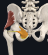
superior gemellus
- attachments: external surface of ischial spine via obterator internus tendon > greater trochanter
- action: laterally rotates hip
- innervation: sacral plexus (L5-S2)

inferior gemellus
- attachments: proximal ischial tuberosity via obturator internus tendon > greater trochanter
- action: laterally rotates hip
- innervation: sacral plexus (L4-S1)

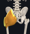
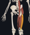
gluteus maximus
- attachments: aponeurosis of erector spinae, sacrum, sacrotuberous ligament, posterior gluteal line (innominate) > greater trochanter, gluteal tuberosity of femur, iliotibial tract
- action: hip extension, lateral rotation
- innervation: inferior gluteal nerve (L5-S2)
gluteus medius
- attachments: external iliac surface > oblique ridge on lateral aspect of greater trochanter, gluteal aponeurosis
- action: abducts and medially rotates hip, keeps pelvis level when opposite leg is raised
- innervation: superior gluteal nerve (L4-S1)

gluteus minimus
- attachments: external iliac surface and margin of greater sciatic notch > anterolateral aspect of greater trochanter
- action: abducts and medially rotates hip, keeps pelvis level when opposite leg is raised
- innervation: superior gluteal nerve (L4-S1)
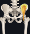
obturator internus
- attachments: anterolateral wall of pelvis and obturator membrane > medial surface of greater trochanter
- action: laterally rotates hip
- innervation: nerve to obturator internus (L5-S2)
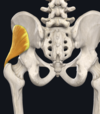
piriformis
- attachments: anterolateral sacrum, posterior inferior iliac spine > upper border of greater trochanter
- action: abducts and laterally rotates hip
- innervation: nerve to piriformis (S1-2)

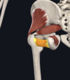
quadratus femoris
- attachments: ischial tuberosity > quadrate tubercle of femur
- action: laterally rotates hip
- innervation: nerve to quadratus femoris (L4-S1, sacral plexus)
iliacus (iliopsoas)
- attachments: iliac fossa, iliac crest, sacral ala, sacroiliac ligament > lesser trochanter of femur
- action: flexes and stabilizes hip joint
- innervation: femoral nerve (L2-4)
psoas major (iliopsoas)
- attachments: anterior transverse processes, vertebral bodies and discs (T12-L5)
- action: flexes and stabilizes joint
- innervation: ventral rami L1-4 (from lumbar plexus)

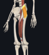
rectus femoris
- attachments: anterior iliac spine, groove superior to acetabulum > base of patella
- action: flexes hip, extends knee
- innervation: femoral nerve (L2-4)

sartorius
- attachments: anterior superior iliac spine > medial aspect of proximal tibia
- action: flexes/abducts/laterally rotates hip, flexes/assists medial rotation of knee
- innervation: femoral nerve (L2-3)

tensor fasciae latae
- attachments: anterior superior iliac spine, external lip iliac crest > iliotibial tract
- action: abducts/flexes/medially rotates hip, assists in maintaining knee extension
- innervation: superior gluteal nerve (L4-S1)
vastus intermedius
- attachments: anterior aspect of proximal 2/3 of femoral shaft > lateral border patella
- action: extends knee
- innervation: femoral nerve (L2-4)

vastus lateralis
- attachments: intertrochanteric line, greater trochanter, gluteal tuberosity, linea aspera > base and lateral border of patella
- action: extends knee
- innervation: femoral nerve (L2-4)
vastus medialis
- attachments: intertrochanteric line, spiral line, linea aspera, medial supracondylar line > base and medial border of patella
- action: extends leg
- innervation: femoral nerve (L2-4)

articularis genu
can be blended with vastus intermedius
- attachments: distal anterior shaft of femur > proximal portion of synovial membrane of knee joint
- action: pulls articular capsule proximally
- innervation: femoral nerve (L2-4) - branch of nerve from vastus intermedius

abductor digiti minimi
- attachments: calcaneal tuberosity > lateral side of proximal phalangeal base of 5th digit
- action: abducts and flexes 5th digit
- innervation: lateral plantar nerve (S1-2)

abductor hallucis
- attachments: medial calcaneal tuberosity > medial side of proximal phalangeal base of great toe
- action: flexes and abducts great toe
- innervation: medial plantar nerve (L4-S1)

adductor hallucis
- attachments: 2-4th metatarsal bases (oblique head), distal ends of 3-5 metatarsal (transverse head) > lateral base of proximal phalanx of great toe
- action: adducts great toe
- innervation: lateral plantar nerve (S1-2)
extensor degitorum brevis
medial slip is extensor hallucis brevis
- attachments: anterolateral calcaneus > dorsal aspect of base of proximal phalanx (digit 1 for EHB), lateral side of tends of extensor digitorum longus (digits 2-4 for EDB)
- action: extensions of digits 2-5 (MP and IP, EDB), extension of digit 1 (EHB)
- innervation: deep fibular nerve (L4-S1)

extensor hallucis brevis
- attachments: extensor digitorum brevis > dorsal aspect of base of proximal phalanx of hallux
- action: extension of great toe at MP joint
- innervation: deep fibular nerve (L4-S1)
quadratus plantae
- attachments: medial surface of calcaneus, lateral process of calcaneal tuebrosity > long flexor tendons
- action: assists flexor digitorum longus
- innervation: lateral plantar nerve (S1-2)
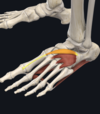
flexor digitorum brevis
- attachments: medial process of calcaneal tuberosity > sides of intermediate phalanges of digits 2-5
- action: flexes toes (proximal IP)
- innervation: medial plantar nerve (L4-S1)
flexor digiti minimi brevis
- attachments: 5th metatarsal base > proximal phalangeal base
- action: flexion of 5th digit (MP)
- innervation: lateral plantar nerve (S1-2)
flexor hallucis brevis
- attachments: cuboid, tendon of tibialis posterior > base of proximal phalanx of great toe
- action: flexion of great toe (MP)
- innervation: medial plantar nerve (L4-S1)
dorsal interossei
- attachments: metatarsal shafts > proximal phalangeal bases, dorsal digit expansions of digits 2-4
- action: abducts and flexes MP joint of digits 2-4
- innervation: lateral plantar nerve (S1-2)

plantar interossei
- attachments: plantar surface of base of metatarsals > dorsal digital expansions of digits 3-5
- action: adducts and flexes MP digits 3-5
- innervation: lateral plantar nerve (S1-2)

lumbricals
- attachments: tendons of flexor digitorum longus > medial sides of dorsal expansions of digits 2-5
- action: flexes proximal phalanges (MP), extends IP joint
- innervation:
- 1st: medial plantar nerve (L4-S1)
- 2nd-4th: lateral plantar nerve (S1-2)

