MSK cadavers upper limb Flashcards


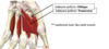
What is the common flexor origin in the forearm for superficial & intermediate flexors
medial epicondyl of humerus
what is the innervation of the superficial flexors
the superficial flexors are innervated by the median nerve (FCR, palmaris longud and pronator teres), apart from flexor carpi ulnaris- ulnar nerve
muscle in the intermediate flexor compartment of forearm & its innervation
flexor digitorum superficialis
Median nerve


A. Flexor carpi ulnaris
B. Palmaris longus
C. Flexor carpi radialis
D. Pronator teres
Action & innervation of brachioradialis

Flexion of the elbow
Innervation = radial nerve
label deep group of anterior forearm muscles & their innervation

FDP- lateral half= median n
medial half= ulnar n
FPL- median n
Pronator quadratus- median n
inserion and origin for FDS and FDP
FDS=Palmar surfaces of middle phalanges of medial 4 digits
FDP= Palmar surface of distal phalanges of medial 4 digits
neurovascular structures of the forearm


Describe Allens test
To determine the patency of the arteries in the distal forearm (prior to sampling of arterial blood, or insertion of arterial lines), Allen’s test is used.
The reason that this is performed is because in some individuals a unilateral circulation exists in the distal forearm. Therefore, if any cannulation is performed there is a risk of causing ischaemia (due to reduced blood flow) to the hand
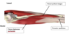
Contens of the anticubital fossa from lateral to medial
Radial nerve, Brachial tendon, Brachial artery, Median nerve
Really Need Beer, To Be At My Nicest

What 4 carpal bones does the flecor retinaculum anchor to
scaphoid, trapezium, hamate and pisiform
Name the contents of the carpal tunnel & label diagram

Contents of Carpal Tunnel
4 tendons of flexor digitorum profundus
4 tendons of flexor digitorum superficialis
1 tendon of flexor pollicis longus
Median Nerve


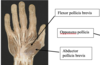
what are the four mucles in the deep part of the central component of the palm
- Flexor digitorum superficialis
- Flexor digitorum profundus
- Flexor tendon sheaths: flexor pollicus longus
- lumbricles
what is the function of the lumbricals
flex the fingers at the metacarpophalangeal joints and extend the interphalangeal joint of 2nd to 5th digits.
- bend a straightened finger
what do the lumbricals arise from
Medial and lateral aspects of the FDP tendon, between the 1st to 5th metacarpals
label this diagram


label these


what type of joint is the proximal and dstil radio-ulnar joint


what carpal bones does the radius articulate with
scaphoid and lunate bones

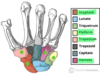
A. ulna
b. lunate
c. triqetrum
d. hamate
e. capitate
f. trapezoid
g. trapezium
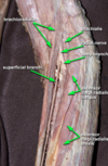
label posterior compartment


label these two nerves


what is the function of the Posterior interosseous nerve
main motor supply of the posterior forearm muscles- deep branch of the radial nerve
where does the superficial branch of the radial nerve run?
As it branches at the cubital fossa it runs deep to brachioradialis and enters the hand passing over the anatomical snuff box
It is purely sensory and distributed to skin on the dorsum of the hand.
label anatomical snuff box


what four muscles attach onto the dorsal digital expansion + label the diagram

- Lumbricals of the hand
- dorsal interossei muscles
- Palmar interossei muscles
- Extensor indicis muscle

Complete the diagram


Complete diagram

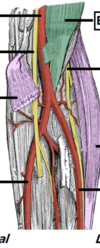
a) Abductor pollicis brevis
b) Opponens pollicis
c) Ulnar artery
d) Ulnar nerve
e) Flexor carpi ulnaris
f) Palmaris longus
g) Flexor carpi radialis

Complete diagram


Complete diagram


Complete the blanks


Complete the diagram


Fill in the blanks


Fill in the blanks


Complete the diagram

Complete the diagram


Complete the diagram


Complete the diagram


Complete the diagram


Complete the diagram


Complete the diagram


Complete the diagram


Complete the diagram


Complete the diagram


Innervation of flexor digitorum profundus vs superficialis
FDP= Ulnar & median nerves
FDS= median nerve
Relationship of the ulnar nerve and ulnar artery
After passing posterior to the medial epicondyle of the humerus the ulnar nerve enters the forearm by passing between the heads of flexor carpi ulnaris muscle.
Both the ulnar nerve and ulnar artery descend close to each other down the medial aspect of the forearm.
The ulnar nerve lies medially to the ulnar artery at the level of the wrist
