MSCT Week 5: Cutaneous Infections Flashcards

1
Q

Types of Cutaneous Infections and Infestations

5 Listed

A
2
Q

Question 1

A

Impetigo (staphylococcus or streptococcus)

3
Q

Impetigo Infection Type and Pathogen

A

Superficial Bacterial Infection

Staphylococcus aureus

Streptococcus pyogenes

4
Q

Impetigo Location, morphology and description

A

commonly around the mouth or perineum

  • crusted
  • glazed
  • eroded papule to plaques
  • peripheral rim of scale
  • Honey Crusted
  • May be tender or asymptomatic
  • uncommonly bullous
5
Q

“Honey Crusted” is descriptive of?

A

Impetigo

6
Q

Impetigo Treatment

A

Topical or oral antibiotics

7
Q

Impetigo Overview

A
8
Q

Identify

A

Impetigo

9
Q

Bullous
Impetigo

A

True Blistering Impetigo

10
Q

Question 2

A

Cellulitis

11
Q

Cellulitis Description

A

Common but serious bacterial skin and soft tissue infection

12
Q

Cellulitis Morphology and appearance

5 listed

A
  • Edematous
  • erythematous
  • warm
  • sometimes taut/shiny localized plaque
  • usually unilateral
13
Q

Cellulitis Etiology

A
  • May be initiated by a skin injury
14
Q

Cellulitis Treatment

A
  • Rest
  • elevation
  • topical or systemic antibiotics
15
Q

Cellulitis Overview

A
16
Q

Question 3

A

Culture the Nasopharynx

because where is the problem and what is the bacteria doing to cause the problem

staph aureus toxin epidermolytic Toxin is coming from bacteria somewhere else

staph aureus lives in nasopharynx or perineum regions

17
Q

Necrotizing Fasciitis Description & Pathogen

A
  • Rare “Flesh-eating bacteria”
  • deeper tissue injury
  • usually anaerobic bacteria or Grp A Streptococcus pyogenes
18
Q

Necrotizing Fasciitis Crepitus

A

Creates Gas

Gas bubbles throughout the skin

crackling of gas upon palpation

19
Q

Necrotizing Fasciitis Morphology and appearance

A
  • purple dusky necrotic color
  • can be ulcerous and bullae
  • crackling of gas upon palpation
20
Q

Necrotizing Fasciitis Symptoms and treatment

A
  • Associated severe pain
  • systemic symptoms
  • Surgical emergency
  • IV antibiotics and other interventions
21
Q

Necrotizing Fasciitis Overview

A
22
Q

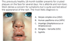
Staph Scalded Skin Syndrome (SSSS) Description and Pathogen

A

Epidermolytic-toxin produced by S. aureus

23
Q

Staph Scalded Skin Syndrome (SSSS) caused by and seen in?

A
  • Cleavage/split of epidermis (basically just peels off)
  • Toxin comes from a bacteria somewhere else (usually nasopharynx or perneum)
  • Typically affects infants and younger children in immunocompromized and physiologically decreased renal function
24
Q

Staph Scalded Skin Syndrome (SSSS) Pertinant negative

A

not affecting mucosa just perioral and eyes

25
Staph Scalded Skin Syndrome (SSSS) Diagnosis
* Must culture the primary site like the nasopharynx or perianal * no true muscosal involvement
26
Staph Scalded Skin Syndrome (SSSS) Treatment
* Systemic anti-staphylococcal antibiotic (PO or IV)
27
Identify
Staph Scalded Skin Syndrome
28
Question 4
Herpes Simplex Virus (HSV)
29
Herpes Simplex Virus Histology
individual keratinocytes infected by herpes simplex virus have mutiple nuclei have steely blue look with margination molding nuclei fit together and mold together 3 Ms Margination Multinucleation Molding
30
Herpes Simplex Virus Histology
* individual keratinocytes infected by herpes simplex virus * have multiple nuclei * have a steely blue look to the nucleus with margination * molding nuclei fit together and mold together * 3 Ms * Margination * Multinucleation * Molding
31
Herpes Simplex Histology
* individual keratinocytes infected by herpes simplex virus * have multiple nuclei * have a steely blue look to the nucleus with margination * molding nuclei fit together and mold together * 3 Ms * Margination * Multinucleation * Molding
32
Herpes Simplex Virus Description morphology and appearance
Fever blisters cold sores genital erosions the initial infection may be asymptomatic to fulminant stomatitis individual lesions may last days to weeks
33
Herpes Simplex Virus Treatment
* systemic antiviral if indicated
34
Herpes Simplex Virus Recurrence
once infected recurrence is common
35
Herpes Simplex Virus Overview
36
Question 5
Chicken Pox (Varicella)
37
Varicella Zoster Virus Symptoms, prevalence
Chicken Pox can have systemic symptoms Largely reduced via immunization Very concerning in the immunocompromized
38
Varicella Zoster Lesion Description
Widespread disseminated papules and vesicles
39
Varicella Zoster Virus Lesion Morphology
* widely distributed vesicles and papules that are discreet from one another (disseminated papules and vesicles
40
Varicella Zoster Virus Reactivation
Zoster or shingles Dermatomal distribution of papules, vesicles, and plaques
41
Varicella Zoster Virus Concerning Presentations 2 listed
* Ophthalmic involvement (can cause blindness) * Ramsay-Hunt Syndrome: Facial Palsy and Ear pain
42
Varicella Zoster Virus Histology
Identical to HSV
43
Varicella Zoster Overview
44
Question 6
Human Papilloma Virus (HPV)
45
Human Papilloma Virus (HPV) Description
Verruca (warts)
46
Human Papilloma Virus (HPV) Subtypes 4 listed but there are more
* Vulgaris * Palmoplantar * Plana * Condyloma acuminata
47
Vulgaris means
Common
48
Plana means
Flat
49
Condyloma acuminata
Genital
50
Human Papilloma Virus (HPV) considerations
if extensive consider immunosuppression
51
Human Papilloma Virus (HPV) Treatment 3 listed
* Cryotherapy (freezing) * Chemical destruction * Topical immunomodulators or irritants
52
Human Papilloma Virus (HPV) Overview
53
Verruca Vulgaris Histology
* Rete bends in towards the middle * papillomatous epidermal acanthosis * hyperkeratosis * hypergranulosis
54
Verruca Vulgaris Histology
Church spires papilomatous architecture hemorhage within the cornified layer
55
Verruca Vulgaris Histology
Keratinocytes infected have HPV change called a koilocyte (keratinocytes have a white halo around them and granular cytoplasm) indicative of HPV infection black dots are hemorrhage dried blood
56
Koilocyte
keratinocytes have a white halo around them and granular cytoplasm Indicative of HPV infection
57
Question 7
Molluscum Contagiosum
58
Molluscum Contagiosum Histology
59
Molluscum Contagiosum Pathogen
Pox Virus
60
Molluscum Contagiosum Characteristic lesion description and location
* dome shaped papules with a waxy surface * single or multiple * may be pruiritic * 5mm * trunk, face, axillae, genital area * spread by scratching
61
Molluscum Contagiosum spread by?
scratching (self-inoculation)
62
Molluscum Contagiosum center of lesion
a curd-like core can be expressed from center
63
Molluscum Contagiosum Course
spontaneous remission 2-3 yrs
64
Molluscum Contagiosum Treatment
* watchful waiting * curetting after topical anesthetic * especially in poorly controlled eczema
65
Question 8
Tinea Pedis (athlete's foot) can be diagnosed by KOH Stain
66
Tzanck Stain diagnoses
Herpes Simplex Virus PCR is usually used now though
67
Tinea Pedis AKA
Athletes foot
68
Mollusca Contagiosum sources of contagion
* SexuallTransmitteded * skin-on-skin
69
Tinea Pedis Pathogen
Dermatophyte infection 40+ species of fungi that feed on dead skin * Trichophyton * Microsporum * Epidermopyton
70
Tinea Pedis Diagnosis
* KOH Stain * Fungal culture if inconclusive
71
Tinea Pedis pathogen and diagnosis overview
72
Tinea Pedis Histology
* neutrophils sitting in the cornified layer * pink staining hyphae in the cornified layer
73
Tinea Pedis Histology Fungal Hyphae
74
Tinea Infections Examples 7 listed
+ Scalp Nails Versicolor
75
Annular Eruption should remind of
Ringworm Tinea Corporis
76
Tinea infections of the scalp and nails
77
Tinea Versicolor
* Pityosporum not feeding on dead skin but sebum and oils neck chest and back * see hypopigmented round or cersinate well demarcated * can be hypopigmentation or hyper or pink
78
Question 9
Secondary Syphilis (Treponema pallidum)
79
Syphilis description and pathogen
* Sexually Transmitted Disease * Treponema pallidum * have primary, secondary and teritary syphilis
80
Primary Syphilis Presentation
solitary or multiple **painless** genital "Chancres"
81
Secondary Syphilis
Rash & condyloma lata, systemic symptoms (febrile, fatigued etc.)
82
Tertiary Syphilis
* Gummas * Aortitis * neurosyphilis
83
Congenital Syphilis
* Stillbirth * Acral bullae/erosions * Rhinitis * Rhagades * Deafness * wet moist white epidermis lifting off erosive change
84
Condyloma lata definition
is a cutaneous condition characterized by wart-like lesions on the genitals
85
Syphilis histology
Plasma cells in the skin are big indicator because they're not usually found there
86
Identify
Syphilis Plasma cells in the skin not usually seen in the skin
87
Identify
Syphilis Plasma cells in the skin which aren't usually found there
88
Identify
Secondary Syphilis
89
Secondary Syphilis Histological Characteristics
90
Question 10
Scabies
91
Scabies Etiology
* Skin infestation by human itch mite (Sarcoptes scabiei) * Adult female burrows into epidermis & lays eggs
92
Scabies transmission
Transmitted by close skin-skin contact
93
Scabies lesion description
Moth-eaten papules, burrows on hands, feet, waistline, genitals
94
Scabies diagnosis
Mineral Prep oil and scraping to remove papule
95
Scabies Transmission
Extremely itchy Self-innoculation transmitting to others
96
Scabies treatment
Permethrin Cream Topically & again in 7 days when eggs have had a chance to hatch
97
Scabies Histology
98
Scabies Overview
99
How does permethrin cream work?
Permethrin is not ovicidal so it kills the mites but the eggs survive and treated again in 7-10 days to kill all new mites that were born
100
Overview
101
MSSA
methicillin-susceptible S. aureus (MSSA) infections
102
MRSA
methicillin-resistant S. aureus (MSSA) infections
103
HPV
Human Papilloma Virus
104
HSV
105
Dermatophytes
106
Cellulitis Symptoms
* fever/afrebile * edema * erythematosus * warm * red * shiny tight skin with an abscess * pain and tenderness
107
Mollusca Contagiosum Most common in?
Children
108
Herpes Simplex Virus Types
* Type 1 is more common orally * Type 2 is more common on the genitals
109
Tinea Pedis AKA
Athlete's Foot
110
Tinea Corporis AKA
Ring Worm
111
Tinea Manuum AKA
Fungal infection of the hands
112
Tinea Cruris AKA
Jock Itch
113
Tinea Onychomycosis
Dermatophytosis of the nails
114
Tinea Capitis AKA
Dermatophytosis or fungal infection of the Scalp
115
Tinea Versicolor Pathogen
Pityrosporum orbiculare
116
Pathology?
Chancre Primary Syphilis
117
Scabies Lesion in Children
* palms and soles pinpointer ovoid white vesicle * look moth-eaten ill-defined raggedy pink and brown hemorrhagic crusting
118
Scabies lesion in adults
slightly scaly papules in the web space in adults