MSCT Week 4: Lower Limb Flashcards

1
Q

Major Functions of the Lower Limb

2 listed

A
  • Support the body weight
  • Move the body weight
2
Q

Gluteal Region

A

between the iliac crest and the gluteal fold that defines the lower limit of the buttocks

3
Q

Each of the two pelvic bones of the gluteal region is formed by?

A

childhood fusion of 3 bones

  • Ilium
  • Ischium
  • Pubis
4
Q

Anterior Thigh boundaries

A

between the inguinal ligament and the knee joint

5
Q

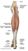
Posterior Thigh Boundaries

A

Between the Gluteal Fold and the Knee

6
Q

Thigh bone is?

A

Femur

7
Q

Leg Boundaries

A

between the knee and ankle joints

8
Q

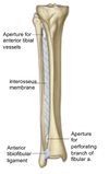
The bones of the leg?

A
  • Tibia
  • Fibula
9
Q

Bones of the foot

3 listed

A
  • Tarsals
  • metatarsals
  • phalanges
10
Q

How is muscular energy reduced to maintain a standing position

A

The ligaments of the hip and knee joints and the shapes of the articular surfaces “locks” the joints in position to reduce the muscular energy required to maintain a standing position

11
Q

Identify Anatomical Parts

A
12
Q

Identify

A
13
Q

Identify Movements

A
14
Q

The Hip Joint Description & Function

2 parts

A
  • a synovial joint between the head of the femur and cup-shaped acetabulum on the lateral surface of the pelvic bone
  • it is a multi-axial ball and socket joint designed for stability and weight bearing
15
Q

Acetabular Labrum Description & Function

2 listed

A
  • the fibrocartilaginous collar on the rim of the acetabulum on the lateral surface of the pelvic bone
  • it deepens the acetabulum and prevents the femoral head from moving inferiorly
16
Q

Acetabular labral tears

A
17
Q

Ligament of the head of the femur Description

A

connective tissue that attaches the head of the femur to the acetabular fossa

18
Q

The femoral head is supplied by?

A

A branch of the obturator artery supplies femoral head through a branch inside the head of the femur ligament

19
Q

The synovial membrane around the hip joint attaches to?

A

The articular surfaces of the femur and acetabulum

20
Q

Identify

A
21
Q

Identify

A
22
Q

Fibrous capsule or membrane of the hip joint Description & Function

A
  • surrounds the synovial membrane
  • holds the femoral neck in the acetabulum
23
Q

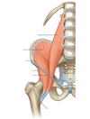
How many ligaments around the fibrous capsule of the hip? what are they called?

A

3

  • Iliofemoral ligament
  • Pubofemoral ligament
  • Ischiofemoral ligament
24
Q

Iliofemoral Ligament location

A

Anterior

25
Pubofemoral ligament location
Anteroinferior
26
Ischiofemoral ligament Location
Posteroinferior
27
Identify
28
Largest synovial joint in the body
The knee
29
The knee joint is formed by the articulations of?
* The medial and lateral femoral condyles * the medial and lateral tibial condyles * the patella
30
The stability of the knee joint relies on?
* Quadriceps * hamstrings * ligaments
31
Ligaments of the knee joint 4 listed
* Medial or tibial collateral ligament (MCL or TCL) * Lateral or fibular collateral ligament (LCL or FCL) * Anterior Cruciate Ligament (ACL) * Posterior Cruciate Ligament (PCL)
32
Medial collateral ligament origin and insertion
* extends from the medial epicondyle of the femur and attaches on the medial aspect of the tibia * it is attached to the capsule and medial meniscus
33
Medial collateral ligament Function
prevents lateral displacement of the tibia under the femur
34
Identify
35
What prevents lateral displacement of the tibia under the femur
Medial collateral ligament
36
Lateral collateral ligament Origin and Insertion
extends from the lateral epicondyle of the femur and attaches to the head of the fibula
37
Lateral collateral ligament Function
Prevents medial displacement of the tibia under the femur
38
Cruciate ligaments description and properties 3 listed
* cross each other in the intercondylar region of the knee * they are inside the capsule but outside the synovial membrane * they connect the tibia and femur and restrict anterior to posterior movement of the knee joint
39
Anterior Cruciate Ligament Origin and Insertion
it extends from the lateral femoral condyle to the anterior tibia
40
Anterior Cruciate Ligament Function
* Keeps the tibia from moving anteriorly on the fixed femur * it resists hyperextension and is weaker than the PCL
41
Posterior Cruciate Ligament Origin and Insertion
it extends from the medial femoral condyle to the posterior tibia
42
Posterior Cruciate Ligament Function
* keeps the tibia from moving posteriorly on the fixed femur * Tension on it is greatest when the knee is flexed
43
Identify
44
Identify
45
Medial and Lateral Menisci description and function
C-shaped fibrocartilage between the articulating condyles that help make the surfaces more congruent and serve as shock absorbers
46
Most common menisci injury
The medial meniscus is more frequently injured than the lateral due to its attachment to the MCL
47
Synovial membrane of the knee joint description & function 2 listed
* attaches to the margins of the articular surfaces and menisci * it forms pouches or bursa that provide low-friction movement for tendons such as the (Suprapatellar bursa and subpopliteal recess)
48
Fibrous capsule of the knee Description & Function 3 listed
* encloses the articular cavity * it is partly formed and reinforced by muscle tendons * the upper end of the popliteal muscle is enclosed by the fibrous membrane
49
Patellar Ligament Description
* The patellar Ligament is the continuation of the quadriceps tendon * it connects the patella to the tibia
50
Identify
51
Most commonly injured structures in the knee
Called the Unhappy Triad of O'Donahue * MCL * Medial Meniscus * ACL Resultant from a blow to the lateral aspect of the knee with the foot planted on the ground The injury is often accompanied by an audible snap and immediate pain and swelling
52
Unhappy Triad of O'Donahue
* Called the Unhappy Triad of O'Donahue * MCL * Medial Meniscus * ACL * Resultant from a blow to the lateral aspect of the knee with the foot planted on the ground * The injury is often accompanied by an audible snap and immediate pain and swelling
53
The Ankle Joint Description
* Is a synovial hinge joint between the talus, tibia and fibula. * the distal ends of the fibula are anchored together by medial (deltoid) and lateral ligaments to create a deep socket for the upper body of the talus
54
Lateral Ligament of the ankle description
Composed of 3 ligaments is the most commonly sprained by foot inversion * Posterior talofibular ligament * Anterior talofibular ligament * Calcaneofibular ligament
55
Most common sprain of the ankle
Anterior talofibular ligament due to foot inversion
56
Medial Deltoid Ligament of the ankle inferior attachment sites
is divided into four parts based on the inferior points of attachment * posterior tibitalar * tibiocalcanela * tibionavicular * anterior tibiotalar parts
57
Sprain of the Medial Deltoid Ligament of the ankle usually occurs how?
most often occurs by foot eversion
58
Identify
59
Identify
60
Lachman Test Procedure, Structure Tested and Positive Result
61
Anterior Drawer Test Procedure, Structure Tested and Positive Result
62
Posterior Drawer Test Procedure, Structure Tested and Positive Result
63
Valgus Stress Test Procedure, Structure Tested and Positive Result
64
Varus Stress Test Procedure, Structure Tested and Positive Result
65
McMurray Test Procedure, Structure Tested and Positive Result
66
Patellar Apprehension and Patellofemoral Grind Test Procedure, Structure Tested and Positive Result
67
Valgus
when the foot is more lateral (knock-kneed)
68
Varus
When the foot is more medial (bow-legged)
69
The muscles in the gluteal region do what?
* Abduct * extend * laterally rotate the femur relative to the pelvic bone
70
Deep Muscles of the Gluteal Region 4 listed
are lateral rotators of the femur at the hip joint * Piriformis * obturator internus * the gemelli (twins) * quadratus femoris
71
Superficial Muscles of the Gluteal Region
Abduct (gluteus minimus and medius) extend the hip (gluteus maximus) the tensor fasciae latae muscle stabilizes the knee in extension by acting on the iliotibial tract (a band of deep fascia that passes down the lateral side of the thigh to attach to the proximal end of the tibia)
72
Iliotibial tract
(a band of deep fascia that passes down the lateral side of the thigh to attach to the proximal end of the tibia)
73
Identify
74
Identify
75
Nerves enter the gluteal region from?
* The pelvis through the greater sciatic foramen * most are in the plane between the superficial and deep muscles
76
Superior Gluteal Nerve innervates and location
* gluteus medius * gluteus minimus * tensor fascia latae Passes superior to the piriformis muscle
77
The nerve to the quadratus femoris also supplies?
Gamellus Inferior
78
The nerve to the obturator internus supplies and location & roots
* Gemellus superior & the obturator internus * passes through the lesser sciatic foramen to innervate * L5-S2 Spinal Nerves
79
The posterior femoral cutaneous nerve of the thigh innervates
innervates the skin of the gluteal region, the posterior thigh, and posterior leg
80
The inferior gluteal nerve innervates
gluteus maximus muscle
81
The piriformis muscle is innervated by branches of?
L5, S1 and S2 spinal nerves
82
The Pudendal Nerve is the nerve of the?
Perineum
83
Pudenal nerve roots
S2-S4
84
Inferior gluteal nerve roots
* L5 * S1 * S2
85
Obturator internus nerve roots
* L5 * S1 * S2
86
Quadratus Femoris nerve roots
* L4 * L5 * S1
87
Superior Gluteal Nerve Roots
* L4 * L5 * S1
88
Trendelenburg sign/gait
an abnormal gait (as with walking) caused by weakness of the abductor muscles of the lower limb (gluteus medius and gluteus minimus people with injury of the superior gluteal nerve have weakness of abducting the thigh at the hip
89
people with an injury of this nerve have weakness of abducting the thigh at the hip
Superior Gluteal Nerve
90
the upper end of the popliteal muscle is enclosed by?
The Fibrous Capsule of the Knee
91
Identify
92
Describe the Trendelenburg Sign
93
The Superior and Inferior Gluteal Arteries supply?
The gluteal region
94
The Superior and Inferior Gluteal Arteries originate from
from the internal iliac artery which is a terminal branch of the aorta
95
Identify
96
How many compartments does the thigh have?
3 Anterior compartment Medial compartment Posterior compartment
97
The compartments of the thigh are separated by?
intermuscular fascial septa
98
Anterior compartment of the thigh functional contents and muscle action
* Femoral Nerve * Branches of the femoral artery * Extension of the Leg
99
Medial Compartment of the Thigh Functional Contents & muscle Action
* Obturator Nerve * Branches of the deep femoral artery and obturator artery * Adduction of the thigh * Gracilis also flexes the knee
100
Posterior Compartment of the Thigh Functional Contents
* Tibial division of sciatic nerve (common fibular division of sciatic nerve to short head of biceps femoris) * Perforating branches of deep femoral artery * Flexion of the leg * Extension of the thigh
101
Anterior compartment of the thigh muscles
Quadriceps femoris * Rectus Femoris * Vastus Lateralis * Vastus Medialis * Vastus intermedius Sartorius Psoas Major Ilicus (iliopsoas)
102
Rectus femoris function
* flexes the thigh at the hip joint * extends the leg
103
Vastus Lateralis Function
Extend the leg
104
Vastus Medialis Function
Extend the leg
105
Vastus intermedius Function
Extend the leg
106
Sartorius Function
* Flexes the thigh * Flexes the leg * laterally rotate the thigh * medially rotate the leg
107
Psoas major Function
Flex the thigh
108
Iliacus AKA
Iliopsoas
109
Iliacus Function
Flex the thigh
110
Muscles of the medial compartment of the thigh
* Gracilis (not shown) * Pectineus (not shown * Adductor Longus * Adductor Brevis * Adductor Magnus * Obturator Externus
111
Gracilis Function
* Adduct the thigh * medially rotate the thigh * flex the knee
112
Pectineus Function
* Adduct the thigh * medially rotate the thigh
113
Adductor Longus Function
* Adduct the thigh * medially rotate the thigh
114
Adductor Brevis Function
* Adduct the thigh * medially rotate the thigh
115
Adductor Magnus Function
* Adduct the thigh * medially rotate the thigh
116
Obturator Externus Function
* Laterally Rotates the thigh
117
Posterior compartment muscles
Hamstrings * Biceps femoris * Semitendinosus * Semimembranosus
118
The posterior compartment muscles mainly
Extend the thigh at the hip and flex the leg at the knee and rotate both joints
119
Biceps Femoris heads
Has 2 heads long and short the long head crosses the hip and knee joints the short head does not
120
Biceps Femoris Function
* Flex the leg at the knee joint * extend the leg at the hip joint
121
Semitendinosus Function
* flex the knee * extend the hip
122
Semimembranosus Function
* flex the knee * extend the hip
123
Pes Anserinus AKA
Goose's Foot
124
Pes Anserinus is what?
is the common insertion of the tendons of the sartorius, gracilis and semitendinosus muscles of the proximal tibia
125
The common insertion of the sartorius, gracilis and semitendinosus muscles
Pes Anserinus
126
Identify
127
Identify
128
Identify
129
The compartments of the leg are separated by?
Interosseous membrane and intermuscular fascial septa
130
The Anterior Compartment of the leg grossly contains
* 4 muscles * deep fibular nerve * Anterior Tibial Artery * Dorsiflexion and inversion of Foot * Extension of toes
131
The Lateral Compartment of the Leg contains
* 2 muscles * Superficial Fibular Nerve * Fibular Artery * Eversion and plantar flexion of foot
132
The posterior compartment of the leg contains & action
* 3 superficial muscles * 4 deep muscles * Tibial nerve * Posterior tibial artery * Plantar flexion * inversion of foot * flexion of toes
133
Identify
134
Muscles of the anterior compartment of the leg
* Tibialis Anterior * Extensor hallucis longus * Extensor digitorum longus * fibularis tertius
135
Anterior compartment of the leg muscles action All have in common 2 listed
* dorsiflex the foot * extend the toes
136
is the most commonly sprained by foot inversion
Lateral ligaments of the ankle * Posterior talofibular ligament * Anterior Talofibular ligament * Calcaneofibular Ligament
137
Lateral compartment muscles of the leg
* Fibularis Longus * Fibularis Brevis
138
Lateral compartment muscles action all have in common
everts foot
139
Posterior compartment superficial and deep muscles of the leg
Superficialis muscles * Gastrocnemius * Soleus * Plantaris Deep Muscles * Tibialis posterior * Flexor Digitorum longus * Flexor hallucis longus * popliteus
140
Identify
141
Identify
142
Identify
143
Identify
144
Compartment Syndrome Description
occurs when increased pressure within a fascial compartment compromises the blood supply, innervation and function of the tissues within that space
145
Symptoms of Compartment Syndrome
* pain * numbness * swelling
146
Compartment syndrome properties 5 listed
* may be acute, following trauma * may be chronic, seen most often in athletes * most common in the anterior leg but can occur in other compartments of leg, forearm, hand, foot * Acute compartment syndrome (ACS) following injury is a surgical emergency * ACS is relieved by cutting the skin and fascia (fasciotomy)
147
Identify
148
Acute Compartment syndrome is treated with
149
Lumbosacral plexus is formed by?
the anterior (ventral) rami of L1-L4 Spinal nerves
150
The femoral nerve is formed by?
The ventral rami of L2-L4
151
Femoral Nerve Motor to?
* Motor to the anterior compartment of the thigh and the pectineus
152
The saphenous nerve travels through
the adductor canal (opening in the aponeurosis of the adductor magnus)
153
An aponeurosis is?
A flat tendon
154
Obturator nerve is formed by?
L2-L4 Ventral Rami of spinal nerves
155
Obturator nerve Motor Functions
* motor to the medial compartment of the thigh and the Adductor Magnus * innervates part of pectineus along with femoral nerve
156
The Sacral plexus is formed by?
Ventral Rami of L4-S5 Spinal nerves
157
The sciatic nerve tibial division
has a tibial division that supplies the posterior compartment of the thigh, except the short head of the biceps femoris
158
The Biceps Femoris Short head is innervated by?
The common Fibular Division of the Sciatic nerve
159
Sciatic nerve divisions
* Tibial division * Common Fibular (peroneal) nerves
160
Common Fibular (peroneal) nerves location
proximal to the popliteal fossa (the space behind the knee)
161
The tibial nerve innervates
the posterior compartment of the leg and is sensory to overlying skin and the plantar surface of the foot (and the 5th toe)
162
The common fibular nerve splits into
* The superficial fibular nerve * The deep fibular nerve
163
The superficial fibular nerve innervates?
The lateral compartment of the leg and is sensory to overlying skin of the leg and foot
164
The deep fibular nerve innervates?
the anterior compartment of the leg and overlying skin of leg and between the great and 2nd toe
165
Identify
166
Describe the major branches of the lumbosacral plexus
167
Four major routes by which nerves and vessels pass into the lower limb
* Obturator Canal * Greater Sciatic Formation * Lesser Sciatic Foramen * Inferior to inguinal ligament
168
The Obturator Canal is
an opening in the obturator foramen through which the obturator nerve passes
169
The Greater Sciatic Foramen main channels
* Below the piriformis muscle * Above the Piriformis muscle
170
The Sciatic Nerve Below the piriformis muscle 5 listed
* Sciatic nerve * inferior gluteal nerve * pudendal nerve * posterior femoral cutaneous nerve * nerves to obturator internus and quadratus femoris
171
The greater Sciatic Foramen Above the piriformis muscle
Between the gluteus medius and minimus * Superior gluteal nerve
172
Lesser Sciatic Foramen nerves that pass
Pudendal nerve passes from the gluteal region into perineum
173
Inferior to inguinal ligament nerves that pass 3 listed
* Femoral nerve * femoral branch of genitofemoral nerve * the lateral cutaneous nerve of thigh
174
Identify
175
Identify
176
Identify
177
Identify
178
Identify
179
Sensory Endpoint of Obturator
medial thigh above knee
180
Sensory Endpoint of Femoral
Medial Malleolus
181
Sensory Endpoint of Tibial
lateral malleolus (sural nerve)
182
Sensory Endpoint of Superficial fibular
Dorsum of the foot
183
Sensory Endpoint of Deep Fibular
Between the first two toes
184
Deep Fibular Nerve * Common entrapment regions * Muscle Weakness * Sensory loss
185
Superficial Fibular Nerve * Common entrapment regions * Muscle Weakness * Sensory loss
186
Tibial Nerve * Common entrapment regions * Muscle Weakness * Sensory loss
187
Femoral Nerve * Common entrapment regions * Muscle Weakness * Sensory loss
188
Obturator Nerve * Common entrapment regions * Muscle Weakness * Sensory loss
189
Femoral artery
* the major artery supplying the lower limb originating from the external iliac artery when it passes under the inguinal ligament to enter the femoral triangle in the anterior thigh * It supplies most of the thigh and all of the leg and foot
190
Borders of the femoral triangle
* inguinal ligament superiorly * sartorius muscle laterally * adductor longus muscle medially
191
The femoral nerve, artery, vein, and lymphatics pass from the abdomen into the lower limb under the?
Inguinal ligament and in the femoral triangle
192
Femoral Sheath
The femoral vein, artery and lymphatics are enclosed by fascia called the femoral sheath
193
Femoral pulse
The femoral pulse can be palpated in the femoral triangle just inferior to the inguinal ligament and midway between the anterior superior iliac spine and the pubic symphysis
194
The superior and inferior gluteal arteries supply
the gluteal region
195
The obturator arteries supplies
the medial compartment of the thigh
196
197
Collateral circulation to the hip joint
anastamoses between
198
Identify
199
Collateral circulation of the knee
anastomoses between
200
Identify
201
Identify
202
Identify
203
Identify
204
Tensor Fascia Lata
the tensor fasciae lata muscle stabilizes the knee in extension by acting on the iliotibial tract (a band of deep fascia that passes down the lateral side of the thigh to attach to the proximal end of the tibia)
205
Anterior Compartment of the thigh Muscle Actions
206
Posterior Compartment of the thigh Muscle Actions
207
Medial Compartment of the thigh Muscle Actions
208
Muscles of the Anterior Compartment of the thigh
Sartorius Vastus medialis Iliopsoas Rectus femoris vastus lateralis vastus intermedius (not shown) pectineus iliacus
209
Muscles of the medial compartment of the thigh 4 listed
* Adductor Longus * Adductor Brevis * Adductor Magnus * Gracilis
210
Muscles of the Posterior Compartment of the Thigh
211
Superficial muscles of the posterior leg
212
Tension on it is greatest on the PCL when
The knee is flexed
213
Hyperextension of the knee is resisted by?
The ACL
214
Femoral Nerve Sensory Branches 2 listed
Sensory branches are the anterior cutaneous nerve of the thigh and the saphenous nerve which goes through the adductor canal opening in the aponeurosis of the adductor magnus to the medial leg and malleoulus
215
Obturator Nerve Sensory Functions
Sensory branch (a cutaneous branch) to the skin above the medial side of the knee
216
The Biceps Femoris Long head is innervated by?
Tibial Division of the Sciatic Nerve