LOCO Revision3 Flashcards
Where does the axillary artery run between? [2]
lateral border 1st rib to inferior border of teres major

Brachial plexus
What are the anterior cord nerves [3]
What are the posterior cord nerves [2]
Anterior cord (lateral to medial)
* Musculocutaneous
* Median
* Ulnar
Posterior cord
* Axillary
* Radial

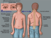
How do you differentiate between fractured clavicle or dislocated shoulder?
Look for rounded profile of the shoulder:
Shoulder dislocation:
* Shoulder squared off – can see acromioclavicular joint
Clavicular fracture:
* Rounded profile of shoulder - SCM pulls medium section upwards. Tenting of the joint
Clavicle on left; shoulder dislocation on right

Which muscle is causing this fractured clavicle to shorten the width of the shoulder? [1]
Pectoralis major (major adductor – brings arm closer) have lifted the shoulder up and shorten the width

Which structure in the shoulder is at risk of damage from shoulder dislocation that increases liklihood of refracture [1]
Glenoid labrum
Describe scapulo-humeral rhythm [2]
Scapula and humerus move in a 1:2 ratio.
When the arm is abducted 180 degrees, 60 degrees occurs by roation of the scapula and 120 degrees by rotation of the humerus at the shoulder joint
Which nerves [2] and muscles [2] are at risk of from axillary lymph node clearance? [2]
Injury to thoracodorsal nerve; Latissimus dorsi
Injury to long thoracic nerve; serratus anterior - causes winging scapula

How does spinal accessory nerve damage present? [4]
- Weak shoulder abduction not as prominent winging of scapula
- Loss of adduction - DOUBLE CHECK
- Atrophy of trapezius
- Shoulder falls
Sternoclavicular joint:
What is the type of joint? [1]
Which structure is present for shock absorption? [1]
Which type of movement does the joint permit? [1] ?
joint type: synovial, saddle joint
shock absoption: articular disc
movements: around 60 degrees when elevate scapula

If you break your clavicle, which structures act and cause different movements of the clavicle? [3]
What do u need to help bring bones back together to heal? [1]
- Sternocleidomastoid muscle: pulls medial aspect of clavicle up
- Pectoralis major muscle pulls arm and clavicle medially
- Gravity will pull down
- need an internal fixation

label these xox
which of these labels are joining points for muscles? [3]

which of these labels are joining points for muscles? [3]
- coracoid process
- supraspinous fossa
- infraspinous fossa

what are two prominent structures / features of the the glenohumeral joint? [2]
Why is this clinically significant [1]
what are two prominent features of the glenohumeral joint?
glenoid cavity accomodates approx/ 1/3 of the humeral head: means that should can have wider range of movement
inferior joint capusule is lax. allows elevate above head. but means is much weaker than superior portion

Which structures deepen the shallow glenoid fossa? [2]
what is the shallow glenoid fossa deepened by? [2]
- glenoid labrum (fibrocart. ring that surrounds articular surface). helps deepen the socket and support the joint
- long head of the biceps - attaches to superior aspect of labrum
label A-E

A: acromioclavicular joint
B: corocoid process
C: clavicle
D: acromion
E: glenoid fossa

Which muscle initates the first 10 degrees of abduction? [1]
supraspinatus


Which of the following is the glenoid labrum?
A
B
C
D
E

Which of the following is the glenoid labrum?
A
B
C
D
E
Which of the following is the acromion?
A
B
C
D
E

Which of the following is the acromion?
A
B
C
D
E
Which of the following is the scapula?
A
B
C
D
E

Which of the following is the scapula?
A
B
C
D
E
Which of the following is the supraspinatus muscle?
A
B
C
D
E

Which of the following is the supraspinatus muscle?
A
B
C
D
E

Which of the following is the glenoid labrum
A
B
C
D
E

Which of the following is the glenoid labrum
A
B
C
D
E
Which of the following is the glenoid cavity
A
B
C
D
E

Which of the following is the glenoid cavity
A
B
C
D
E
What is muscle D? [1]
What is bone E? [1]

D: supraspinatus
E: Acromion
Which of the following is the articular capsule
A
B
C
D
E

Which of the following is the articular capsule
A
B
C
D
E
Which of the following is the articular capsule
A
B
C
D
E

Which of the following is the articular capsule
A
B
C
D
E
which arteries, found in the hand, do the ulnar [1] & radius [1] arteries supply?
radial artery gives rise to deep palmar arch
ulnar artery gives rise to superficial palmar arch


where do each of the following arrive from?
- Muscularcutaneous
- axillary
- median
- radial
- ulnar nerve
where do each of the following arrive from?
- Muscularcutaneous: C5-C7
- axillary: C5-C6
- median: C5-T1
- radial: C5-T1
- ulnar nerve; C8-T1
What is the best way to id the different nerves of the brachial plexus?
Musculocutaneous, median & ulnar are anterior to axillary artery & form M shape
Behind axillary artery: **
superior - axillary, inferior - radial**

Musculocutaneous nerve
Which group of muscles does the musculocutaneous nerve supply? [1]
Name the muscles [3]
Where is the sensory area that the musculocutaneous arm supplies? [1]
musculocutanous supplies the flexor compartment motor supply:
- bicep brachialis
- brachialis
- corachbrachialis
musculocutanous supplies the flexor compartment sensory supply:
- skin of lateral forarm


What is Erb’s palsy caused by damage to? [1]
which nerves? [3]
what does it result in? [3]
Erbs palsy:
- Damage to the superior trunk of the brachial plexus.
- This happens quite commonly in difficult birth (pulling the head away from the upper limb) results in nerve palsy.
- Damaging C5 and C6 mainly affects musculocutaneous, axillary and medial nerve. It results in:
- Adducted shoulder
- Medially rotated arm
- Extended elbow
Label A-D

A: Latissimus dorsi muscle forming the posterior axillary fold
B: Subscapularis muscle
C:Deltoid
D: External abdominal oblique muscle
Label A-D

A: Brachialis
B: Long head of biceps brachii muscle
C: Deltoid muscle (insertion on humerus)
D: Short head of biceps brachii muscle
Label A-E

A: bicep brachii
B: triceps brachii
C: brachioradialis
D: subscapularis
E: Axillary artery
Label A

Brachioradialis muscle
Label A-C

A: Coracobrachialis muscle
B: Teres major
C: Medial head of triceps brachii muscle
Label 1-5

1: bicep brachii
2: brachialis
3: Pronator teres muscle
4: Brachioradialis muscle
5: Pronator quadratus muscle
Which of the following is the median nerve
A
B
C
D
E

Which of the following is the median nerve
A
B
C
D
E
Label E [1]

Pronator teres
Label A [1]

supinator muscle

A - musculocutaneous nerve
B - median nerve
C - axillary nerve
D - ulnar nerve

what is 1 & 2?

2 coracobrachialis
1. brachialis

what muscle is this

brachialis
Bicep brachii
Where do the 2 heads of the bicep brachii attach? [2]
The two heads join together and attach onto which bone? [1]
Describe the movements of the bicep brachii muscle [2]
Location of 2 heads:
- short head - found medially. attaches to corocoid process
- long head - found laterally (L4L) attaches to supraglenoid tubercle
Join together to form one muscle, together inserts onto radius at the radial tuborisity
movements:
- powerful supinator
- flexor and shoulder and elbow

A: short head of bicep brachii
B: radial nerve
C: brachial artery
D: tricep (long head)
E: musculocutaneous nerve

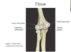
capitulum articulates with which bone? [1]
trochlea articualtes with which bone? [1]
capitulum articulates with which bone? [1]
- radius
trochlea articualtes with which bone? [1]
- ulnar
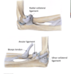
State which ligaments are found around the elbow joint [2]
Which movements do they help cause movement of? [1]
Ligaments:
- annular ligament
- radial collateral ligament
- ulnar collateral ligaments
together reinforce hinge movement
which ligament is commonly torn in young girls?
Ulnohumeral joint
Radio humeral joint
Proximal radioulnar joint
why?
which ligament is commonly torn in young girls?
Ulnohumeral joint
Radio humeral joint
Proximal radioulnar joint
bc annular ligament if loosely attached to the ulnar in infants
What are the 3 joints of the elbow?
Ulnohumeral joint is where movement between the ulna and humerus occurs.
Radio humeral joint is where movement between the radius and humerus occurs.
Proximal radioulnar joint is where movement between the radius and ulna occurs
what are the boundaries of the cubital fossa?
Boundaries of the cubital fossa:
- superior border: line from the lateral to the medial epicondyle
- flexor muscles of the forearm and the bracioradialias acting as borders also.


median nerve !

Describe the role of the:
annular ligament [1]
Which joint does it help to create? [1]
Which movements does this joint allow? [2]
Annular ligament: encircles the head of the radius and keeps in the radial notch of the ulnar: creates proximal radioulnar joint
Permits:pronation and supination of the formarm
Label 20-22 & A-C

20 Site of humero-ulnar joint
21 Site of humeroradial joint
22 Site of proximal radio-ulnar
A = humerus
B = radius
C = ulna
Label 1-3

1: ulnar collateral ligament
2: olecranon
3: radius
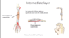
Which is the muscle of intermediate layer of forearm?
Where does it run to from the medial epidcondyle?
What movements does it cause? [3]
which is the muscle of intermediate layer of forearm?
* flexor digitorum superficialis
where does it run to from the medial epidcondyle?
* middle phalanx
Movements:
- flexes the wrist
- flexes the MCP
- flexes the PIP
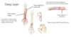
What are deep layer muscles of the forarm? [3] where run to ? what movements?
flexor digitorum profundus:
- makes way to fingers and distal phalanx !
- causes flexion of the wrist, MCP, PIP and DIP joints
flexor pollicis longus
- forearm to distal phalanx of thumb
- thumb flexion
pronator quadratus
- between ulnar and radius
- initiates pronation of the forearm
Pronation of the forearm is undertaken by pronator quadratus (deep layer) and pronator teres muscle (superficial layer).
which of these is the main muscle involved in pronation and which assists pronation?
pronator quadratus (deep layer): main
pronator teres muscle (superficial layer): assists

lumbricals: flex MCP
dorsal interrossei: abduct fingers (DAB)
palmar interrossei adduct the fingers (PAD)

During total 180 degrees of abduction, [] degress is from glenohumeral joint and [] degrees from scapulothoracic joint
120: glenohumeral joint
80: scapulothoracic joint
Label B & C

B = thoracodorsal
C = long thoracic
Label A-C

A: Pec major
B: serratus anterior
C: long thoracic nerve
Which nerve is at risk here? [1]

median
