L35: GI 1 & 2 MICROANAT Flashcards
What is this structure?

Parietal Cells
- Large central nucleus
- Acidophilic cytoplasm
- “Fried-egg” look
What is this structure?

Parietal Cells and HCL
- Plentiful mitochondria
- Acid secretion needs ATP
-Folded cell membranes increases:
SA, microvilli, intracellular canaliculus
-More folds in active partietal cells
What are these structures? (not circled by under)

Chief Cells
- Found in base of glands in clusters
- Apical granules
- Initiates protein digestion
- Secrete pepsinogen into the lumen
- Lifespan of 75 days
What is this structure?

Rugae
- Located in submucosa
- Formed by CT
- Flatten when full
- *Inc SA
What is this structure?

Serosa
- Another name= peritoneum
- Thin CT layer
- Covered by mesothelium of cuboidal cells
- Secretes small amounts of serious fluid into peritoneal
- Cavity for lubrication
What is this structure?

Reticulum
- Mucosal lining is cornified
- Stratified squamous keratinized epithelium
What is this structure?

Mucosal Epithelium
- Simple columnar with microvilli: brush border
- EVAGINATES into villi (SA)
- INVAGINATES into short intestinal glands
- Microvilli, villi, gland increases SA for absorption
- Contains goblet cells, which secrete mucous
What is this structure inside mucosal epithelium?

Enterocytes
- Absorptive function
- TALL microvilli
- Increase tight junctions- containment of luminal contents
- Increase lateral infoldings
- Digestive function
- Glycocalyx enzymes (sugar coat to inc absorp. even more)
- 5 day lifespan (esp in proximal SI due to acids)
What is this other structure inside mucosal epithelium?

Goblet Cells
- Protective
- Apical mucinogen granules
- Mucus prevents self-digestion
- 5 day lifespan
What is this structure?

Lamino propria
- Provides host defense
- GALT= Gut-associated Lymphoid Tissue (lymphatic nodules)
- Aggregated nodules- Peyer’s patch (only ileum)
What are two structures found on this transverse section of villus? What do they do?

This is in the lamina propia.
- Fenestrated capillaries- Transport of protein, carbs
- Lymphatic capillaries- Called lacteals (transport dietary fats)
What is the muscle found on muscularis mucosa?

Smooth muscle
- Contract intestinal glands
- Some muscle extends up villi
- Contractions help move lymph (arterial system then to liver)
What is this the blue line indicating in this structure?

Submucosa
What is this structure and what is significant about it?

Plicae circulares
- Found in jejunum & ileum
- Evaginated submucosa covered with mucosa
- Unlike rugae in stomach, plicae circulares cannot flatten*
- Fold increase SA
Recognize parts. Where would Plicae circulares be more found or less found in ileum?

- For absorptive capacity
- Found more in proximal end and less & less as gets deeper to ileum because as move towards, most absorption has already happened
What is this structure of? Significance?

Muscularis externa
- Composed of inner circular (segmentation) & outer logitudinal (tube shortening)
- Functions in peristalsis
In the muscularis externis, what is the arrow pointing to and what is it innervated by?

- Neuronal cell bodies
- *Innervation by myenteric plexus
What does this indicate?

Serosa- portions of SI suspended in peritoneal cavity (jejunum & ileum)
Adventitia- portions of tract fixed to abdominal wall (only around duodenum)
What is the blue arrow indicating?

Mucosa
-Epithelium, Lamina propria, Muscularis mucosa
What is the red arrow indicating?

Submucosa
- Supporting layer
- Blood & lymphatic vessels
- Folds
- Submucosal glands
What structure is indicated by the blue arrow? What two sublayers make up this?

Muscularis externa
1) Inner circular
2) Outer longitudinal
What is indicated in the black box? Function?

Myenteric plexus
-For innervation; occurs btwn CM and LM
What is this and what are the “lego pieces” needed?

Esophagus
- Transport food from oral cavity to stomach
- Lego pieces include:
1. Stratified squamous epithelial for protection
2. Mucous glands for lubrication
3. Skeletal and smooth muscle
What is this? Describe acrynoms for the MUCOSA layer & listen function (some)

Esophagus: mucosa
- Epithelium: Stratified Squamous (SS)
- -Lamina Propia (LP)*: diffuse lymphoid tissue
- Muscularis Mucosa (MM): smooth muscle
What is this? Describe acrynoms for the SUBMUCOSA layer & listen function (some)

Esophagus: Submucosa
- Esophageal Gland (EG): Excretory duct (D), mucus for lubrication
- Blood and lymphatic vasculature
What is the boxed structure? Describe function and acrynoms.

Esophagus: muscularis externa
- Inner circular and outer longitudinal sub-layers
- Skeletal muscle proximal, smooth muscle distal
- Where is contraction voluntary? Involuntary? (Sm vs Sk)
- -*Myenteric plexus between sub-layers
What junction is this a part of ?

Esophagogastric junction
What are the blue structures indicating on this SEM?

Rugae
- Secretion of “gastric juice”
- Increase surface area so we can eat more (full)
- Formation of chyme
What do these structures indicate?
Gastric Pits
-Invagination of epithelium forms gastric glands
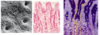
What is indicated by the yellow highlighted portion? Dark purple highlighted portion? Functions?

Yellow: Parietal cells- secrete HCl
Purple: Chief cells- protein digestion
What is this structure? (simple labeling)

Mucosa
What is this structure? (simple labeling)

Mucosal epithelium
What is this structure? (simple labeling and one significance)

Large intestine- reduced SA
What is this structure which houses microfauna to ferment and break down cellulose? They also have ____ cells and do not have ____ expansion

Cecum
Goblet cells
Do not have villar expansion
What is this structure? (simple labeling)

Lamina propria
What is this structure in box? (simple labeling)

Muscularis mucosa
What is this structure? What happens when cancer cells descend into here?

Submucosa
Metastasis occurs rapidly
What is this structure? (simple labeling)

Muscularis externa
What is this structure? (simple labeling)

Anal canal
What is this in the dog and cats? simple labeling- also what is A and what is S?

Anal sacs
A: apocrine sweat glands
S: sebaceous glands
