HEMODYNAMIC DISEASE, THROMBOEMBOLIC DISORDERS AND SHOCK Flashcards
List 2 possible causes for the findings shown in the gross specimen photograph attached.

The image shows a mural thrombus in the aortic lumen.
Causes include:
- ulcerated atherosclerotic plaque
- aneurysmal dilation
imaging test of choice in pulmonary embolism
CT pulmonary angiography
How do septic infarcts occur?
occur when infected cardiac valve vegetations embolize or when microbes seed necrotic tissue
See the attached image. List 2 other conditions associated with edema due to the same underlying mechanism responsible for the finidngs seen in the image.

Th image depicts lymphedema in filariasis
Underlying mechanism: Lymphatic obstruction
2 other conditions:
Lymphedema after a modified radical mastectomy and radiation therapy
Breast lymphedema (inflammatory carcinoma), due to blockage of subcutaneous lymphatics by malignant cells
See the attached image.
Cause and mechanism?

Acute LVF: blood backs up into the lungs –>transudate in alveoli–>pulmonary edema
2 differences between an antemortem thrombus and a postmortem clot
An antemortem thrombus is adherent to the vessel wall and is composed of Lines of Zahn.
A postmortem clot is not attached to the vessel wall and does not exhibit lines of Zahn.
See attached image.
List 3 possible causes

Diagnosis: Fat embolism
Causes:
a. fractures of long bones
b. soft tissue trauma
c. burns
What word best fits the description provided?
Accumulation of fluid within tissues when the net rate of fluid movement exceeds the rate of lymphatic drainage
Edema
2 consequences of acute decompression sickness
- bends- rapid formation of gas bubbles within skeletal muscles and supporting tissues in and about joints
- chokes - respiratory distress due to formation of gas bubbles in the pulmonary vasculature leading to edema, hemorrhage, and focal atelectasis or emphysema
Morphology of lung in shock
- diffuse alveolar damage
- alveolar hyaline membranes

two of the most important causes for arterial thrombosis
Atherosclerosis
Endocardial injury
Why do patients with Hashimotos and Grave’s disease develop myxedema?
T cell cytokines –>stimulate fibroblasts–>synthesize excess hyaluronic acid
Virchow’s triad of Thrombosis
(1) endothelial injury
(2) stasis or turbulent blood flow
(3) hypercoagulability of the blood
Is this an arterial or venous thrombus?
- Predominant component: friable meshwork of platelets, fibrin, red cells, and degenerating leukocytes.
- begin at sites of turbulence or endothelial injury
- tend to grow retrograde
Arterial thrombosis
What is the mechanism responsible for the findings shown?
History of chronic alcohol abuse
See attached image
LIver biopsy: Bridging fibrosis

- Decreased oncotic pressure
- Increased hydrostatic pressure within the portal vein due to extensive fibrosis
Forgot the 2nd one, didn’t ya? Gotcha!
What do you mean by organization of a thrombus?
ingrowth of endothelial cells, smooth muscle cells, and fibroblasts into a thrombus and eventual incorporation into the vessel wall.
How does turbulent blood flow lead to thrombosis?
- Disrupts laminar flow –>platelets come in contact with endothelium
- Prevents dilution and washout of activated clotting factors
Give one term that best fits the description provided below:
Venous thrombus passage through an Inter atrial or inter ventricular septal defect–> venous embolus–>passes through the defect–> reaches systemic circulation
Paradoxical embolism
Give one word that best fits the description
Passage of any material that can lodge in a blood vessel and obstruct its lumen through venous or arterial circulation
Embolism
Predominant component of venous thrombi
More enmeshed red cells (and relatively few platelets) – hence aka red/stasis thrombi as they mainly occur at sites of stasis
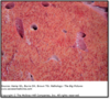
In which condition would you be likely to see the findings shown in the attached image?

Waterhouse Friedrichsen syndrome assoc with meningococcal septicemia
3 important clinical consequences of an arterial thrombus
a. Critical vessel occlusion
b. formation of a mural thrombus
c. Embolization
Identify the pattern of shock described below:
CVP- Decreased
PCWP- Decreased
CO-Increased
SVR - Decreased
mVO2 - Increased
Septic shock
List 4 important possible consequences of a pulmonary embolism
- Acute right heart failure–>sudden cardiac death (cor pulmonale)
- pulmonary hemorrhage
- pulmonary infarction
- Chronic thromboembolic pulmonary hypertension
Identify the pattern of shock described below:
CVP- Increased
PCWP- Increased
CO-Decreased
SVR - Increased
mVO2 - Decreased
Cardiogenic shock
What pattern of infarction is likley to occur in the organs listed below?
Spleen, kidney, heart
Pale/ white
Why is pulmonary infarction a rare occurrence following pulmonary embolism?
The lung is supplied by both the pulmonary arteries and the bronchial arteries, and the intact bronchial circulation is usually sufficient to perfuse the affected area. Understandably, if the bronchial arterial flow is compromised (e.g., by left-sided cardiac failure), infarction may occur.
Arterial or venous thrombosis?
calf tenderness associated with forced dorsiflexion of the foot
Clinical image attached

Venous thrombosis
4 fates of a thrombus
- Propagation
- Embolization
- Dissolution
- Organization and recanalization
Which condition is this radiographic finding associated with?

Saddle embolism – Occlusion of the main pulmonary artery by an embolus that straddles the pulmonary artery bifurcation

List 3 examples for edema developing due to decreased oncotic pressure
- Liver cirrhosis
- Protein malnutrition
- Nephrotic Syndrome
- Protein losing gastroenteropathy
How do you establish the diagnosis of protein losing gastro-enteropathy?
Increase in alpha 1 antitrypsin clearance
What is the mechanism responsible for the morphologic findings shown in the attached image?

Increased hydrostatic pressure in the pulmonary capillaries
Morphologic features of chronic passive lung congestion
alveolar septal fibrosis
hemosiderin-laden macrophages aka “heart-failure cells.”
brown induration

Give one microscopic term that best fits this appearance of the liver biopsy in a patient with acute right ventricular failure
- central vein and sinusoids are distended.
- centrilobular hepatocytes may undergo ischemic necrosis
- Periportal hepatocytes – fatty change
Acute liver congestion

Most frequent cause of septic shock
gram-positive bacterial infections
Identify the pattern of shock described below:
CVP- Decreased
PCWP- Decreased
CO-Decreased
SVR - Increased
mVO2 - Decreased
Hypovolemic shock
Hyperemia or congestion?
a. passive process
b. results from reduced outflow of blood from a tissue
c. Affected tissues take on a dusky reddish-blue color (cyanosis) due to red cell stasis and the presence of deoxygenated hemoglobin
Congestion
Why does edema develop in right heart failure?
Increased PH in the venacava–>transudate enters the interstitial tissue around the ankle and lower legs–>pitting edema
Microscopic morphology of kidney in shock
acute tubular necrosis

When does transudation occur in a vessel (with reference to PH and PO ) ?
When PH >PO
What is the mechanism of development of edema in a person with cellulitis?
Increased permeability of venules due to inflammation
Consequence of thrombosis of the superior mesenteric vein
hemorrhagic small-bowel infarction
Gross morphologic appearance of chronic passive congestion of the liver
centrilobular regions are grossly red-brown and slightly depressed (because of cell death) and are accentuated against the surrounding zones of uncongested tan liver (nutmeg liver)

Hyperemia or congestion?
- active process
- arteriolar dilation –> increased blood flow
- Affected tissues turn red (erythema) because of increased delivery of oxygenated blood.
Hyperemia
Pathophysiologic mechanism of amniotic fluid embolism
tear in the placental membranes or rupture of uterine veins–>infusion of amniotic fluid/fetal tissue into the maternal circulation
Cerebral atrophy or cerebral edema?
Gross specimen of brain showing narrowed sulci and widened gyri
Cerebral edema

Consequence of chronic decompression sickness
multiple foci of ischemic necrosis
Identify the pattern of shock described below:
CVP- Decreased
PCWP- Decreased
CO-Decreased
SVR - Decreased
mVO2 - Decreased
Neurogenic shock
What term best fits the description provided?
Accumulation of fluid within the body cavity when the net rate of fluid movement exceeds the rate of lymphatic drainage
Effusion
Diagnosis?
History of prolonged immobilization following surgery
Sudden onset dyspnea, Pleuritic chest pain, Tachypnea,Tachycardia
EKG: “S1Q3T3” pattern
Pulmonary embolism
What is the mechanism of development of edema common to all the clinical conditions listed below?
- Congestive heart failure
- Constrictive pericarditis
- Ascites (liver cirrhosis)
- Venous obstruction or compression
- Thrombosis
- External pressure (e.g., mass)
- Lower extremity inactivity with prolonged dependency
Increased hydrostatic pressure
When does reabsorption occur in a vessel (with reference to PH and PO ) ?
When PO > PH
Trace the route taken by a thrombus arising in the deep veins and causing pulmonary embolism
DVT–>fragmented thrombus–>larger veins–>right heart–>pulmonary arterial tree
Most common site for venous thrombi
lower extremity veins (90%)
List 2 clinical conditions assoc with sodium and water retention, leading to edema
Chronic renal failure
Congestive heart failure
Why does edema develop in left heart failure?
Increased PH in the pulmonary capillaries——> transudate enters the alveoli & interstitium of the lungs–> pulmonary edema
What’s your diagnosis?
lungs are two to three times their normal weight
Sectioning yields frothy, blood-tinged fluid
Pulmonary edema

How does pulmonary edema cause deleterious clinical consequences?
impede oxygen diffusion, creates a favorable environment for bacterial infection
Give one word that best fits the description provided below:
grossly and microscopically apparent laminations
pale platelet and fibrin deposits alternating with darker red cell–rich layers
Lines of Zahn

List important conditions favoring deep vein thrombosis
- Vascular stasis
- Injury and inflammation
- Hypercoagulability
- Advanced age
- Sickle cell disease
Morphologic feature of amniotic fluid embolism
presence of squamous cells shed from fetal skin, lanugo hair, fat from vernix caseosa, and mucin derived from the fetal respiratory or gastrointestinal tract in the maternal pulmonary microvasculature

5 underlying possible mechanisms for red/hemorrhagic infarct
- venous occlusion
- in loose, spongy tissues
- tissues with dual circulations
- in tissues previously congested by sluggish venous outflow
- when flow is reestablished to a site of previous arterial occlusion and necrosis
What is the pathophysiology?
- sudden onset of tachypnea
- dyspnea
- tachycardia
diffuse petechial rash
Microscopic image of lung attached

Diagnosis: Fat embolism
Pathophysiology: Injuries–>rupture vascular sinusoids in the marrow/small venules–>marrow/adipose tissue herniates into the vascular space–>lung
Give one word that best fits the description provided
an area of ischemic necrosis caused by occlusion of either the arterial supply or the venous drainage
Infarct
What’s the most likely underlying mechanism?

arterial occlusion in a solid organ with end-arterial circulation resulting in a pale infarct
List 2 possible causes for the findings shown in the gross specimen photograph attached.

The image shows a mural thrombus in the heart chambers.
Causes include:
- Arrhythmias
- dilated cardiomyopathy
- myocardial infarction
- myocarditis
What’s the diagnosis?
sudden onset of dyspnea
tachypnea
pleuritic chest pain
and hemoptysis
see attached clinical image

Pulmonary infarction (hemorrhagic/red infarct)
What pattern of infarction is likley to occur in the organs listed below?
Testes, ovaries, lung, small bowel
Red/hemorrhagic infarct
List 2 conditions where turbulent flow may be encountered
- Ulcerated atherosclerotic plaque
- Aneurysms
- Mitral stenosis secondary to RHD assoc with LA dilation and atrial fibrillation
- Polycythemia vera
- Sickle cell anemia
What is meant by recanalization of a thrombus?
Formation of capillary channels that help re-establish continuity of a vessel that has been occluded by a thrombus.
Why do you get Cortical cell lipid depletion in the adrenal gland in shock?
Because of conversion of the relatively inactive vacuolated cells to metabolically active cells that utilize stored lipids for the synthesis of steroids