Gametes - Foetal Physiology Flashcards
First organ system developed - necessary to sustain viable embryo
circulatory system
Critical period for development of circulatory system
day 20 - day 50
wk 3 - begins development
wk 4 - functioning heartbeat
How far does the foetus need to be from blood supply to become hypoxic
150 um from blood supply
formation of new BVs through angiogenesis
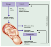
Function of ductus venosus
Links umbilical vein with IVC - allows blood to bypass foetal liver
How is flow through ductus venosus regulated
By sphincter
50-80% of blood can avoid hepatic sinuses
If there is enough pressure on sphincter it will open (if there is an overload - uterine contractions compress BVs and more blood to foetal heart - overload)
Function of foramen ovale
Links RA with LA
blood flow: RA → LA, then upwards to ascending aorta
makes sense - most oxygenated blood goes to brain & spinal cord, avoids oxygen rich blood going to pulmonary circulation
What does ductus arteriosus link
how does it control blood flow
Links pulmonary artery with descending aorta
Decreased blood flow to non-functioning lungs
10% of foetal blood travels via lungs - growth and development of lungs
Overview of foetal circulation

Site of oxygenation in foetus
Placenta
How does oxygen traverse the placental membrane
Difference in partial pressure
proportion of blood that bypasses the immature foetal liver
80%
Where is there mixing of blood
in RA
Speed ensures only a small amount of mixing
What happens to the foramen ovale at birth
Removal of placenta results in decreased venous return - causes decreased RA pressure
Neonate takes their 1st breath - once opened there will be a decrease in pulmonary resistance - this contributes to decrease in RA pressure
=> more blood flow to pulmonary circulation - increase in LA blood flow

Most common atrial septal defect
Patent foramen ovale
Alone - no haemodynamic importance as pressure in LA > RA so keeps it closed
With other defects e.g. cyanosis of skin and mucus membrane
Closure of ductus arteriosus at birth - depends on
Oxygen
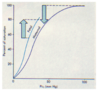
pO2 in foetal ductus arteriosus
15-20 mmHg
by the time the blood goes to maternal sinuses - the pO2 will have dropped to about 15
pO2 in neonatal ductus arteriosus
100 mmHg
Critical point pO2 in relation to closure of DA
50 mmHg (pO2 is normally 100 mmHg in artery)
Bradykinin from lungs and PGs E2/F2
=> VasoC
Primary function of DA
Bypass pulmonary circulation bevause oxygenation is not happening there
Problems associated with patent ductus arteriosus (1 in 5500)
Infants - few problems
Adults - increased re-circulation, increased cardiac output
Decreased cardiac and respiratory reserves
(Less O2 blood being circulated - in an attempt to get enough O2 to tissues there is increased cardiac output - increased BP, decreased cardiac and resp reserves because HR and stroke vol will be increased so there won’t be reserve to increase it more during times of stress/exercise)
When does the ductus venosus close
how does pressure in portal system change as a result
within 1-3 hours
Pressure in portal system increases by 6-10 mmHg to force blood through the liver
NEWBORN
- BP
- Pulse rate
- CO (L/min)
- Cardiac Index (L/m2/min)
- 70/45
- 140
- 0.6
- 2.5-3
ADULT
- BP
- Pulse rate
- CO (l/min)
- Cardiac index (L/m2/min)
- 120/80
- 70
- 5
- 2.5-3
Newborn BP
70/45
Newborn pulse rate
140
newborn CO
0.6 L/min
newborn CI
2.5-3 L/m2/min
Adult pulse rate
70
Adult CO
5
Adult Cardiac Index
2.5-3 L/m2/min
When do foetal respiratory movements decrease
In 3rd trimester
PROTECTIVE MECHANISM
- Decreased foetal growth
- Decreased foetal waste in amniotic fluid
- Decreased resp movements - decreased swallowing of wastes
Blood flow related to maternal sinuses
Uterine arteries to uterine vein
Placenta - organ type
Foeto-maternal organ
What are the foetal capillaries
Chorionic villi - dip into maternal sinuses but no direct contact
Gas exchange across capillary wall
pO2 maternal sinuses
50 mmHg
pO2 foetal vein
30 mmHg
pO2 foetal artery
20 mmHg
by the time blood has come back around again (offloaded O2 on its way)
pCO2 maternal sinuses
45 mmHg
pCO2 foetal vein
45 mmHg
pCO2 foetal artery
46 mmHg
this small difference in pCO2 (compared with maternal sinuses - 45 mmHg) drives CO2 from foetal side into maternal circulation
this change in CO2 causes a localised change in pH which allows further O2 to be carried by foetal blood
What difference drives O2 into foetal circulation
pO2 maternal sinuses (50 mmHg) - pO2 foetal vein (30 mmHg)
What happens in adults if pO2 drops to 60 mmHg
Peripheral chemoreceptors are triggered - causes hyperventilation to bring it back up to pO2
ADULT Hb
- Name
- Polypeptides
- 2,3-DPG
- Conc
- Hb-A
- α, β
- 2,3-DPG binds well
- 14.8g/100ml
FOETAL Hb
- Name
- polypeptides
- 2,3-DPG
- Conc
- Hb-F
- α, δ
- 2,3-DPG binds poorly
- 16.8g/100ml
How does foetus survive at low pO2
Oxygen dissociation curve shifts to left - little binding of 2,3-DPG
=> at any given pO2, foetal Hb carries 20-30% more O2 than adult Hb
Oxygen dissociation curve for foetal vs maternal blood

Uterine blood flow
Increased maternal CO
Increased up to 500ml/min to uterus
How do oestrogen and progesterone affect blood flow
Oestrogens increase uterine vasodilation
Progesterone increase uterine venoconstriction - diminishes rate at which blood leaves the area - pooling of blood
Double Bohr effect
Increase in pH shifts O2 dissociation curve to left
Alkaline conditions
Binds more O2 at any given pO2
Acidic conditions
Binds less O2 at any given pO2
What happens when CO2 dissolves to maternal circulation
Localised decrease in foetal pCO2 and increase in pH
Foetal O2 dissociation curve shifts to left
=> at any pO2 foetus binds more O2 than mother
What happens when CO2 dissolves to maternal circulation
Localised increase in maternal PCO2 and decrease in pH
Maternal O2 dissociation curve shifts to the right
At any pO2 mother binds less O2 than foetus
What happens with the removal of placenta (foetus’ supply of O2)
Hypoxia (foetus has been surviving in hypoxic conditions) and hypercapnia
HYPERCAPNIA stimulates first breath
Slight asphyxiation (lack of O2) at birth as neonate travels through birth canal
Increase in pCO2 via central chemoreceptors - stimulation of first breath
(air moves in // diaphragm contracts, volume in cavity increases, pressure decreases - Boyle’s law - external ICs contract to pull ribcage out)
Pressure volume curve for baby’s first/second breath

Neonate resp rate
40 breaths per min
Neonate TV
16 ml/min
Neonate minute vol
640 ml/min
Neonate FRC
1/2 adult value
FRC of neonate - consequence of a decrease in FRC
Rapid changes in blood gases can occur if resp is altered
Define FRC
Vol of lungs in lungs at the end of a normal exp - prevent fluctuation of gases - no yo yo like effect
How does surfactant affect the lungs
Increased compliance of lungs
Decreased surface tension in alveolus
Makes lungs compliant - easier to bring in air - reduces surface tension attraction of hydrogen molecules that want to collapse alveoli
When is surfactant produced
What type of cells compose surfactant
At 28 weeks
Type II alveolar epithelial cells
Foetal cortisol stimulates production of surfactant
Function of phospholipids in surfactant
How are they organised
PLs = amphipathic compounds
Hydrophilic portion - reduces attraction of H+ to each other
2 parallel hydrophobic tails
Line up in alveoli - tails face lumen of alveolus - repel water molecules
What hormone predominates in the last weeks of pregnancy
What does it cause
Oestrogen reaches a peak
causes myometrial weakness and irritability
weak braxton hicks contractions may take place
What causes uterine contractions
As birth nears, oxytocin and PGs cause uterine contraction
oestrogen allows for gap junction
Effect of emotional and physical stress on body
Activates hypothalamus
Sets up a positive feedback mechanism to release more oxytocin
Initiation of parturition
Oestrogen from placenta mostly (less so from foetal ovaries)
Increased rate of contractility - positive feedback - more PGs produced, increased sensitivity

Foetal and mother hypothalamic activities

What does cortisol stimulate
PG production by chorion/placenta
Weak androgens (DHEAS) - upregulate PGs
Lactation

Milk ejection reflex
