6. Pharyngeal Arches Flashcards

1
Q

Components of the pharyngeal apparatus

A

Arches, pouches, grooves, & membranes

2
Q

Pharyngeal apparatus contributes to formation of what structures?

A

Nasal cavities, mouth, larynx, pharynx, and neck

3
Q

When do pharyngeal arches develop?

A

4th week from NCC

4
Q

1st pair of arches

A

Primordial jaws (lateral to developing pharynx)

5
Q

Rudimentary and not visible on surface of embryo

A

5th/6th arches

6
Q

Embryonic layers of the pharyngeal arches

A

Core of mesoderm and mesenchyme / external ectoderm / internal endoderm

7
Q

Functions of the pharyngeal arches

A

Support the lateral walls of primordial pharynx and give rise to prominence of tissue that contribute to craniofacial development

8
Q

Connective tissue in the head (dermis and SM)

A

NCC-derived mesenchyme

9
Q

populates each arch to form PA musculature

A

paraxial mesoderm

10
Q

angioblasts differentiate into endothelium

A

Lateral plate mesoderm

11
Q

Extraocular musculature

A

Prechordal plate mesoderm

12
Q

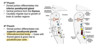
Structures of pharyngeal arches

A

Cartilaginous rod, muscular component, cranial nerves, and arch artery

13
Q

Forms malleus and incus

A

Meckel’s cartilage (cartilage of PA1)

14
Q

Perichondrium forms anterior ligament of malleus and sphenomandibular ligament

A

Meckel’s cartilage (cartilage of PA1)

15
Q

Ventral parts form primordium of mandible

A

Meckel’s cartilage (cartilage of PA1)

16
Q

Where do the bones form with relation to Meckel’s cartilage?

A

Lateral to the cartilage

17
Q

Dorsal region contributes to stapes and styloid process of the temporal bone then disintegrates

A

Riechert’s cartilage (cartilage of PA2)

18
Q

Perichondrium forms stylohyoid ligament

A

Riechert’s cartilage (cartilage of PA2)

19
Q

Ossification of ventral end forms lesser cornu/horn of hyoid bone

A

Riechert’s cartilage (cartilage of PA2)

20
Q

Ossification forms greater cornu of hyoid bone

A

Third arch cartilage

21
Q

What forms the body of the hyoid bone?

A

Hypopharyngeal eminence

22
Q

Prominence in the floor of embryonic pharynx (from PA3 and PA4)

A

Hypopharyngeal eminence

23
Q

Laryngeal cartilage + epiglottis

A

Fourth arch cartilage

24
Q

Where is the epiglottis derived from?

A

NCC

25
Laryngeal cartilage (only)
Sixth arch cartilage
26
Where are the muscular components of the PAs derived from?
Paraxial mesoderm and prechordal plate
27
Muscles of the 1st pharyngeal arch
Muscles of mastication (CN V)
28
Muscles of the 2nd pharyngeal arch
Stapedius m. & muscles of facial expression (CN VII)
29
Muscles of the 3rd pharyngeal arch
Stylopharyngeus m. (CN IX)
30
Muscles of the 4th pharyngeal arch
Cricothyroid m., levator veli palatini m., and constrictors of pharynx m. (CN X)
31
Muscles of the 6th pharyngeal arch
Intrinsic muscles of the larynx (CN X)
32
What innervates the 1st pharyngeal arch?
Trigeminal nerve (CN V)
33
What innervates the 2nd pharyngeal arch?
Facial nerve (CN VII)
34
What innervates the 3rd arch?
Glossopharyngeal nerve (CN IX)
35
What innervates that 4th-6th arches?
Vagus nerve (CN X)
36
What innervates the 4th pharyngeal arch (specific)?
Superior laryngeal branch of CN X & its recurrent laryngeal branch
37
(1) How many pairs of arteries form the aortic arch artery system? (2) Where do they arise from?
(1) 5, (2) aortic sac
38
Basket-like structure arising from dorsal aorta
Aortic sac (forms great arteries of thorax and head/neck)
39
Arteries from the (1) ______ are bilateral; arteries from the (2) _____ are asymmetrical.
(1) 1st - 3rd aortic arch arteries, (2) 4th & 6th aortic arch arteries
40
Where are pharyngeal grooves located?
Between pharyngeal arches
41
What are the pharyngeal grooves covered by?
Ectoderm
42
What does the 1st pharyngeal groove become?
External acoustic meatus
43
Where do the grooves of 2-4 lie?
Cervical sinus
44
When pharyngeal endoderm lines the pharynx internally, what is created?
Pharyngeal pouches
45
Where are the 5 pharyngeal pouches found?
Craniocaudal sequence between the arches (ie: 1st pair lies between 1st and 2nd arches)
46
* Remnants of cervical sinus and/or 2nd groove * Persist and form a slowly enlarging, painless, free-lying cyst in the neck (inferior to the angle of the mandible) * Accumulation of fluid and cellular debris derived from desquamation of their epithelial linings
Cervical cysts
47
* Failure of the 2nd PA groove and cervical sinus to obliterate * Detected due to discharge of mucus * Typically bilateral and commonly associated with auricular sinuses
Cervical sinus
48
* Abnormal canal that opens into the **tonsillar sinus** and externally in the side of the neck * Persistence of parts of 2nd groove and pouch
Cervical fistula
49
Where pouch endoderm contacts ectoderm of pharyngeal grooves
Pharyngeal membranes
50
Pharyngeal membranes are quickly infiltrated by \_\_\_\_\_\_.
Mesenchyme
51
What forms the tympanic membrane?
1st pharyngeal membrane and intervening mesenchyme
52
From what arch does the **thymic primordium** develop?
Pharyngeal arch 3 endoderm (comprised of epithelial cords)
53
3 functions of the epithelial cords
(1) Grow into surrounding mesenchyme \> thymic lobules, (2) become arranged around central point \> thymic corpuscles, (3) form epithelial reticulum for housing lymphocytes
54
Are growth and development of the thymus complete at birth?
No.
55
When is the thymus most active?
During childhood (then it involuted)
56
What is the first endocrine gland to develop?
Thyroid gland (From thyroid primordium)
57
What does the thyroid gland form from?
Median endoderm thickening in floor of the primordial pharynx \> descends in neck w/ tongue growth then passes ventral to the developing hyoid bone and cartilages
58
Connects tongue and thyroid gland
Thyroglossal duct
59
Malformation of eyes, ears, mandible, and palate
First arch syndrome
60
What does 1st arch syndrome result from?
Insufficient migration of NCC into the 1st arch during the 4th week
61
* Malar hypoplasia w/ down-slanting palpebral fissures * Defects of the lower eyelids * Deformed external ear (sometimes middle and internal ears)
Treacher-Collins syndrome (mandibulofacial dysostosis)
62
Treacher-Collins syndrome mutation
Autosomal dominant, mutation in TCOF-1 (Treacher Collins-Franceschetti syndrome 1)
63
Functions of TCOF1 gene
* Encodes for TREACLE (involved in ribosome biogenesis) * Truncated in TCS leading to increased apoptosis of cranial NCC
64
* Typically occurs *de novo* * Associated with hypoplasia of the mandible, cleft palate, & duvets of the eyes and ears
Pierre Robin sequence
65
Micrognathia
Small mandible (initiating defect in Pierre Robin sequence)
66
Physical features of Pierre Robin sequence
**Micrognathia** \> posterior displacement of the tongue \> obstruction of full closure of palate \> **bilateral cleft palate**
67
Absence of thyroid gland or one of its lobes (rare)
Agenesis of thyroid gland
68
Unilateral failure of thyroid formation (left lobe more commonly absent); likely mutation in receptor for TSH
Thyroid hemiagenesis
69
* Breakdown of signaling from PA endoderm to NCC * Agenesis of thymus and parathyroid glands * Congenital hypoparathyroidism * Shortened philtrum of upper lip, low-set, and notched ears * Nasal clefts and thyroid hypoplasia * Cardiac abnormalities
DiGeroge Syndrome
70
PA1 artery
Terminal branch of maxillary artery
71
PA2 artery
Stapedial artery (embryonic) and caroticotympanic artery (adult)
72
PA3 artery
CCA, root of ICA
73
PA4 artery
Arch of aorta (left), right subclavian artery, original sprouts of pulmonary arteries
74
PA6 artery
Ductus arteriosus and roots of definitive pulmonary arteries
75
PA1 muscles
Muscles of mastication
76
PA2 muscles
Muscles of facial expression
77
PA3 muscles
Stylopharyngeus m.
78
PA4 muscles
Constrictors of pharynx, cricothyroid, levator veli palatini m.
79
PA6 muscles
Intrinsic muscles of the larynx (except cricothyroid)
80
PA1 ligaments
Anterior ligament of malleus and spheno-mandibular ligament
81
PA2 ligament
Stylohyoid ligament
82
PA1 skeletal elements
83
PA2 skeletal elements
84
PA3 skeletal elements
85
PA4 skeletal elements
86
PA6 skeletal elements
87
Where does the arch artery arise from? What is its route?
(1) **truncus arteriosus** of the heart (2) passes around primordial pharynx to enter the dorsal aorta
88
1st pouch expands into \_\_\_\_
tubotympanic recess \> tymanic cavity & mastoid antrum
89
1st pouch elongates to form \_\_\_\_\_-
pharyngotympanic tube
90
Portion of the 2nd pouch forms \_\_\_\_.
tonsillar sinus
91
When endoderm of 2nd pouch proliferates, ____ is formed.
tonsillar epithelium
92
When mesenchyme of the 2nd pouch differentiates, _____ of palatine tonsil is formed.
lymphoid nodules
93
1. Dorsal portion of the 3rd pouch differentiates into \_\_\_\_. 2. Ventral portions of the 3rd pouch form \_\_\_\_.
1. inferior parathyroid gland 2. thymus
94
Describe migration of the 3rd pouch.
caudal migration due to growth of brain and cardiac regions
95
Dorsal portion of the 4th pouch differentiates into \_\_\_.
superior parathyroid glands
96
(1) ____ fuses with thyroid gland to give rise to (2) \_\_\_\_\_
1. Ultimobranchial body 2. Parafollicular cells