13. Development of Orofacial Structures Flashcards

1
Q
  • derived from mesenchyme
  • gives rise to bones that enclose the brain
  • cartilaginous and membrane components
A

neurocranium

2
Q
  • derived from mesenchyme
  • gives rise to bones that comprise facial skeleton
  • cartilaginous and mebranous components
A

viscerocranium

3
Q
  • mesenchymal origin without artilage formation
  • neurovascularization occurs
  • osteoblasts deposit osteoid (form bone and osteocytes)
A

intramembranous ossification

4
Q
  • pre-existing cartilaginous model (long bones)
  • primary ossification centers appear in diaphysis
  • chondrocytes hypertrophy (matrix calcifies)
A

endochondral ossification

5
Q

cartilaginous parts of the neurocranium

A
  • occipital bone
  • body of sphenoid bone
  • ethmoid bone
  • petrous and mastoid parts of the temporal bone
6
Q

membranous parts of the neurocranium

A

calvaria: frontal and parietal bones

7
Q

embrologyic origin of cartilaginous viscerocranium

A

NCC > bones and connective tissue

8
Q

membranous viscerocranium

A
  • maxillary prominence
  • squamous part of temporal bone
  • maxilla
  • zygomatic bone
9
Q

premature fusion of cranial structures

A

craniosyntosis

10
Q
  • premature fusion of cranial suture: sagittal
  • long, narrow wedge shaped cranium
  • most common
A

scaphocephaly

11
Q
  • premature fusion of cranial suture: entire coronal suture
  • high, tower-like cranium
A

brachycephaly

12
Q
  • premature fusion of cranial suture: one side of coronal suture
  • twisted and asymmetric
A

plagiocephaly

13
Q

premature fusion of cranial suture: frontal (metopic) suture

A

trigonocephaly

14
Q

derivatives of fronal nasal prominence

A

forehead & dorsum/apex of nose

15
Q

derivative of the lateral nasal prominence

A

alae of nose

16
Q

derivatives of medial nasal prominence

A

nasal septum, ethmoid bone, and cribriform plate

17
Q

derivatives of maxillary prominence

A

upper cheek ad upper lip

18
Q

derivatives of mandibular prominence

A

chin, lower lip, cheek

19
Q

What drives shape/rate of growth of the head?

A

brain development

(“brain develops rapidly - especially forebrain - which influences head shape”)

20
Q
  1. Name the facial primordia.
  2. when do they first appear?
A
  1. 5 facial primordia: 2 maxilary prominences, 2 mandibular prominences, 1 frontonasal prominence
  2. Appear during week 4
21
Q

Where is the facial primordia located?

A

surrounding the stomodeum

separated from primordial pharynx by oropharyngeal membrane (ruptures around day 26)

22
Q
  1. Which is the first facial prominence to form?
  2. Describe process.
A
  1. Mandibular prominence (lower jaw and lip)
  2. Oropharngeal membrane disintegrates > merging of medial ends of mandibular prominences
23
Q

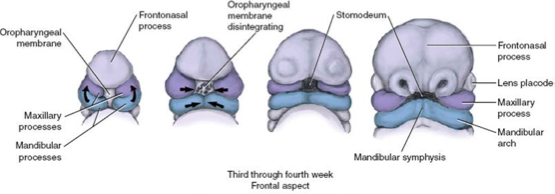
incomplete fusion of the medial ends of the mandibular prominences

A

chin dimple

24
Q
  1. What gives rise to upper lip, maxilla, and secondary palate?
  2. Describe this process.
A
  1. Maxillary prominences
  2. Grow medially > merge laterally w/ mandibular prominences
    • Primordial lips & cheeks invaded by mesencyme from PA 2 > give rise to facial muscles
25
What gives rise to the **forehead, rostral boundary of stomodeum, and nose**?
**frontonasal prominence** [surrounds ventrolaterla part of forebrain]
26
bilateral oval thickenings of surface ectoderm on the inferolateral portion of frontonasal prominence
**nasal placodes**
27
proliferation of nasal placodes cause horse-shaped elevations called \_\_\_\_
medial and lateral nasal prominences
28
formation of the medial and lateral nasal prominences result in formation of (1) ____ which ultimately form (2) \_\_\_\_\_\_
1. nasal pits 2. primordial nares
29
median lateral prominence
* MNP shifted towards midline w/ medial growth of maxillary prominences * regulated by PDGFRA * fusion results in formation of *philtrum*
30
intermaxillary segment
philtrum
31
lateral nasal prominence
* separated from maxillary prominence by nasolacrimal groove * merges with maxillary prominence by **end of week 6**
32
When does the lateral nasal prominence merge with maxillary prominence?
end of week 6
33
Where do the auricular hillocks form?
six auricular hillocks form around the **1st pharyneal groove** [mandibular development \> ears pushed from neck to side of head @ level of the eyes]
34
signaling molecule responsible for mandibular processes and removing webbing from hands/feet
BMP
35
**lateral nasal prominence** (forms ala of the nose)
36
Does facial development occur fast or slow during fetal period?
slow
37
Name 2 changes in proprtion and relative positions of facial components during fetal period.
* nose is initially flat and mandible is underdeveloped **until** 14 eeks * as brain emerges, cranial cavity expands bilterally, orbits move **from lateral to forward-facing orientation**
38
What can cause small appearance of the face prenatally?
* rudimentary upper and lower jaws * un-erupted deciduous teeth * small nasal cavities and maxillary sinuses
39
How are nasal cavities formed?
* **nasal placodes** depress and form nasal pits * _mesenchyme_ from medial.& lateral nasal prominences induces pits to become deeper \> **primordial nasal sacs** * primordial nasal sacs *grow dorsally and ventrally into the forebrain* * oronasal membrane seaprates primordial nasal sacs (rupture end week 6)
40
connection between nasopharynx and nasal cavity
primordial choanae
41
superior, middle, and inferior turbinates
nasal conchae
42
inflammation of the nasal conchae/turbinates
rhinitis
43
Where does the olfactory nerve specialize into?
olfactory epithelium
44
1. Name the 3 stages of **palate formation.** 2. When do the first 2 stages occur? 3. When is the critical period for these stages?
1. Primary, secondary, and final palates 2. Between weeks 6-12 3. Critical Period: week 6 - beginning of week 9
45
events during **primary palate** stage
fusion of median nasal prominences located between maxillary prominences forms premaxillary part of maxilla
46
events during **secondary palate** stage
develops from lateral palatine processes (palatal shelves) mesenchymal projections extending from internal aspect of maxillary prominences
47
What happens in the **final palate** stage?
bone extends from maxillae & palatine bones into the lateral palatien processes to form **hard palate** posterior portions will not ossify \> **soft palate and uvula** are formed **palatine raphe** forms = line of fusion
48
remnant of nasopalatine canal
incisive fossa
49
* downward growth of internal parts of merged medial nasal prominences * later fuses with lateral palatine processes (A to P)
nasal septum
50
most common craniofacial malformation caused by defects in fusion (unilateral or bilateral) over 100 syndroms are associated
cleft lip and palate
51
* genetics: SATB2, SHH, TGF-alpha, TGF-beta3, IRF6 * environmental: anti-epileptic drugs, smoking, binge drinking, folate deficiency, obesity
risk factors for cleft lip and palate
52
bony abnormalities of pterygoid plates and midfacial growth abnormalities presents: upper airway obstruction, noisy breathing, cyanosis (**worse with feeding, improves with crying**) can be part of treacher-collins, CHARGE, Kallman, orVACTERL syndromes
choanal atresia
53
2 parts of the tongue
* oral: anterior 2/3 * pharyngeal: posterior 1/3
54
formation of the **oral part of the tongue**
* **median lingual swelling** appears at the end of the _4th week_ * 2 **lateral tongue swellings** (distal tongue buds) develop on either side of **median lingual swelling** * ​induced by _mesenchyme from pharyngeal arch 1_
55
What forms the **pharyngeal part of the tongue**?
* **copula**: ventromedial parts of PA2 * **hypopharyngeal eminence:** ventromedial parts of PA3/4
56
Where do oral and pharyngeal parts of the tongue fuse?
hypopharyngeal eminence overgrows the copula @ **terminal sulcus**
57
Where is tongue musculature derived from?
myoblasts of occipital myotomes (CN XII)
58
**Innervation of the tongue**
59
bifid tongue
glossoschissis
60
* short frenulum * can present with problems breastfeeding and speaking
ankyloglossia
61
associated with Beckwith-Wiedemann and Down Syndromes
macroglossia
62
What induces **odontogenesis**?
communication between **NCC** and **oral epithelium**
63
U-shaped bands of oral epithelium that follow curves of primitive jaw
dental laminae
64
Where are the tooth buds from?
* anterior mandibular * anterior maxillary * posterior
65
states of **odontogenesis**
1. cap stage 2. bell stage 3. root evelopment 4. eruption
66
What happens during the **cap stage of odontogenesis**?
tooth bud becomes cap-shaped when _invested by mesenchyme_
67
What does a **tooth bud** consist of?
* enamel organ * ectodermal cells from dental lamina: **enamel** * outer cell layer lined by outer enamel epithelium * inner cell layer lines papilla lined by inner enamel epithelium * dental papillae * internal part of tooth: **dentin and pulp** * dental follicle/sac * mesenchyme surrounding dental papilla and enamel organ * **cementum**
68
What occurs during the **bell stage of odontogenesis**?
enamel organ becomes bell shaped due to differentiation of enamel
69
originate from dental papilla cells next to inner enamel epithelium predentin calcifies to become dentin
odontoblasts
70
inner enamel epithelium differentiates in response to dentin production produces enamel
ameloblasts
71
What makes up the **root**?
* epithelial root sheath * pulp * cementum * periodontal ligament
72
What forms epithelial root sheath?
fusion of inner and outer enamel epithelium
73
central dental papilla
pulp
74
inner cells of dental sac form cementoblasts
cementum
75
outer cells of dental sac
periodontal ligament
76
Describe **tooth eruption**.
* occurs at different stages * root of tooth and crown erupt through oral epithelium * mandibular teeth erupt first * deciduous root is resorbed by odontoclasts (osteoclasts) * crown and upper root are shed
77
1. How many teeth does **permanent dentition consist of**? 2. When do deciduous permnanet teeth appear? 3. What happens to non-deciduous molars? 4. When do tooth buds for permanent teeth appear?
1. 32 2. 10 weeks 3. Develop as buds from posterior extensions of dental laminae 4. Different times; mostly during **fetal period** * Buds for 2nd/3rd permanent molars develop after birth