16. Ear Anatomy and Histology Flashcards

1
Q

external ear

A

auricle to the tympanic membrane

2
Q

middle ear

A

tympanic membrane to oval window

3
Q

inner ear

A

oval window to internal acoustic meatus
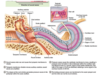
4
Q

Label external ear.

A
5
Q

external ear blood supply

A

external carotid artery

[posterior auricular a. & superficial temporal a.]

6
Q

cellular makeup of external ear

A

cartilage covered by integument

7
Q

external ear sensory innervation

A
8
Q

sensation to the external ear canal

A
9
Q

histology of the external acoustic meatus

A
  • outer 2/3: soft connective tissue & certilage
  • inner 2/3: skin and bone
10
Q

only place in the body where skin, periosteum, and bone exist directly on top of each other

A

external acoutic meatus (external auditory canal)

11
Q

cell type of outside layer of tympanic membrane

A

stratified squamous epthelium

12
Q

cell type of inside layer of tympanic membrane

A

simple cuboidal epithelium

13
Q

describe tympanic membrane structure

A
  • has concavity, cone-shaped oriented toward extenral acoustic meatus called umbo
  • has flaccid part and tense part
  • moves with sound and transmits to ossicles
14
Q

Where is the middle ear (tympanic cavity) located?

A

petrous portion of temporal

15
Q

2 parts of the middle ear

A
  • tympanic cavity proper (mesotympanum)
  • epitympanic recess (attic)
16
Q

walls of the middle ear (tympanic cavity)

A
  • roof: tegmental wall and jugular wall
  • lateral: membranous wall
  • medial: labyrinthine wall
  • posterior: mastoid wall
  • anterior: carotid wall
17
Q

prominences of the anterior wall of middle ear

A
  • lateral semicircular canal
  • facial nerve
  • labyrinth wall (promontory)
18
Q

connects tympanic cavity (middle ear) with nasopharynx (back of the throat)

A

pharyngotympanic tube

19
Q

cell type of pharyngotympanic tube

A

posterior lateral part (2/3) is bone but remainder (1/3) is cartilaginous

[note this is opposite of the external auditory meatus]

20
Q

function of pharyngotympanic tube

A

equalize pressure [ear popping]

21
Q

muscles that expend the pharyngotympanic tube

A
  • levator veli palatini m.: contracts lognitdunally which pushes against one wall
  • tensor veli palatini m.: pulls against another wall
22
Q

Name the 3 auditory ossicles.

A
  1. malleus
  2. incus
  3. stapes
23
Q

bridge the tympanic membrane with the oval window of the cochlea

A

auditory ossicles

24
Q

tensor tympani m.

[origin, insertion, innervation, action]

A
25
stapedius m. [origin, insertion, innervation, action]
26
* cavity within the petrous region of the temporal bone * lined w/ endosteum and is separated from membranous labyrinth by perilymph
**bony labyrinth**
27
* made of small sacs and tubes within bony labyrinth * forms continuous space enclosed with epithelium * 2 different sections perform 2 different jobs * contain sensory cells
28
2 different sections of the membranous labryinth perform 2 different jobs
* cochlear duct * utricle, saccule, semicircular ducts
29
different structures within **bony labyrinth** and **membranous labyrinth**
30
hair cells
31
spiral space w/in bone containing cochlear duct
cochlear canal
32
divides canal into 3 parallel soft tissue-lined compartments
cochlea
33
middle compartment of the cochlear duct contains endolymph lateral wall is *stria vascularis* (SV)
scala media
34
lateral wall of scala media source of endolymph
stria vascularis (SV)
35
contains perilymph moved by stapes at oval window continuous with scala tympani
scala vestibuli
36
connected to round window
scala tympani
37
on floor of scala media resting on basilar membrane
organ of corti
38
inner spiraling bon shelf serving as support for tectorial membrane and spiral ganglia
cochlea
39
cell types within **organ of corti**
hair cells (sensory) inner and outer phalangeal cells (support for hair cells) pillar cells (support between basilar membrane and tympanic lip)
40
* 2 rows of outer hair cells and 1 row of inner hair cells * hair cell sterocilia embedded into overlying tectorial membrane extending from spiral lamina * basilar membrane vibrates with sound causing deflection of stereocilia attached to tectorial membrane opeing K+ channels in hair cells * bipolar neuron cell bodies located in spiral ganglion within modiolus
41
Describe function of the **external ear**.
42
functions of the **middle ear**
43
function of the **inner ear**
44
path of sound waves within the **inner ear**
45
path of sound waves within the **inner ear**
46
**inner ear:** balance - semicircular canals and ampulla
47
**inner ear**: balance - utricle and saccule
48
hair cells and otoliths
49
hair cells
50
* between bony and membranous labyrinths * composition like CSF * drains via perilymphatic duct into subarachnoid * originates from periosteum
perilymphatic space
51
* within membranous labyrinth * high in K+ but low in Na+ * originates from *stria vascularis* * drains via endolymphatic duct into venous sinuses of dura mater
endolymphatic space
52
space within the organ of Corti
corticolymphatic space
53
What kind of cartilage is the auricle made of?
elastic cartilage
54
How is elastic cartilage supplied with blood and nutrients?
perichondrium
55
What happens if the perichondrium is sheared off the elastic cartilage?
Hematoma \> necrotic cartilage
56
What would necrotic cartilage result in?
Chondrogenesis aka new cartilage growth
57
damage to tympanic membrane can be due to \_\_\_\_
medial ear pressure b/c of fluid or barotrauma **or** external trauma
58
treatment for damage to tympanic membrane
most heal *but* need follow up with ABX (avoid Gentamicin)
59
infection of mastoid cells which can spread into cranial fossa via petrosquamous cranial suture; treated with ABX
mastoiditis
60
* ear ache with possible fluid or pus in middle ear due to inflammation or infection * TM appears red and bulged * fluid maybe visible through membrane * untreated can impair hearing and scarring of auditory ossicles
otitis media
61
Meniere's syndrome
62
similar symptoms to above; usually resolves within a week
viral labyrinthitis
63
hearing loss
64
innervation of the external ear
* great auricular nerve * auriculotemporal nerve
65
innervation of the TM
**external surface**: auriculotemporal nerve (V3) w/ small auricular branch of the vagus (CN X) **internal surface**: glossopharyngeal (CN IX)
66