Week 5: Parasitology Part II Flashcards
(127 cards)
learning objectives
For the parasitic organisms identify
- the geographical region of risk
- mode of transmission
- Life-cycle
- clinical symptoms
- clinical complications of infection
- laboratory diagnostic tests
- appropriate treatment
- side effects of treatment
Case 1
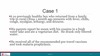
Question

D. Schistosoma japonicum
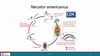
Schistosomiasis Life-cycle
- Infected person (Reservoir) releases trematode eggs in urine or stool
- Eggs contaminate freshwater
- Eggs hatch and infect snails (intermediate host) and divide
- Free swimming cercariae released into the water and penetrate human skin
People defecate oocytes which enter freshwater and eggs hatch and infect snails and divide, the snails release free-swimming cercariae into the water which penetrate human skin
Cercariae lose tails during penetration and become schistosomulae and enter the circulation and migrate to the portal blood in liver and mature into adult worms that perform sexual reproduction
Paired adult worms migrate to mesenteric venules of bowel/rectum laying eggs that circulate to the liver and are shed in stools to infect more snails

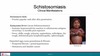
Schistosomiasis pathophysiology
eggs can migrate to the liver to be shed in stools or to other tissues such as wall of intestine or liver (mansoni or japonicum), bladder wall (haematobium) -> granulomas -> fibrosis
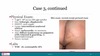
Liver Schistosomiasis
- Schistosoma mansoni
- Schistosoma japonicum
Bladder wall fibrosis Schistosomiasis
Schistosoma haematobium
Clinical manifestations of Schistosomiasis
- Swimmer’s itch: Cercariae penetrating skin (pruritic papular rash after penetration)
- Katayama fever (Acute schistosomiasis)
- systemic hypersensitivity reaction to Schistosoma antigens occurring 1-2 months post-exposure
- Fever, chills, cough, urticaria, angioedema, arthralgias, dry cough, abdominal pain, lymphadenopathy, and splenomegaly
- Massive eosinophilia
- Deposited eggs cause granulomas & fibrosis
- periportal fibrosis/portal hypertension
- Hematuria and bladder cancer
Would you predict that a patient with Schistosomiasis would have eosinophilia?
Yes, during swimmer’s itch phase and especially during the Katayama fever.
The granulomatous stage doesn’t have a big immune response
What laboratory test to diagnose Schistosomiasis
- Stool test if it is in the liver and biliary system
- Urine for bladder
Schistosoma species
- Schistosoma mansoni
- Schistosoma haematobium
- Schistosoma japonicum
Schistosoma mansoni region
- Africa
- South America
- Carribean
Schistosoma mansoni eggs found in
- Stool
Schistosoma haematobium region
- Africa
- Middle East
Schistosoma haematobium eggs found in
Urine
Schistosoma micro egg exam
Schistosoma mansoni micro egg exam

Schistosoma japonicum region
- Indonesia
- China
- SE Asia
Schistosoma japonicum eggs found in
Stool
Schistosoma japonicum micro exam of eggs

Schistosomiasis treatment
Praziquantel
Praziquantel MOA
Kills adult schistosomes and causes them to become dislodged from mesenteric, hepatic or pelvic veins -> phagocytosed in liver
Kills adult worms (does not kill eggs or treat fibrosis)
Praziquantel best used when?
During acute schistosomiasis (single dose 6-8 weeks after last exposure to potentially contaminated freshwater)