Week 4: Infections of the liver & biliary tree Flashcards
Case 1


Clinical findings of infections of the biliary system
- Charcot’s triad (RUQ pain, fever and chills, jaundice) continuous pain may radiate to right infrascapular region
- Murphy’s Sign: inhibition of inspiration by pain when the area of the gallbladder fossa is palpated
Laboratory findings of infections of the biliary system
- Leukocytosis
- Elevated alk phos & DB if the common bile duct is involved
Causative organisms of infections of the biliary system
- GNR: E coli, Klebsiella
- GPC: Enterococcus
- Anaerobes also but not that common
Types of infections of the biliary system
- Acute cholecystitis
- Cholangitis
What is acute cholecystitis
- obstruction of biliary drainage
- up to 50% complicated by infection
Complications of acute cholecystitis
- emphysematous cholecystitis
- pyogenic liver abscess
- bacteremia
- perforation
- peritonitis
What is cholangitis?
- Infection of the common bile duct
- stasis from obstruction favors the growth of bacteria
- increased pressure predisposes bacteremia
Complications of cholangitis
may be associated with hypotension and altered mental status
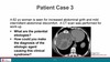
Case 2

Routes of hepatic invasion causing pyogenic liver abscess
- Cholangitis
- Hepatic artery
- Portal vein
- Direct extension from contiguous focus of infection
- cholecystitis
- subphrenic abscess
- perinephric abscess
- Penetrating trauma
Common presentation of pyogenic liver abscess
- middle-aged
- 50% solitary
- Right-sided

Diagnosis of pyogenic liver abscess
- Fine needle aspiration for gram-stain and culture (aerobic & anaerobic)
- Blood cultures (aerobic & anaerobic) positive 50%
Laboratory findings of pyogenic liver abscess
- peripheral leukocytosis
- elevated alk phos
- Culture: polymicorbia;
- GNR: E coli, Klebsiella
- GPC: Enterococcus, Strep viridians group
- Anaerobe: Bacteroides fragilis
Treatment of pyogenic liver abscess
- Long-term antibiotics (4-6 weeks) PLUS drainage
Gram-positive cocci flowchart

gram-negative bacilli flowchart

Bacteroides fragilis type
- Anaerobic GNR
- GI flora
- Major component of polymicrobial abdominal infections

Case 2 continued


Cause of amebic liver abscess
- Ingestion of contaminated food/water with cysts of Entamoeba histolytica
Stages in human amebic liver abscess
- Excystation in intestinal lumen
- Trophozoites migrate to colon and adhere to epithelium
- Trophozoites multiply by binary fission
- Infectious cysts released in stool
Signs & Symptoms of Amebic Liver Abscess
- 10% symptomatic colitis (DDx of bloody diarrhea)
- <1% liver abscess
- Entamoeba histolytica induces apoptosis in hepatocytes & neutrophils, forming large, non-purulent, “anchovy paste” abscesses

Diagnosis of Amebic Liver Abscess
- imaging & serum Ab
- Trichrome stain for cysts in stool not specific since look like nonpathogenic species
Treatment of amebic liver abscess