(LESSON 16) Blood Vessels Flashcards
Structure of Arteries, Veins, and Capillaries
Image

Lumen
the central blood-filled space of the blood vessel
Tunica Intima
innermost tunic of a vessel wall in intimate contact with the blood in the lumen.
- Internal layer of simple squamos epithelium
- Forms a smooth surface that minimizes friction of blood flow
- Subendothelial layer lies just external to endothelium
- In vessels larger than 1mm in diameter
- Loose connective tissue
Tunica Media
- Middle tunic
- consists primarily of circular smooth muscles fibers with circular sheets of elastin and collagen fibrils between
- Thicker in arteries than veins
- Maintains blood pressue
-
Vasoconstriction**
- Contraction of the smooth muscle cells with decreases the diameter of the vessel
-
Vasodilation
- Relaxation of the muscle cells that increases vessel diameter
- Both activities are regulated by vasometer nerve fibers.
Tunica Externa
- The outermost layer of the vessel wall
- a layer of connective tissue that contains many collagen and elastic fibers
- fibers run longitudinally
- protects the vessel, strengthens wall, anchors vessel to surrounding structures
Arteries
- Vessels that carry blood away from the heart.
- In systemic circuit blood is oxygen-rich
- in pulmonary circuit blood is oxygen-poor
- Blood proceeds from elastic arteries to muscular arteries, to arterioles
Elastic arteries
- the largest arteries near the heart. ie aorta and major branches
- from 2.5 cm to 1 cm in diameter
- AKA conducting arteries
- High elastin content dampens the surges of blood pressure

Muscular Arteries
- AKA distributing arteries
- Distal to elastic arteries
- supply groups of organs, individual organs, and parts of organs
- Constitute most of the named arteries
- 1cm-0.3mm in diameter
- thicker tunica media can regulate the amount of blood going to certain organs, according to needs

Arterioles
- Smallest arteries
- 0.3mm-10wm? in diameter
- contain only 1-2 layers of smooth muscle cells
- Nervous system and local factors determine diameter

Capillaries
- The smallest bust most important blood vessels
- 8-10wm in diameter
- Renew surrounding tissue fluid of all body cells with oxygen and nutirents
- remove CO2 and Nitrogenous waste
- Just large enough to allow erythrocytes to pass through in single file
- composed of one layer of endothelial cells surrounded by a basement membrane (tunica intima)
- Some capillaries perform site-specific functions
- Lungs: oxygen enters blood through capillaries
- Small intestine: receive digestive nutrients
- endocrine glands: pick up hormones
- Kidneys: remove nitrogenous waste
Capillary Bed
- A network of the body’s smallest vessels
- run through almost all tissue, especially loose connective tissue
- When precapillary sphincters relax, blood fills the true capillaries
- when sphinctes contract, they force most blood to flow straight from metarterioles to thoroughfare channels, bypassing the true capillaries
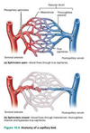
Metarteriole
A vessel that is structurally intermediate between an arteriole and a capillary-from which branch true capillaries.
Terminal arteriole-metarteriole-thoroughfare channel-postcapillary venule
Thoroughfare channel
A vessel structurally intermediate between a capillary and a venule. True capillaries merge into this, which then join the venule
Precapillary sphincters
- Smooth muscle that wraps around the root of each true capillary where it leaves the metarteriole.
- regulates bood flow to surrounding tissue according to needs for oxygen and nutrients.
Structure of Capillries Cut in Cross section
image
A. Continuous Capillary
B. Fenestrated Capillary
C. Sinusoidal Capillary
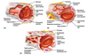
Intercellular Clefts
- Gaps of unjoined membrane
- small molecules exit and enter cavity here
Pericyte
- spider shaped cells that strengthen and stabilize capillary
- Thin processes form a network that is widely spaced to not interfere with spillary permeability
- External to endothelial cells
Continous VS Fenestrated Capillaries
Fenestrated: Have pores (fenestrations) spanning the endothelial cells. Occur only in areas of exceptionally high rates of exchange between blood and surrounding tissue fluid.
- small intestine, kidneys, synovial membrane of joints.
Continuous: No pores. More common, occuring in most organs of the body
- Skeletal muscle, skin, and central nervous system
Routes of capillary permeability
- Direct diffusion through endothelial cell membrane
- C02/Oxygen
- intercellular clefts
- most exchange of small molecues
- pinocytotic vesicles that invaginate from plasma membrane and migrate across the endothelial cell.
- transport dissolved gases, nutrients, and waste
- Fenestrations in fenestrated capillaries
Low Permeability: Blood-Brain Barrier
- Complete tight junctions
- no fenestrations or intercellular clefts
- vital molecules for the brain are ushered through endothelial cells.
- CO2, Oxygen, and some anesthetics may also diffuse unhindered
- Prolonged emotional stress can cause tight junctions in brain to open, allowing toxic substances through
- Gulf War Syndrome
Sinusoids
or
Sinusoidal Capillaries
- Wide, leaky capillaries
- Twisted course and large diameter ensure that blood slows to allow time for many exchanges to occur
- Occur with extensive exchange of large materials
- proteins
- cells
- Occur in
- Bone Marrow
- spleen
- Usually fenestrated
- fewer cell junctions
- in some, intercellular clefts are wide open
Veins
- The blood vessels that conduct blood from capillaries toward the heart.
- Systemic circuit: carry oxygen-poor blood
- Pulmonary circuit: carry oxygen rich blood returning from lungs
- Blood pressure much lower than in arteries
- blood pressure declines substantially passing through arterioles/cap. beds
- Walls of veins are much thinner
- At any time veins hold 65% of body’s blood
Venules
- The smallest veins
- 8-100wm in diameter
- Join to form veins
Postcapillary venules
- The smallest venules
- consist of endothelium on which lie pericytes
- Function like capillaries
- during inflammatory responses more fluid and leukocytes leave the circulation through these than through capillaries
*
Valves
- Prevent backflow of blood away from the heart
- Counteracts low venous blood pressure
- Each has several cusps formed by tunica intima
Mechanisms that counteract low venous blood pressure
- Valves
- normal movement of the body ensures blood moves only in the proper direction
- skeletal muscular pump
- contracting muscles press against thin-walled veins, propelling blood toward the heart (image)

Varicose Veins
- Valves in vein weaken and fail
- Vein twists and swells with pooled blood
- Factors:
- Hereditary
- Obesity
- Pregnancy
Hemorrhoids
varicosities of the anal canal
Vascular Anastomoses
- The interconnected or united vessels
- provide alternate pathways (collateral channels) for blood to reach certain regions
- occur around joints
- more often in veins than arteries
Vasa Vasorum
- Little vessels that nourish living cells in blood vessels
Atherosclerosis
- Aorta and coronary arteries are the vessels most affected by this degenerative disease, but all arteries are susceptible
- The following typifies the sequence of events leading to atherosclerosis
- damage to intima (can be caused by chemicals, virus, stress, etc.)
- injured tissue releases growth factors
- sequesters and oxidizes LDLs
- attracts monocytes to the area
- monocytes migrate beneath intima
- monocytes become macrophages which become foam cells
- other molecules congregate creating plaques
Lymphatic System
- Main Function is to return excess tissue fluid back to the vascular system
- Protexts our bodies from foreign organisms by fighting infection and conferring immunity to disease.
- Components
- lymphocytes
- lymphoid tissue
- lymphoid organs
- spleen
- lymph nodes
- thymus
Lymphatic Vessels
- Collect excess tissue fluid from the loose connective tissue around blood capillaries
- Return it to blood stream
- Also returns leaked blood proteins back into the blood stream

Lymph
- Clear Water
- the fluid inside of lymphatic vessels
- Only flows towards the heart
Edema
- When a body region swells with excess tissue fluid due to blockage of lymphatic vessels
- Can go away with exercise to area.
- bouncing and wiggling while sitting actually performs important function of moving lymph up legs.
Lymphatic Capillaries
- Highly permeable vessels that collect the excess tissue fluid
- located near blood capillaries in loose connective tissue
- Single layer of endothelial cells

Lymphatic Collecting Vessels
- where lymph goes after lymphatic capillaries
- accompany blood vessels
- Narrow and delicate. Not seen in dissecting.
- Same tunics as blood vessels (intima, media, externa) but much thinner
- Superficial lymphatic collecting vessels in skin travel with superficial veins
- Deep lymphatic collecting vessels of trunk and digestive viscera travel with deep arteries.
Lacteals
A unique set of lymphatic capillaries.
- Located in vili of small intestine
- Absorb digestive fats from intestine
- chyle: Lymph that becomes milky white from fats and is carried to blood stream
Areas absent of Lymphatic Capillaries
- bone
- teeth
- bone marrow
- entire CNS
- excess tissue fluid here drains through the nervous system into the cerebrospinal fluid.
- cerebrospinal fluid returns tissue fluid to blood at superior sagittal sinus
Lymphangiography
- Radiographic Process
- Lymph Valves bulge at the base creating a distinctive appearance
- vessels are injected with radiopaque dye
- the distinctive appearance allows phyisicians to recognize vessels on X-Ray film
Lymphatic Valves
- Vessels are not connected to heart so fluid flows under low pressure
- For this reason Lymphatic collecting vessels contain more valves than do veins
- Valves direct flow of lymph
Mechanisms that aid the flow of lymph
- Contracting skeletal muscles
- pulsations of nearby arteries
- muscular tunica media contracts
- normal movement of limbs and trunk
*Despite these mechanisms, propulsion is sporadic and slow. This explains while people who stand on their feet at work a long time can develop severe edema around ankles by the end of the workday.
Lymph Nodes
- Bean shaped organs situated along lymphatic collecting vessels
- cleanse the lymph of pathogens
- These are not glands
- 500 in the body
- 1 to 25mm in diameter
- Large clusters of nodes in these areas:
- cervical
- axillary
- inguinal

Cervial Nodes
Superficial, along jugular veins and carotid arteries, and receive lymph from head and neck
Axillary Nodes
in the armpit
Inguinal Nodes
In the superior thigh, filter from upper and lower limbs respectively.
Tracheobronchial Nodes and other nodes in the mediastinum
Recieve lymph from the thoracic viscera
Aortic Nodes
deep nodes along the abdominal aorta. filter lymph from posterior abdominal wall
iliac nodes
Deep nodes along the iliac arteries. filter lymph from pelvic organs and the lower limbs.
Structure of a lymph node
image

Capsule
dense connective tissue that surrounds node
trabeculae
fibrous strands that extend inward from the capsule to divide the node into compartments
afferent lymphatic vessels
convex aspect of the node through which lymph enters the node
hilium
indented region through which lymph exits node and passes through efferent lymphatic vessels.
lymph sinuses
large sinuses within the node between the afferent and efferent vessels.
- Subscapular
- cortical
- medullary
Reticular tissue in human lymph node
image

Lymph Trunks
- after leaving nodes, the largest lymphatic collecting vessels converge
- Drain large areas of the body
- large enough to be found by a skilled dissector

Five Major Lymph Trunks
From inferior to superior
- Lumbar Trunks
- Intestinal Trunk
- Bronchiomediastinal Trunks
- Subclavian Trunks
- Jugular Trunks
Lymph Ducts
- Trunks drain into ducts
- Largest lymphatic vessels
- Some individuals have two, others just have one
Thoracic Duct
- Present in all individuals
- Most inferior part (cisterna chyli) at union of lumbar and intestinal trunks
- empty at junction of subclavian and internal jugular veins
- Drains 3/4 of the body:
- head
- neck
- thorax
- left upper limb
- entire lower half of body
Right lymphatic duct
- 20% of people have it
- when present this empties into the neck
- drained by right jugular, subclavian, and bronchimediastinal trunks
- More common these trunks open independently into the neck veins
Lymph Organs
- Thymus
- Lymph Nodes
- Spleen
- Tonsils
Thymus
- Infants and children require greater activity of this gland than adults.
- hormone production
- activation of T-lymphocytes
- thymic lobules
- cortex and medulla
- atrophies with age
Spleen
- largest lymphoid organ
- immune response, blood cleaner, erythrocyte producer (fetus), and erythrocyte graveyard
- red pulp (RBCs):
- white pulp (WBCs)
Valves are only found in
veins
Tonsils
exterior surface is covered by squamous epithelium
Elephantiasis
- Wuchereria bancrofti is the parasitic culprit.
- Edema is the consequence.
- Inguinal lymph nodes are sites of blockage.
- treated with antibiotics
Crypts
- Trap bacteria in tonsils
- Trapped bacteria works its way through epithelium to underying lymphoid tissue
- Lymphocytes are activated
- Leads to infection to tonsils during early childhood
- Also generates great variety of memory lymphocytes for long-term immunity.
