Lecture 18- GI emergencies 2/2 Flashcards
Acute mesenteric ischemia
Symptomatic reduction in blood supply to GI tract
- Risk factors of acute mesenteric ischemia
*
More common in females (75%) and if you have a history of peripheral vascular disease
causes of acute mesenteric ischaemia
- Acute occlusion (70%)
- Non-occlusive mesenteric ischaemia (20%)
Acute occlusion (70%)
Arterial embolism in the SMA (50%)- midgut

Non-occlusive mesenteric ischaemia (20%)
- Low CO
- Mesenteric venous thrombosis (5-10%)
- Systemic coagulopathy and malignancy
most cases of acute mesenteric ischaemia are in
lderly patients. With CVD risk factors.
- Symptoms of acute mesenteric ischaemia
- Can be difficult to diagnose because symptoms can be fairly non-specific
- Abdominal pain disproportionate to the clinical findings
- Classic pain- comes on 30 mins after eating and lasts 4 hours
- Nausea and vomiting
- Abdominal pain disproportionate to the clinical findings
- Can be difficult to diagnose because symptoms can be fairly non-specific
Pain on left hand side
- why Pain on left hand side in acute mesenteric ischaemia-
- blood supply to the splenic flexure (point where transverse colon turns into the descending colon) is more fragile

AMI Investigations
*
- Blood tests
- Metabolic acidosis / increased lactate levels- ischaemic drugs
- Erect chest x-ray- check for perforation (gas under the diaphragm)
- CT angiography (90% sensitivity)- intravenous contrast
Treatment AMI
- Surgery- resection of ischaemic bowel–> bypass graft
- Thrombolysis/angioplasty
Prognosis of AMI
*
- Mortality is high (arterial thrombosis’s up to 70% mortality)
- Often older pts with comorbidities
2 examples of major upper GI bleeding
- peptic ulceration
- oesophageal varices
peptic ulceration causes …….% of acute upper GI bleeding
20-50%
what is peptic ulceration
- Disruption in gastric/duodenal mucosa that extends through the muscularis mucosa
- Greater than 5mm diameter
which type of ulcer is most common
- Duodenal ulcers most common
- First part of duodenum
- Gastro-duodenal artery lies behind first part of duodenum
where are peptic ulcers found in the stomach
- Lesser curve and antrum common sites
gastric ulcer erosion is especially dangerous if it erosed the
splenic artery

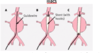
what are oesophageal varices
porto-systmeic anastomosis
- 12-14% of acure upper GI bleeding
explain how oesophageal varices occur and how they can lead to major upper GI bleeding
- an example of porto-systemic anastomosis
- they are often caused by portal hypertension
- back flow of blood from the portal vein –> left gastric –> which drains the distal portion of the oesophagus
- oesophagus also draine dby the azygous vein
- left gastric vein and azygous anastomose
- these anastomes distend with blood – rupture –> bleed

causes of oesophageal varices
portal hypertension
- Caused by anything that slows blood flow through portal vein
- Pre-hepatic (portal vein thrombosis)
- Hepatic (cirrhosis schistosomiasis)
- Post hepatic causes (hepatic vein thrombosis, RHF)
- Normal pressure in portal vein
- 5-10mm Hg
- Problem happens around 10 mmHg
- porto-systemic anastomoses are areas that
have venous drainage through portal vein and systemic veins e.g. the distal portion of the oesophagus
Treatment of oesophageal varices
- Fluid resus
- If bleeding not controlled by banding
- Transjugular intrahepatic portosystemic shunt (TIPS)
-
Drug treatment
- Terlipressin- reduces portal venous pressure
Transjugular intrahepatic portosystemic shunt (TIPS)
- expandable metal is palced within the liver
- bridge the portal vein to the hepatic vein
- decrompresses protal vein pressyre
- reduction in variccal pressure
- reduction in ascites