Lecture 18- GI emergencies 1/2 Flashcards

1
Q

Causes of peritonitis

A
  • Ascites
  • Cirrhosis
  • Perforated appendicitis
  • Perforated peptide ulcer
  • Perforated diverticulitis
  • Volvulus
  • Cancer
2
Q

what is peritonitis

A

Inflammation of the serosal membrane that lines the abdominal cavity

3
Q

the peritoneal cavity is usually

A

sterile

4
Q

primary peritonitis is

A

spontaneous

5
Q

secondary peritonitits

A
  • Breakdown of peritoneal membrane leading to foreign substances entering cavity
6
Q

inflammation in both primary and secodnary peritonitis is

A

uniform

7
Q
  • Peritonitis can be
A

infectious or sterile

8
Q

Peritoneal cavity-

A

space between the visceral and parietal layers of the peritoneum

9
Q

viceral and parietal components of the periotneum are

A

continous

10
Q

visceral periotneum

A

any part of the serosal memrbane that is not lining the abdominal wall

11
Q

parietal peritoneum

A

any part of the serosal memrbane that is lining the abdominal wall

12
Q

peritioneal cavity contains

A

no viscera, only a small amount of fluid

13
Q

peritoneal cavity can be divided itno 2 sec tions

A

greater and lesser sac

14
Q

greater and lesser sac conencted by the

A

foramen of winslow

15
Q

what is a mesentry

A

The mesentery attaches the intestines to the abdominal wall, and also helps storing the fat and allows the blood and lymph vessels, as well as the nerves, to supply the intestines.

16
Q

primary peritonitis is also called

A

spontaneous bacterial peritonitis (SBP)

17
Q

spontaneous bacterial peritonitis (SBP) is most commonly seen in

A
  • patients with end stage liver disease (pts with cirrhosis)
18
Q
  • what is Spontaneous bacterial peritonitis (SBP)
A

is an infection of ascitic fluid that cannot be attributed to any intra-abdominal, ongoing inflammatory, or surgical correctable condition

19
Q

Ascites

A

Pathological collection of fluid within peritoneal cavity

20
Q
  • In cirrhosis what causes ascites
A
  • caused by a combination of
    • Portal hypertension
      • Causing increase hydrostatic pressure in veins draining the gut
    • Decreased liver function resulting in less albumin production
      • Decreased intravascular oncotic pressure
    • The result is net movement of fluid into the peritoneal cavity
21
Q

symptoms of Primary peritonitis- spontaneous bacterial peritonitis (SBP)

A
  • Abdominal pain, fever and vomiting
  • Usually symptoms are mild (slightly milder than regular peritonitis)
22
Q

Diagnosis of SBP

A

Aspirating ascitic fluid- neutrophil count >250 cell/mm3

23
Q

Secondary peritonitis

A

Secondary (surgical) peritonitis is a result of an inflammatory process in the peritoneal cavity secondary to inflammation, perforation, or gangrene of an intra-abdominal or retroperitoneal structure

24
Q

pathophysiology of secondary peritonitis

A
  • Peritoneal cavity is normally sterile
  • If viscera perforates then the contents will enter the peritoneal cavity
25
Q
  • Common cause of secondary bacterial peritonitis include
A
  • Peptic ulcer
  • Appendicitis
  • Diverticulitis
  • Post surgery
26
Q
  • Non-bacterial causes of secondary peritonitis
A
  • Tubal pregnancy that bleeds (peritoneal cavity is not enclosed in females)
  • Ovarian cyst
  • Blood is highly irritant to peritoneal cavity
27
Q

Symptoms of secondary peritonitis

A
  • Abdominal pain
    • Lying very still
    • Knees flex
    • Shallow breathing
  • May come on gradually or acutely
  • Diffuse abdominal pain common in perforated viscera
28
Q

Treatment of secondary peritonitis

*

A
  • Control infectious source- surgery
  • Eliminate bacteria and toxins- Abx
  • Maintain organ system function
    • Intensive care
29
Q

define bowel obstruction

A

Mechanical or functional problem that inhibits the normal movement of gut contents

  • Can effect large and small bowel
  • All ages affected
30
Q

Common causes of bowel obstruction in children

A
  • Intussusception
  • Intestinal atresia- failure to recanalize bowels during development
31
Q

Common causes of bowel obstruction in adults

A
  • Adhesion
  • Incarcerated hernias
32
Q

Small bowel obstruction

Symptoms

A
  • Nausea and vomiting (bilious) are most common early symptoms
  • Abdominal distension
  • Absolute constipation (late)
33
Q

causes of small bowel obstruction

A
  1. Intra-abdominal adhesion
  2. hernias
  3. IBD
34
Q
  • Intra-abdominal adhesion
    *
A

abnormal fibrous bands between organs or tissues or both in the abdominal cavity that are normally separated

  • Arises after more than 50% of abdominal surgeries
    • Greater omentum involved in 90%, bowel in 50%
  • Damage to mesothelium (direct trauma, post op infection)
    • Capillary bleeding leading to exudation of fibrinogen
35
Q

Other consequences of adhesions

A

Abdominal pain

Infertility

36
Q

small bowel obstruction caused by which sort of hernia

A

incarcerated groin hernias

37
Q

IBD and small bowel obstruction

A
  • Crohns- repeated episodes of inflammation causing narrowing
38
Q

Diagnosis of small bowel obstruction

A
  • History- abdominal pain is crampy intermittent- colic pain
  • Physical exam- abdominal distension, increased /absent bowel sounds, presence of hernia
  • Imaging
39
Q

how would smalll bowel obstruction look on CT

A
  1. 3cm or more= distended small bowel
  2. central position of bowel
  3. plica circulares clear- lines across hole of bowel
40
Q

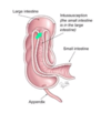
Intussusception

A

When one part of the gut telescopes into an adjacent section

41
Q

causes of intussusception

A
  • Not understood well
  • Potential motility issue
  • Lead point (a mass that precipitates the telescoping action)
    • Mechels diverticulum
    • Enlarged lymph nodes
42
Q
  • Intussusception can extend quite far –
A

even prolapse out of the rectum

43
Q

when do you get oedema in intussusception

A
  • As soon as the lymphatic and venous drainage is impaired you get oedema
    • Enough oedema can impede arterial supply (infarction)
44
Q
A
45
Q

symptoms of intussusception

A

Symptoms

  • Abdominal pain
  • Vomiting
  • Haematochezia- bright red blood passed PR
46
Q

Treatment of intussusception

A
  • Air enema- pressure can pop the bowel into the right place
  • surgery
47
Q

Large bowel obstruction

Typically affects

A

older people

48
Q

common causes of large bowel obstruction

A
  • Colon cancer – mechanical obstruction
  • Diverticular disease (20%)- sigmoid colon
  • Volvulus- sigmoid and caecal (5%)
49
Q

symptoms of large bowel obstruction

A

Symptoms often appear gradually if caused by cancer but abrupt with volvulus

  • Change in bowel habit (cancer)
  • Abdominal distension
  • Crampy abdominal pain
  • Nausea/vomiting (later)
50
Q

how would a large bowel obstruction look on a CT

A
  • more peripheral
  • can seehuastra which doesnt go the whole way across
  • 3/6/9 rule
51
Q

-3/6/9 rule

A

. The upper limit of 3 cm applies to the small intestinal loops, 6 cm for the colon and 9 cm for the cecum (blind intestine).

52
Q

Volvulus

A
  • Derived from latin ‘to twist’*
  • Part of the colon twists around its mesentery
53
Q

where are volvulus most common

A
  • Most common is sigmoid colon (60%) and caecum (can technically be anywhere)
54
Q

volvulus can result in

A

obstruction

55
Q

obstruction of the sigmoid colon

A
  • Can result from overloaded sigmoid colon (constipation)
    • Extra mass elongates the sigmoid (relatively small mesenteric attachment)
    • High fibre diet can also lead to sigmoid and twisting
  • Results in large and small bowel obstruction
56
Q
  • Sign of sigmoidal volvulus
A

Coffee bean sign

57
Q

Small vs large bowel obstruction

Age

*

A
  • Small bowel obstruction more common in younger age group
  • Large bowel obstruction in older age group
58
Q

in bowel obstruction what is important

A

Competence of the ileo-caecal valve is of great importance

  • If it is competent then the colon cannot decompress proximally
    • Closed loop obstruction- ischaemia and perforation more likely
59
Q

abdominal pain in small bowel obstruction

A

colicky 3-4 mins

60
Q

abdominal pain in large bowel obstruction

A

colicky 10-15 mins

61
Q

vomiting in small bowel obstruction

A

early

62
Q

vomiting in large bowel obstruction

A

late

63
Q

constipation in small bowel obstruction

A

late

64
Q

constipation in large bowel obstruction

A

early