Exam 2: Platelets and Coagulation Flashcards

1
Q

Platelet

Functions

A
  • Limit bleeding after injury to a blood vessel
  • Promote vessel repair
  • Regular maintenance of an intact endothelium
2
Q

Platelet Count

A

Normal range: 150,000 - 450,000 platelets/microliter

3
Q

Thrombocytopenia

A
  • Abnormally low platelet count
    • < 150,000/microliter
  • Characterized by:
    • Easy bruising
    • Nose bleeds
    • Petechial rash
    • Spontaneous bleeding
      • Occurs below 20,000 platelets/microliter
4
Q

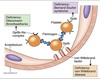
Idiopathic Thrombocytopenic Purpura

(ITP)

A

Autoimmune disease where anti-platelet Ab destroy platelets.

5
Q

Platelet

Lifecycle

A
  • Derived from bone marrow cells called megakaryocytes
  • Lifespan ~ 8-10 days
  • Removed by sleen or via clotting process
6
Q

Platelet

Morphology

A

Small, flat, disc-shaped membrane-enclosed bits of cytoplasm.

Hyalomere

  • Clear outer region
  • Contains bundles of microtubules
    • Helps maintain discoid shape
  • Contains actin & myosin
    • Involved in shape change of activated platelets

Granulomere

  • Central region with basophilic stippling
  • Contains usual cytoplasmic organelles
  • Contains at least 3 types of granules
    • Alpha, Delta, and Lambda

2 systems of membrane bound channels:

  • Open canalicular system
    • Invaginations of the plasma membrane
    • Facilitates rapid exocytosis of granules
  • Dense tubular system
    • Stores Ca2+ needed for exocytosis
    • Not continuous with the plasma membrane
7
Q

Alpha (α) Granules

A

Contains:

Platelet-derived growth factor (PDGF) ⇒ mitogen for vessel repair

von Willebrand Factor (vWF) ⇒ mediates platelet adhesion to endothelium (collagen and laminin)

8
Q

Delta (δ) Granules

A

Contains:

Ca2+, ATP, ADP ⇒ all enhance platelet aggregation

Serotonin ⇒ vasoconstriction

(Picked up by platelets in circulation)

9
Q

Lambda (λ) Granules

A

Contains:

Lysosomal enzymes ⇒ clot resorption

10
Q

Vessel Repair

Process

A

Adhesion

  • vWF binds to components of the damaged basement membrane (collagen, laminin)
    • vWF can be secreted by many cells including platelets
  • vWF attracts platelets which have surface receptor for vWF
  • Single layer of platelets forms @ site of endothelial damage

Aggregation

  • Adhered platelets secrete fibrinogen
  • Other platelets attracted to the site via cell surface receptors for fibrinogen
  • Fibrinogen links the platelets together
  • Forms a multilayered primary hemostatic plug
    • Fills the defect in the vessel wall

Activation

  • Aggregation causes platelet activation
  • Results in:
    • Secretion of granule mediators
    • Synthesis and release of aracidonic acid derivatives
      • Thromboxane A2 (TXA2)
    • ​Change of platelet shape
      • Mediated by the hyalomere
  • Released mediators cause:
    • Further platelet aggregation
      • TXA2, serotonin, and Ca2+
    • Vasoconstriction (limits bleeding)
      • Serotonin and TXA2
    • Blood coagulation
      • Platelets release several coagulating factors from α-granules
      • Meshwork of fibrin formed which stabilizes the platelet plug forming the secondary hemostatic plug

Clot Retraction

  • After ~ 1 hr, platelets contract due to actin-myosin interaction
    • Platelet plug ↓ in size & flattens against vessel wall
    • Helps to re-establish smooth blood flow

Clot Resorption

  • Mediated partially by lysosomal enzymes from λ-granules

Vessel Repair

  • Mediated by platelet-derived growth factor (PDGF) from α-granules
  • PDGF is strongly mitogenic for cells needed to rebuild the vessel wall including:
    • Endothelial cells
    • Fibroblasts
    • Smooth muscle
11
Q

Platelet Activation

Regulation

A
  • Platelets activated upon binding to collagen and laminin of damaged basement membrane
    • Not exposed in healthy vessels
  • Healthy endothelial cells produce factors that inhibit platelet aggregation
    • Ex. Prostacyclin I2 (PGI2) from arachidonic acid
12
Q

Aspirin

A

Inhibits cyclooxygenase activity

  • ↓ platelet function
  • Prolongs bleeding times
13
Q

Thrombosis

A

Formation of a clot (thrombus)

  • Can cause serious damage
  • Can lead to death if vessel occluded
14
Q

Thrombus

Charactertistics

A
  • Meshwork of fibrin + plug of activated platelets
  • Clotting occurs in association with membranes
  • Pathology:
    • In coronary arteries ⇒ MI
    • In cerebral arteries ⇒ ischemic stroke
    • In peripheral arteries ⇒ claudication (leg pain with exercise) or amputation
    • In deep veins ⇒ DVT
15
Q

Embolus

A

A mass traveling through the circulation.

Can be a thrombus broken off a site of coagulation.

If thrombus lodges in the lung ⇒ pulmonary embolism (PE)

16
Q

Blood Clotting

Overview

A

Initiated on the membranes of endothelial cells and platelets:

  1. Formation of a fibrin clot
  2. Formation of a platelet plug
  3. Vasoconstriction (eicosanoids, PGs, Txs)
  4. Limits to the process (anticoagulation)
  5. Clot dissolution (fibrinolysis)
  6. Wound repair

Functions through cascade of proteolytic cleavage or conformational changes.

17
Q

Fibrin Clot Formation

Charactertistics

A
  • Intrinsic and extrinsic pathways converge on the final common pathway
  • Major factors
    • Named by Roman numerals & common names
      • Factor IX = Christmas factor
    • Are glycoproteins synthesized primarily by the liver
  • Functions using cascades
    • Activation primarily by proteolytic cleavage
    • Successive proteins are serine proteases
      • Cleaves peptide bond on carboxyl side of Arg or Lys
    • Activation can also be caused by conformational changes
    • Facilitates acceleration and amplification of process
  • Non-proteolytic proteins also needed ⇒ accessory proteins (cofactors)
18
Q

Clotting Effectors

A

Presence accelerates the rate of certain steps in fibrin clot formation:

  • Negatively charged phospholipids (PS, PI)
    • Normally found on inner leaflet of plasma membrane
    • Exposure signals injury
  • Ca2+
    • Binds negatively charged γ-carboxyglutamate (Gla) residues on certain clotting proteins
    • Facilitates binding of these proteins to exposed negatively charged phospholipids
19
Q

Gla Proteins

A

Factors II, VII, IX, X

  • Contains negatively charged γ-carboxyglutamate (Gla) residues
  • γ-carboxylation
    • Post-translational modification
      • 9-12 Glu residues @ N-terminus carboxylated to Gla residues by glutamyl carboxylase
    • Occurs in lumen of RER in hepatocytes
    • Vit K required as co-enzyme
      • Oxidized in reaction to epoxide
      • Must be reduced back to hydroquinone form to continue
        • Done by Vitamin K epoxide reductase (VKOR)
20
Q

Dicumarol

&

Warfarin (Coumadin)

A

⊗ reduction of Vit K by VKOR

  • Inhibits clotting by depleting pool of Vit K
  • Oral administration
    • Slow onset
    • Long half-life
  • Polymorphisms in cytochrom P450 and VKOR isozymes results in varied dosing
21
Q

Tissue Factor

(TF)

A
  • Transmembrane glycoprotein
    • Abundant in vascular subendothelium
  • Released by damaged tissue ⇒ extravascular
  • Key protein in the extrinsic pathway
    • Pathway is quickly shut down by tissue factor pathways inhibitors (TFPI)
22
Q

Extrinsic Pathway

A

“Tissue Factor Pathway”

  1. Vascular injury exposes extravascular TF (Factor III) to Factor VII
  2. TF binding causes conformation change of VII activating it to VIIa
    • VII can also be activated by Factor XIIa from intrinsic path or thrombin (IIa) from common path
  3. VIIa is a serine protease which activates factor X
  4. Factor Xa enters the common pathway

Extrinsic pathway is quickly shut down by tissue factor pathway inhibitors (TFPI).

23
Q

Intrinsic Pathway

A

“Contact Pathway”

All protein factors are found in the blood ⇒ intravascular

Contact Phase

Results in the activation of factor XII XIIa

  1. Contact of blood with a negatively charged surface ⇒ conformational change & activation of factor XII → XIIa
    • In vitro ⇒ glass blood vials
      • Sodium citrate/oxalate added to chelate Ca2+ and prevent clotting
    • In vivo ⇒ ⊖ PL on damaged endothelium or an abnormal surface
      • E.g. mechanical heart valve, stent, knee/hip replacements
  2. Amplification of contact phase
    • Factor XIIa cleaves Prekallikrein-HMWK at an anionic surface producing Kallikrein
      • HMWK = High molecular weight kininogen
    • Kallikrein can then proteolytically cleave additional Factor XII ⇒ amplification

No known bleeding disorders associated with factor deficiencies of the contact phase.

X Activation Phase

  1. Factor XIIa cleaves XI-HMWK at an anionic surface to produce Factor XIa
    • Factor XI can also be activated by Thrombin from common pathway
    • Defective Factor XI → Hemophilia C
  2. Factor XIa cleaves Factor IX (Christmas factor)IXa
    • Defective Factor IX → Hemophilia B
    • Can also be cleaved by Factor VIIa from extrinsic pathway
  3. Factor IXa combines with Factor VIIIa
    • Interaction with VIIIa ↑↑↑ rxn rate
    • Factor VIII found in the blood bound to von Willebrand factor (vWF)
      • vWF protects VIII from degradation
      • Thrombin from common pathway cleaves vWF off VIII activating it
    • Defective Factor VIII → Hemophilia A
  4. IXa:VIIIa complex cleaves Factor XXa
    • Factors VIIIa, IXa, 10, and Ca2+ on membrane ⇒ Tenase complex
24
Q

Hemophilia

A
  • Coagulopathy caused by clotting factor deficiencies
    • Factor VIII ⇒ Hemophilia A
      • 6x more common than B
      • Found on chromosome Xq
    • Factor IX ⇒ Hemophilia B
      • Found on chromosome Xq
    • Factor XI ⇒ Hemophilia C
      • Autosomal recessive
  • Manifestations
    • Decreased/delayed ability to clot
    • Formation of abnormally friable clots
  • Severity related to residual activity
    • Effects seen if < 30% activity
    • Severe form if < 1% activity
      • Spontaneous, prolonged bleeding particularly into joints and muscle
  • Treatment
    • Recombinant factor replacement
    • Somatic gene therapy in development
25
Common Pathway
**Factor Xa to Fibrin:** 1. **Factor Xa** from both _intrinsic_ and _extrinsic_ paths associates with **Factor Va**(accessory protein) forming **Xa-Va** ⇒ **Prothrombinase complex** * Binding of Va ⇒ 20x ↑ in Xa activity 2. Binding of Ca2+ to Gla residue on **Prothrombin** (_Factor II_) facilitates binding of Prothrombin to membrane and to Xa-Va complex 3. Xa-Va complex cleaves **Prothrombin** (II) **→ Thrombin** (IIa) * Excises Gla region releasing Thrombin from the membrane * Only time Gla excised * Gla-peptide remains bound to membrane surface * Taken up by the liver 4. **Thrombinplasma** cleaves **Fibrinogen** (Factor I) → **Fibrin** (Ia) 5. Fibrin forms the **fibrin soft clot** 6. **Thrombinplasma** activates **Factor** **XIII** → **XIIIa** 7. **Factor XIIIa** (transglutaminase) cross-links Gln from one fibrin with Lys of another ⇒ _isopeptide bond_ * Converts soft clot into an **insoluble** **hard clot**
26
Fibrin
* **Fibrinogen** * Soluble glycoprotein made by the liver * Dimers of three different polypeptides held together at N-termini by disulfide bonds * N-termini of the chains form "**tufts**" on the central three globular domains (D, E, D) * **Thrombin** _cleaves the negatively charged tufts_ producing **fibrinopeptides**: * Fibrinogen → fibrin monomers * Solubility decreases * Fibrin monomers associate laterally * **Soft fibrin clot** **formed**
27
Roles of Thrombin
28
Blood Clotting Complete Pathway
_Pathway Interconnections:_ * **Factor VIIa** of extrinsic path activates **factor IX** of intrinsic path * **Factor XIIa** of intrinsic path activates **factor VII** of extrinsic path * **Thrombin (IIa)** of common path activates factors of intrinsic and extrinsic paths * Factors 7, 12, 11, 8, and 5
29
Clotting Factors Classifications
30
**Platelet plug** provides...
primary hemostasis
31
**Fibrin net** provides...
secondary hemostasis
32
Platelet Plug Formation
1. **vWF** _binds exposed collagen_ on endothelial surface 2. **Platelets** bind: * _To vWF_ via **GP1b** * Part of the membrane receptor complex (**GP1b/V/IX**) * vWF made by endothelial cells and megakaryocytes * _Directly to collagen_ via **GPVI** 3. Binding stops forward movement of platelets causing **adherence** 4. Platelet activation causes **degranulation** * Thrombin is the most potent activator * Required for platelet activation and fibrin formation * Also **exposes anionic PL** which allows _formation of tenase complex_ on surface of platelets 5. Conformational Δ following activation leads to **platelet** **aggregation** * Forms the **platelet plug** ⇒ _primary hemostasis_ 6. Conformational Δ in **GPIIb/IIIa** receptor exposes fibrinogen binding sites 7. Fibrinogen _binds & links_ activated platelets to one another * Fibrinogen → Fibrin by **Thrombin (IIa)** * Cross linkage by **Factor XIIIa** from plasma and platelets * **Fibrin net** strengthens platelet plug to resist shear forces * Achieves _secondary hemostasis_
33
Platelet Activation
1. **Thrombin** binds to and activates protease-activated receptors (**PARs**) on the surface of platelets & endothelial cells * Type of Gq receptor 2. Receptor activates **PLC** which cleaves PIP2 → **DAG** and **IP3** 3. **DAG** activates **PKC** leading to **degranulation** 4. **IP3** causes release of **Ca2+** from _delta granules_ 5. **Calcium** causes: * **Shape changes in platelet** * Ca2+ activates MLCK which phosphorylates MLC * Favors association with actin * **Activation of PLA2** * Synthesis of **TXA2** * Platelet degranulation * Vasoconstriction * Via serotonin * Activation of additional platelets * By binding to GPCR on platelet membrane
34
Platelet Activation Regulation
* Vascular wall is **separated from blood** by endothelial cells * Components like collagen and laminin are not exposed * Endothelial cells synthesize the **vasodilators** **PGI2** and **NO** * Endothelial cells have a cell **surface ADPase** that converts ADP → AMP
35
Platelet Degranulation
Degranulation releases: * Delta granules * **Serotonin**: causes vasocontriction * **ADP**: activates additional platelets * **Plavix** blocks ADP receptors preventing ability to activate platelets * Alpha granules: * **PDGF**: helps in wound healing * **Factor** **Va** * **vWF** * **Fibrinogen** * Many more
36
Von Willebrand's Disease
* **Deficiency of von Willebrand's factor** * Results in _abnormal platelet adhesion_ * Most common inherited coagulopathy * AR
37
Bernard-Soulier Syndrome
* **Deficiency of platelet receptor for von Willebrand's factor** * GP1b receptor * Results in _abnormal platelet adhesion_
38
Glanzmann's Thrombasthenia
* **Deficiency of platelet receptor for fibrinogen** * GPIIb/IIIa complex * Results in _decreased platelet aggregation_
39
Immune thrombocytopenia
Autoimmune disorder caused by autoantibodies to platelet receptor for fibrinogen.
40
Pathways of Coagulation Interrelationships
* **TF—VIIa** of extrinsic path activates **IX** of intrinsic path * **XIIa** of intrinsic path activates **VII** of extrinsic path * **Thrombin (IIa)** of common path activates components of both the intrinsic and extrinsic paths and also activates platelets * **Tenase complex** of intrinsic path forms on the surface of activated platelets and damaged endothelium * **One fifth of total Va** is released by activated platelets * **XIIIa** and **vWF** present in both plasma and platelets
41
Antithrombin III | (AT-III)
* Made by the liver * **Serine protease inhibitor (serpin)** * _ATIII binds to and inactivates:_ * **Thrombin** (most important) * Binding makes catalytic domain of thrombin inaccessible * **Factor IXa through XIIa** * Inactivation of **X** is key * **Factor VIIa-TF complex** * Complexes removed by liver * AT-III : Thrombin binding is greatly increased by **heparin** * Sulfated GAG released by mast cells in response to injury
42
Heparin
* Therapeutically used to **↓ clot formation** * IV administration * Rapid-onset * Short half-life
43
Activated Protein C Complex | (APC)
**Protein C-Protein S complex inactivates Va and VIIIa by proteolysis.** * **Protein C**(Gla) activated by **thrombin-thrombomodulin** complex * **Thrombomodulin** expressed on the surface of undamaged endothelial cells * Binds thrombin * ↓ thrombin's affinity for fibrinogen * ↑ thrombin's affinity for Protein C * **Protein C** complexes with **Protein S**(Gla) ⇒ **activated Protein C complex (APC)** * **APC cleaves Va and VIIIa** * Protein S helps achor Protein C to the clot
44
Tissue Factor Pathway Inhibitor | (TFPI)
Protein that inhibits the extrinsic pathway shortly after its activation. Intrinsic path becomes dominant.
45
Fibrinolysis
* **Plasminogen** binds to fibrin & _incorporated into clots_ during formation * Physiological activators of plasminogen bind and cleave **plasminogen** → **plasmin** * **Tissue plasminogen activator (t-PA or TPA)** * Made by endothelial cells * Secreted in inactive form in response to thrombin * Becomes active when bound to fibrin-plasminogen * **Urokinase (u-PA)** * Made in the kidney * **Plasmin** _hydrolyzes fibrin clot_ * Bound plasmin and TPA protected from inhibitors * When fibrin clot is dissolved, they are exposed and inactivated * **α-2 antiplasmin** ⇒ ⊗ plasmin * **PAI (plasminogen activator inhibitor)** ⇒ ⊗ TPA
46
Plasmin
* Serine protease which degrades fibrin * Plasminogen made by the liver & released into the blood * Binds to fibrin and is incorportated into clots as they are formed * Activated by TPA or u-PA
47
Therapeutic Fibrinolysis
Treat patients with TPA or u-PA
48
Thrombophilia
**"Hypercoagulability"** _Caused by:_ * **Factor V Leiden** * **G20210A prothrombin gene mutation** * ↑ factor II activity * Most prevalent genetic risk factor for venous thrombosis in Spanish populations * **Deficiencies of proteins C, S, and ATIII** * **Excess lipoprotein A** * **Hyperhomocysteinemia** * **Anti-phospholipid Ab** ⇒ referred to as Lupus anticoagulant
49
Factor V Leiden
* **Mutant form of factor V ⇒ resistant to APC cleavage** * Most commonly inherited thrombophilic condition * Esp. Caucasians, specifically Scandinavians * Heterozygotes have a 7x increased risk of forming clots * Homozygotes have 80x increased risk
50
Virchow's Triad
Three broad categories thought to contribute to thrombophilia:
51
Coagulation Summary
* Tissue injury ⇒ blood vessel damage ⇒ collagen exposure * Platelets bind collagen * Mediated by vWF * Platelet activation * Thrombin is key * Platelet degranulation and synthesis of arachidonic acid products * Vasoconstriction * Platelet recruitment * Support the clotting cascade * Factors of intrinsic, extrinsic, and common paths of clotting cascade + Ca2+ + anionic surface ⇒ fibrin clot formation * Initial platelet plug ⇒ primary hemostasis * Fibrin mesh ⇒ secondary hemostasis * Clotting limited to injured site * Proteins that inactivate thrombin ⇒ AT-III, heparin * Proteins that degrade V & VIII ⇒ APC * After wound is healed, fibrin clot degraded by plasmin * Plasmin inactivated by α-2 antiplasmin * TPA inactivated by PAI
52
Abnormal Clotting Summary
Disorders of vessels, platelets, and coagulation proteins ⇒ deviations in ability to clot. * Vessels * Scurvy * Plaque formation * Hyper-Hcy * ↑ Lp(a) * Platelets * ↓ number ⇒ thrombocytopenia * ↓ function * Deficiency of vWF ⇒ VW disease * Deficiency of GP1b ⇒ Bernard-Soulier syndrome * Deficiency of CPIIb/IIIa ⇒ Glanzmann thrombasthenia * Coagulation proteins * VIII ⇒ Hemophilia A * IX ⇒ Hemophilia B * XI ⇒ Hemophilia C * Factor V Leiden
53
Clotting Clinical Tests
* _Platelets_ * Platelet counts * Platelet function tests * _Extrinsic through common path_ * **Prothrombin time (PT)** * Uses thromboplastin * Combination of PL + TF * Expressed as INR (international normalized ratio) * _Intrinsic through common path_ * **Activated partial thromoplastin time (aPTT)** * Uses partial thromboplastin * Just the PL portion b/c TF isn't needed to activate intrinsic path