Exam 1: Cell Injury and Cell Death Flashcards

1
Q

Response to Stress

A
  • Cells and organs in homeostasis
  • Response to stress ⇒ adaptation or death
  • Response dependent on severity and length of stress
2
Q

Adaptations

A

In response to changes in physiologic and pathologic stimuli to maintain homeostasis.

When stress removed, can recover without harm.

Includes:

  • Hypertrophy
  • Hyperplasia
  • Atrophy
  • Change in phenotype
    • Metaplasia
    • Dysplasia
3
Q

Hypertrophy

A

Increase in cellular size and functional activity.

  • Physiologic
    • Muscle hypertrophy w/ inc. workload
      • ↑ protein synthesis ⇒ ↑ myofilament size ⇒ ↑ force generation
      • Ex:
        • Bodybuilder ⇒ inc. demand
        • Heart ⇒ chronic hemodynamic overload
  • Pathologic
    • Cancer
    • Response to injury
4
Q

Cardiac Hypertrophy

Pathogenesis

A
  1. Mechanical sensors ⇒ production of growth factors and agonists
  2. Activation of signal transduction pathways
  3. Activation of transcription factors
  4. Inc. synthesis of muscle proteins
5
Q

Hyperplasia

A

Increase in cell number.

  • Occurs in response to stimuli
  • Can occur with hypertrophy
  • Only seen in tissues where cells can divide
  • Physiologic vs Pathologic
6
Q

Physiologic Hyperplasia

Examples

A
  • Breast glandular epithelium
    • Puberty or pregnancy
  • Liver regeneration
    • Regenerate after donation
  • Bone marrow
    • Make more RBCs after bleed or hemolysis
7
Q

Pathologic Hyperplasia

Examples

A
  • Endometrial hyperplasia
    • Causes abnormal bleeding
    • Due to excess estrogen ⇒ imbalance between estrogen/progesterone
  • Benign prostatic hyperplasia
    • In response to androgens
    • Increases with age
    • No increased risk for neoplasm
  • Response to viral infection
    • Ex. HPV ⇒ warts
      • Viral factors can interfere with host proteins that regulate cell proliferation
      • Can be a cancer precursor
8
Q

Atrophy

A
  1. ↓ cell size & organelles
    • ↓ metabolic demands
    • Attempt to prolong survival
    • May diminish cell function
    • May lead to irreversible injury and death
  2. ↓ size of organ or tissue
9
Q

Physiologic Atrophy

Examples

A
  • Embryonic development
  • Shrinkage of uterus after delivery
10
Q

Pathologic Atrophy

Examples

A
  • Decreased workload
    • Atrophy of disuse
  • Loss of innervation
  • Diminished blood supply
    • Seen in brain w/ age
  • Inadequate nutrition
    • Marasmus ⇒ cachexia
  • Loss of endocrine stimulation
    • Uterus after menopause
  • Pressure
    • Enlargeing tumor compresses normal tissue
11
Q

Protein and Organelle

Clearance

A

Two pathways to clear damaged proteins and organelles:

  1. ↓ protein synthesis
    • In response to ↓ metabolic activity
  2. ↑ protein degradation
    • Ubiquitin-proteasome pathway
      • Activation of ubiquitin ligases
      • Ubiquitin attached to proteins
      • Degraded in proteasomes
    • Autophagy
      • Cells “eats” it own components
      • Debris may remain as residual bodies ↑ lipofuscin granules
12
Q

Metaplasia

A

One cell type is replaced by another normal cell type.

  • Reversible change
  • Usually in response to stress
  • New cell type more able to withstand stress
  • Stem cells ‘reprogrammed’ to differentiate along new path
    • Signaled by cytokines, growth factors, ECM components
    • Promotes expression of genes that drive differentiation
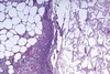
13
Q

Squamous Metaplasia

A
  • Columnar ⇒ squamous epithelium
  • Most common epithelial metaplasia
  • In areas of chronic irritation
    • Ex. bronchi of smokers
14
Q

Barrett’s Esophagus

A

“Intestinal metaplasia”

  • Squamous ⇒ columnar epithelium w/ globlet cells
  • Due to gastric reflux
  • Can lead to cancer
15
Q

Osseous (cartilaginous)

Metaplasia

A
  • Production of cartilage or bone in areas of tissue injury
  • Causes:
    • Chronic irritation
    • Stress
    • Tissue damage
  • Ex:
    • Irritation due to dentures
    • Injury to muscle ⇒ myositis ossificans
16
Q

Cell Injury

A

Reversible vs Irreversible

  • Stimulus
    • Limit of adaptive response exceeded
    • Exposed to injurious agent or stress
    • Deprived of essential nutrients
    • Compromised by mutations that affect essential cellular constituents
  • Time lag between injury and effects
    • Signs of reversible injury takes longer
17
Q

Cellular Injury

Causes

A
  • Oxygen deprivation
    • Hypoxia, ischemia
  • Physical agents
    • Mechanical, temp, radiation, electrical, pressure
  • Chemical agents
    • Pollutants, poisons, drugs, metabolists
  • Infectious agents
    • Virus, bacterial, fungi, parasites
  • Immune reactions
    • Exogenous, autoimmune
  • Genetic derangements
    • Enzymes, structural proteins
  • Nutritional imbalance
  • Proliferation errors (DNA)
18
Q

Reversible Injury

Changes

A
  • Functional changes
    • ↓ oxidative phosphorylation
      • Depletes ATP and glycogen stores
    • ↓ transporter function
      • Loss of membrane activity and integrity
    • Defects in protein synthesis
    • Cytoskeletal damage
    • DNA damage
  • Morphological changes:
    • Cellular swelling ⇒ due to ∆ in ion concentration and water influx
    • Mitochondrial swelling & amorphous densities
    • RER swelling & ribosome detachment
    • Clumping of nuclear chromatin
    • Membrane blebbing & loss of microvilli
    • Cytoplasmic vacuoles
19
Q

Cell Injury

Mechanisms

A
  • Mitrochondrial damage
    • ↓ ATP
    • ↑ ROS
  • Calcium entry
  • Membrane damage
  • Protein misfolding
  • DNA damage
20
Q

Depletion of ATP

Causes

A
  • ATP produced through:
    • Ox Phos of ADP
    • Glycolysis
  • ↓ [ATP] caused by:
    • ↓ O2 supply
    • ↓ nutrient supply
    • Mitochondrial damage
    • Toxins
21
Q

Depletion of ATP

Effects

A

If [ATP]intracellular falls to 5-10% of normal:

  • Na/K pump activity ⇒ cell swelling
  • ∆ cellular metabolism ⇒ shift to anaerobic glycolysis
    • Depletes glycogen stores
    • Produces lactic acid
      • ↓ cellular pH ⇒ ↓ enzyme activity
  • ↓ calcium pump activity ⇒ Ca2+ influx ⇒ damage to many cellular components
  • Ribosome detach from RER & polysomes dissociate ⇒ ↓ protein synthesis
  • ↑ protein misfolding ⇒ accumulation in RER ⇒ activation of misfolded protein response ⇒ cell injury & cell death
  • Irreversible damage to mitochondrial & lysosomal membranes ⇒ necrosis
22
Q

Mitochondrial Damage

A
  • Causes:
    • ↑ [Ca2+]cytosol
    • ROS
    • O2 deprivation
  • Types of mitochondrial damage:
    • Formation of mitochondrial permeability pore ⇒ failure of ox phos ⇒ ↓ [ATP]
    • Abnormal ox phos ⇒ ↑ [ROS]
    • Release of sequestered proteins from intermembrane space ⇒ apoptosis
      • Ex. cytochrome C & BCL proteins that activate caspases
23
Q

Loss of Calcium Homeostasis

A
  • Caused by ↓ ATP ⇒ influx of calcium
  • Key event in cell death
  • ↑ [Ca2+]intracellular effects:
    • Irreversibly poisons mitochondria
    • Inhibits many cellular enzymes
    • Activation of lytic enzymes
      • Phospholipases, proteases, endonucleases, ATPases
    • Initiates free radical formation
    • Denatures cellular proteins
      • Can lead to initiation of unfolded protein response
    • Activation of apoptosis
24
Q

Free Radicals

Characteristics

A
  • Have a single unpaired e- in an outer orbital
  • Extremely reactive with cellular macromolecules
  • ROS are a type of oxygen-derived free radical generated within cells
  • Build up if not scavenged and disposed of properly
  • Initiates autocatalytic rxns ⇒ propagates more free radicals
25
Free Radical Generation
* **Redox rxns during normal metabolism** * Small amount of partially reduced intermediates generated * Superoxide anion, hydrogen peroxide, hydroxyl ions * **Absorption of radiant energy (UV, X-rays) and ionizing radiation** * Hydrolyzes water ⇒ free radicals * **Activated WBCs generate ROS during inflammation (respiratory burst)** * **Enzymatic metabolism of exogenous chemicals/drugs** * **Transition metals (Fe, Cu)** can donate or accept free electrons during rxns * Can catalyze formation of TOS * Binding to storage/transport proteins reduces reactivity * **Nitric oxide** can act as a free radical
26
Free Radical Removal
* Inherently unstable ⇒ **decay spontaneously** * Cells have non-enzymatic & enzymatic mechanisms to remove free radicals * _Non-enzymatic_ * **Antioxidants** ⇒ prevent formation or inactivate/scavange free radicals * Vit E, A, and C * Glutathione * Free Fe and Cu * _Enzymatic_ ⇒ enzymes act as free radical scavening systems * Catalase, superoxide dismutase, glutathione peroxidase
27
Free Radical Pathologic Effects
1. **Lipid peroxidation in plasma/organellar membranes** * Attack double bonds in unsaturated FAs of membrane lipids * Generates peroxide * Results in autocatalytic chain reaction ⇒ propagation * Causes membrane damage 2. **Oxidative modification of proteins** * Damage enzyme active sites * Disrupt conformation of structural proteins * Enhance degradation of misfolded proteins 3. **Lesions in DNA** * Single or double stranded breaks in DNA * Crosslinking of strands * Implicated in cell aging and malignancy
28
Membrane Permeability Defects
* _Mechanisms of injury:_ * **ROS** ⇒ lipid peroxidation * **↓ phospholipid synthesis** * **↑ phospholipid breakdown** * Products accumulate & have detergent effect on membranes * **Cytoskeletal abnormalities** * Due to proteases affected by ↑ cytosolic calcium * _Results in damage to:_ * Mitochondrial membranes ⇒ apoptosis * Plasma membranes * Lysosomal membranes
29
DNA and Protein Damage
* Cell initiates suicide program if: * DNA damage too severe to be corrected * Level of misfolded proteins too high * Results in death by apoptosis
30
Ischemic and Hypoxic Injury
* Ischemia results from hypoxia due to ↓ blood flow * Usually due to blockage in arterial flow * Can be due to reduced venous drainage * Worsens hypoxia by also affecting anerobic energy generation * Reversible if oxygen flow restored in time * Organs respond differently to hypoxia d/t differences in metabolic activity
31
Organ Response to Anoxia
Cell death will begin in: * Neurons ⇒ 3-5 minutes * Myocardia ⇒ 20-30 minutes * Renal tubule cells ⇒ 30-60 minutes * Hepatocytes ⇒ 1-2 hours * Skeletal muscle ⇒ many hours * CT ⇒ many hours
32
Chemical (Toxic) Injury
* Direct toxicity * Chemicals injure cells by combining with molecular components * Ex. mercuric chloride poisoning, cyanide, chemotherapy * Conversion to toxic metabolites * Usually done by cytochrome P-450 mixed function oxidases * Ex. carbon tetrachloride, acetaminophen
33
\_\_\_ leads to cell death.
Irreversible cell injury
34
'Point of no return' between reversible and irreversible damage characterized by...
* Inability to reverse mitochondrial dysfunction * Lack of ox phos and ATP generation * Profound disturbances in membrane function
35
Reversible vs Irreversible Cell Injury
36
Cell Death
* End result of progressive cell injury * Many pathologic causes ⇒ ischemia, infection, toxins * Can be a normal process ⇒ embryogenesis, maintaining homeostasis * Two pathways ⇒ necrosis & apoptosis
37
Necrosis Overview
* **Always pathological** * Ischemia, toxins, infections, trauma * **Results from cell membrane damage** * Loss of ion homeostasis * Cell contents leak into EC space ⇒ **inflammatory response** * Lysosomal enzymes enter cytoplasm & digest cell ⇒ necrosis
38
Necrosis Morphological Changes
Takes many hours for histologic or gross changes to be visible. * _Cytoplasmic changes_ * **↑ eosinophilia** * Loss of RNA * Denatured proteins * **Vacuoles** * **Myelin figures** * From damaged cell membranes * Degraded into FA, may calcify * _Nuclear changes_ * **Karyolysis** ⇒ nuclear fragmentation * **Pyknosis** ⇒ nuclear contration * **Karyorrhexis** ⇒ fragmentation of pyknotic nuclei * _Plasma membrane destruction_ * Enzymes & proteins leak out into ECM and blood * Detectable in blood within 2 hours of necrosis
39
Coagulative Necrosis
* Typically caused by ischemia or infarction * **Dead tissue structure preverved for several days** * Area called an **infarct** * Injury denatures enzymes ⇒ no proteolysis of dead cells ⇒ **see eosinophilic cells without nuclei** * Dead cells eventually cleared by WBCs via phagocytosis and lysosomal digestion * Typically seen with MI's
40
Liquefactive Necrosis
* Results from focal bacterial or fungal infections * PMNs and Mφ degrade necrotic material and pathogen ⇒ pus * Seen in CNS s/p hypoxic injury
41
Gangrenous Necrosis
(Clinical term) * Limb ischemia ⇒ _coagulative necrosis_ ⇒ **dry gangrene** * Superimposed bacterial infection ⇒ _liquefactive necrosis_ ⇒ **wet gangrene**
42
Caseous Necrosis
* 'Cheese-like' central necrotic area of a granuloma * Seen in tuberculosis and some other infectious * Caseation appears pink, amorphous, and granular
43
Fat Necrosis
* **Focal areas of destruction** * Usually specific to the **release of pancreatic lipases** or **trauma to the breast** * Fat reacts with calcium ⇒ **saponification** ⇒ soap * Appears chalky-white grossly * Shows outlines of necrotic fat cells microscopically
44
Saponification
**Deposition of chalky white material in areas of fat necrosis.** * Results from combo of FA's released from intracellular stores + extracellular calcium * Ex. dystrophic calcification * Serum calcium is normal
45
Apoptosis Overview
* **Programmed cell death** ⇒ highly regulated * **Serves many normal functions** * **Activated by either an intrinsic or extrinsic pathway** * Relies on sequential activation of proteases through cleavage or caspase cascades * **No inflmmatory reaction triggered**
46
Physiologic Apoptosis Examples
Serves many normal functions: * **Response to DNA or protein damage** * **Embryologic development** * Ex. Webs between fingers * **Tissue remodeling** * **Normal turnover** * **Involution of hormone-dependent tissues** * **Cell loss in proliferating cell populations** * Ex. lymphocytes that don't express useful antigen receptors * **Elimination of self-reactive lymphocytes** * **Death of neutrophils after activation**
47
Pathologic Apoptosis Examples
* **After exposure to toxins** * **Death of cells with mutations or DNA damage** * Removes potentially cancerous cells * **Improperly folded proteins** * Caused by mutations or free radical damage * Accumulates in ER ⇒ ER stress ⇒ apoptosis * **Death of infected cells** * Viral infections ⇒ adenovirus, HIV * Host immune responses ⇒ hepatitis * **T-cell mediated mechanisms** * Kill tumor cells * Reject transplants * **Atrophy in parenchymal glands when ducts obstructed**
48
Apoptosis Morphological Changes
* Degradation of cellular DNA and proteins * Organelles tightly packed ⇒ **cell shrinkage** * **Chromatin condensation** * Aggregates under nuclear membrane * Nucleus may break up * **Formation of cytoplasmic blebs & apoptotic bodies** * Membranes remain intact * **Phagocytosis of apoptotic cells or cell bodies** before materials leak out ⇒ **no inflammatory response**
49
Apoptosis Mechanism
* **Results from activation of enzymes** * **Caspases** * Inactive proenzymes * Must undergo enzymatic cleavage for activation * Cleaved caspases are a marker of apoptotic cells * **Activation depends on balance between pro- and anti- apoptotic signals** * **Two phases:** 1. **Initiation** ⇒ some caspases become active 2. **Execution** ⇒ other caspases trigger degradation of cellular components
50
Apoptosis Intrinsic Initiation Pathway
**"Mitochondrial pathway"** * Major mechanism * Results from ↑ permeability of outer mitochondrial membrane * **Releases pro-apoptotic molecules into cytosol** * **Cytochrome C** ⇒ initiates apoptosis * **BCL2 family** ⇒ controls release * _Some anti-apoptotic_ ⇒ BCL-2, BCL-XL, MCL-1 * _Some pro-apoptotic_ ⇒ BAX and BAK * _Some are sensors_ ⇒ regulate the balance * **Path activates initiator caspase-9**
51
Apoptosis Extrinsic Initiation Pathway
**"Death Receptor-Initiated Pathway"** * Triggered by activation of death receptors on plasma membrane of some cells * **Death receptors** * Members of TNF receptor family * **'Death domain'** site required * Protein-protein interactions delivers apoptotic signals * **Type I TNF receptor (TNFR1)** ⇒ best known * **Fas (CD95)** **receptor** ⇒ found on many cells * Binds **Fas ligand (FasL)** * Found on T cells that recognize self-Ag * Eliminate self-reactive lymphocytes * Found on some cytotoxic T-cells * Kill virus-infected and tumor cells * Fas-FasL binding brings together Fas molecules * **Forms binding site for FADD** (Fas-associated death domain) * FADD binds inactive **Caspase-8** via death domain ⇒ activation * **Path activates initiator caspase-8 and caspase-10**
52
Apoptosis Execution Phase
Intrinsic and extrinsic initiation pathways converge at the activation of **executioner caspases**: * **Executioner caspases ⇒ caspase-3 and caspase-6** * Acts on cellular components * Results in: * Cleavage of DNA * Degradation of nuclear matrix * Degradation of proteins * Etc. * **Apoptotic bodies removed by phagocytes** * May be recruited by cell surface markers on apoptotic bodies * **Thrombospondin** * **C1q**
53
Decreased Apoptosis
Defective apoptosis ⇒ increased cell survival: * **Survival of abnormal cells** * **TP53 mutations** * Defective DNA repair * ↑ susceptibility to accumulation of mutations * ↑ cancer risk * Most common genetic abnl found in human cancers * **Failure to remove self-reactive and dead cells** * **↑ risk of autoimmune disorders**
54
Increased Apoptosis
Increased apoptosis ⇒ excessive cell death * Neurodegenerative diseases * Apoptosis due to mutations and misfolded proteins * Ischemic injury * E.g. MI and stroke * Death of virus infected cells
55
Apoptosis vs Necrosis
56
Autophagy Overview
* Cell digests its own constiuents * Protects against nutriend deprivation * Performs intracellular "quality control' * Involved in elimination of intracellular infections * Rodels cellular components to meet needs
57
Autophagy Types
1. Chaperone-mediated autophagy 2. Microautophagy 3. Macroautophagy
58
Chaperone-mediated Autophagy
Direct translocation across lysosomal membrane by chaperone proteins.
59
Microautophagy
Inward invagination of lysosomal membrane for delivery.
60
Macroautophagy
Sequester and transport portions of cytoplasm in double-membrane bound autophagic vacuoles (autophagosome) to lysosomes. Primary mode of autophagy.
61
Intracellular Accumulations
* Material accumulates in cytosol, organelles, or nucleus * May be reversible or irreversible * Can cause damage if extreme
62
Metabolic Derangement Pathways
1. **Inadequate removal of normal substances** * Due to defect in packaging or transport * Ex. Steatosis in liver 2. **Accumulation of abnormal endogenous substances** * Due to defective folding, packaging, or secretion 3. **Failure to degade metabolites** * Due to inherited enzyme deficiences * "Storage diseases" * **Deposit and accumulate abnormal exogenous substances** * Due to inability to degrade or excrete substances * Ex. carbon in macrophages
63
Steatosis
"Fatty change" * **Abnormal accumulation of lipids/TAGs in parenchymal cells** * Seen in liver and other organs * Causes: * Toxins * Protein malnutition * DM * Obesity * Anoxia
64
Atherosclerosis
* Smooth muscle cells and macrophages filled with lipid vacuoles * Contains mostly cholesterol and cholesteryl esters * Forms plaques on blood vessel walls
65
Xanthomas
Cholesterol accumulation in macrophages in subepithelial CT of skin and tendons.
66
Cholesterolosis
Accumulation of cholesterol-laden macrophages in lamina propria of the gallbladder.
67
Niemann-Pick Disease Type C
Lysosomal storage disorder leading to abnormal accululation of cholesterol in cells.
68
Protein Accumulations Appearance
Appears as rounded eosinophilic droplets, vacuoles, or aggregates in the cytosol.
69
Protein Droplets
Protein resorption droplets in proximal renal tubules. Found in patients with protein loss via urine.
70
Immunoglobulin accumulation in plasma cells are called...
Russell bodies
71
Alpha 1 Antitrypsin Deficiency
Defective intracellular transport and secretion of critical proteins. Material builds up in ER of hepatocytes.
72
Cytoskeletal Protein Accumulation
* Alcoholic Hyaline * Made of keratin intemediate filaments * Accumulates in the liver * Amyloid * Accumulates inside and between cells * Many organs affected
73
Hyaline Change
* Homogeneous, glassy pink material * Accumulates inside or between cells * Non-specific marker for cell injury * Extracellular hyaline can be seen in walls of arterioles with chronic HTN
74
Glycogen Accumulation
* Stored in healthy cells * Can be deposited in excessive amounts w/ abnormalities in glucose or glycogen metabolism * Appears as clear vacuoles in cytoplasm * Stained with PAS * Accumulates in glycogen storage diseases
75
Exogenous Pigment Accumulation
* Coal dust (anthracotic pigment) * Accumulates in lungs and lymph nodes * Excessive amounts can produce serious disease * Ex. Coal workers pneumoconiosis * Tattoos * Pigments phagocytosed by derml macrophages
76
Endogenous Pigment Accumulation
* Lipofuscin * Melanin * Hemosiderin
77
Lipofuscin
* Golden brown fine granules * 'Wear and tear' pigment * Contains lipid and proteins from membranes * Does not cause injury * Increases with age * Often seen in liver and heart
78
Hemosiderin
* Golden-brown granules * Derived from hemoglobin * Forms where there is local or systemic excess of iron * Normally see small amounts in macrophages * Bone marrow * Spleen * Liver * Local excess can accumulate in areas of bleeding * Systemic excess causes deposition in many tissues ⇒ **hemosiderosis**
79
Abnormal deposition of calcium salts is called...
pathologic calcification
80
Dystrophic Calcification
* Local deposition of calcium in dying tissues * Serum calcium levels normal * See fine, white clumps w/ gritty texture * Basophilic on H&E
81
Metastatic Calcification
* Deposition of calcium salts in normal tissue * See hypercalcemia * May affect GI mucosa, kidneys, lungs, and blood vessels * If very severe, can affect organ function
82
Cellular Aging Overview
**Progressive decline in cellular function and viability.** Results from: * **Genetic abnormalities** ⇒ mechanistic alterations * **Accumulation of cellular and molecular damage** * Due to exposure to exogenous influences
83
Cellular Aging Mechanisms
* **DNA damage** * DNA repair mechanisms unable to correct all errors * Damage accumulates over time * Werner syndrome ⇒ see premature aging * **Cellular senescence** * Normal cells stop dividing after a fixed number of divisions * Telomeres shorten each time * Telomerase absent in most somatic tissues * Cancer cells have reactivated telomerase ⇒ proliferates indefinitely * Become arrested in a non-diving state ⇒ _senescence_ * **Defective protein homeostasis** * **Deregulated nutrient sensing** * Caloric restriction shown to increase lifespan * Associated with IGF-1