Exam 2: Circulatory disturbances Flashcards
Hemodynamic
Homeostasis
Maintaining hemostatic balance requires:
- Normal blood circulation
- Balance between intra- and extracellular fluid compartments
- Normal concentrations of body fluid components
- Proteins
- Electrolytes
Hemodynamic Disturbances
-
Obstructive circulatory disturbances
- Thrombosis
- Hemorrhage
- Edema
- Shock
-
Blood volume and fluid distribution disburbances
- Hyperemia
- Hemorrhage
- Edema
- Shock
-
Water and electrolyte balance disturbances
- Edema
- Dehydration
Thrombus
Blood clot blocking blood flow.
- Serves no useful purpose
- Can cause vessel occlusion
- More significant in arteries
- Can be a site for emboli generation
- More significant in veins

Virchow’s Triad
3 elements which can work together to ↑ risk of pathologic thrombus formation.
- Endothelial injury
- Alterations in normal blood flow
- Hypercoaguability

Endothelial Injury
- Most important factor
-
Lose protective mechanisms of endothelium
- Platelets adhere to exposed collagen
- Common causes:
- MI
- Ulcerated atherosclerotic plaques
- Traumatic injury
- Vasculitis
- Hemodynamic stress due to HTN
- Injury from bacterial endotoxin
- Smoking
Abnormal Blood Flow
- Arteries ⇒ usually due to turbulence
- Veins ⇒ usually due to stasis
- Effects of ∆ blood flow
-
Disrupts laminar flow
- Brings platelets into contact with endothelium
- Prevents dilution of clotting factors by fresh blood
- Retard inflow of clotting factor inhibitors
- Promotes endothelial cell activation ⇒ local thrombosis
-
Disrupts laminar flow
Hypercoaguability
-
Primary ⇒ genetic
- Clotting factor mutations
- Factor V Leiden
- Lack of anticoagulants
- Protein C & S deficiency
- Clotting factor mutations
-
Secondary ⇒ acquired
- Prolonged immobilization
- Tissue damage
- Cancer ⇒ necrotic tumor cells release procoagulant factors
- Prosthetic heart valves
- Atrial fibrillation
- Hormonal imbalances
- Oral contraceptives, estrogen therapy, pregnancy
Postmortem Clot
Gross Appearance
Shows two distinct layers:
Serum ⇒ chicken fat clot
Cells ⇒ currant jelly clot

Antemortum Clot
Gross Apperance
“Thrombus”
- Adherent to the vessel wall
-
Multicolored layers
- Variagated appearance
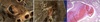
Thrombus
Microscopic Appearance
- See integration of fibrin and cellular layers
- Produces striped appearance ⇒ Lines of Zahn

Arterial Thrombi
- Usually occlusive
- Contains high [fibrin] ⇒ white thrombi
- Common sites
- Coronary
- Cerebral
- Femoral
Venous Thrombi
“Phlebothrombosis”
- Can be occlusive
- Sluggish flow ⇒ high [RBC] ⇒ red thrombi
- Common sites
- Leg veins (90%)
- Periprostatic plexus
- Periuterine veins
Mural Thrombus
Thrombus is attached to vessel wall
Non-occlusive
Typically see in heart or aorta

Occlusive Thrombus
Grows circumferentially in the lumen
Occludes vessel

Propogative Thrombus
Clot has a tail.
See in deep veins of extremities.

Vegetation
- Thrombus builds up on heart valves
- Can also get deposition of blood borne bacteria in vegetation
- See in cancer

Thrombus
Fates
-
Fibrinolysis by plasminogen-plasmin system
- More common with recent thrombi d/t less fibrin polymerization
-
Central softening
- Due to leukocyte action
-
Retraction
- Due to thromabasthenin in platelets
- Organization and recanalization
- Embolization

Thrombus
Organization and Recanalization
Organization ⇒ ingrowth of endothelial cells, smooth muscle cells, and fibroblasts into thrombus
Recanalization ⇒ formation of capillary channels within thrombus which can re-create a lumen

Embolus
Detached intravascular solid (thrombus), liquid (fat), or gaseous mass that is carried by the blood to a site distant from the point of origin.
99% of all emboli are thromboemboli.
Arterial Thromboemboli
- Originate from:
- Mural thrombi
- Diseased heart valves
- Atherosclerotic plaques
- Aneurysms
- Main sites of embolization:
- Legs
- Brain
- Intestines
Venous Thromboemboli
- Originate from:
- Deep veins of lower extremities ⇒ DVT (95%)
- Pelvic venous plexus
- Right side of heart
- Cavernous sinus veins
Pulmonary Thromboemboli
Characteristics
- Usually originate from a DVT
- Very common
- 20-25 per 100,000 hospitalized patients
- Consequences of PE determined by size of clot
- Determines where it will lodge

Pulmonary Embolism
Risk Factors
-
Venous stasis
- CHF or chronic venous insufficiency
-
Injury
- Trauma, surgery, postpartum
-
Hormonal imbalance
- Pregnancy or OCP
- Advanced age
- Immobilization
- Sickle cell disease
Large PE
Consequences
- Usually lodges in the main pulmonary artery
- Can straddle both sides ⇒ saddle embolism
- Results in:
-
Severe hypoxemia
- Interferes with return of blood to left heart
- ↓ CO
- Arterial hypotension
- Shock
-
Acute right heart failure
- See with > 60% obstruction of pulmonary circulation
- Can result in death ⇒ acute cor pulmonale
-
Severe hypoxemia

Small PE
Consequences
- Travels further into the pulmonary circulation
- Causes infarction of a smaller area
- More likely to be asymptomatic
- 60-80% of PEs are clinically silent
- Can leave a “web” in vessel after organization and absorption into vessel wall
- Can be multiple clots simultaneously ⇒ shower of emboli
- Having one increases risk of more
- Can result in severe hypoxemia
- Interfers with blood return to left heart

Paradoxical Embolism
-
Emboli travels from venous → arterial circulation
- Via interarterial or interventricular defects
- Ex. DVT causing CVA
Fat Embolism
-
Common causes:
-
Trauma to fat-containing tissues
- Long bone fx, soft tissue trauma, burns
- Fat is suctioned into ruptured venules
- Hyperlipidemia
-
Trauma to fat-containing tissues
-
Only produces symptoms occasionally
- See sudden onset tachypnea and dypsnea after 24-72 hour latent period
-
Neurological involvment
- Results in irritability and coma
- Brain shows petechiae
- Due to release of free FA and resultant toxic injury to vascular endothelium

Air Embolism
- Common causes:
- Puncture wound of great vessel
-
Decompression sickness → “The bends”
- Nitrogen gas dissolves in tissues during dive
- If depressurize too rapidly ⇒ bubbles form in tissues
- Chronic form ⇒ Caisson’s disease
Atheromatous Embolism
Atherosclerotic plaque breaks off and travels to another site.
Amniotic Fluid Embolism
- Rare but catastrophic complication of pregnancy
- Desquamated cells from fetus enters amniotic fluids ⇒ maternal circulation ⇒ lungs
- See sudden-onset of cyanosis and dyspnea
- Can lead to disseminated intravascular coagulation (DIC)

Ischemia
Decrease or loss of blood supply to a localized area of the body.
Causes loss of vital nutritional substances, especially O2, and accumulation of metabolic products.
Infarction
Localized area of ischemic necrosis resulting from circulatory insufficiency.
Due to either interference of arterial flow or venous obstruction.
Ischemia or Infarction
Progression from ischemia ⇒ infarction depends on:
- Rapidity of development
- Degree of vascular occlusion
- Pre-existing disease in the organ
- Sufficiency of collateral circulation
- Tissue vulnerability to hypoxia
Infarct
Morphology
- Most are wedge shaped with base towards periphery
-
Often has 3 zones:
- Central ischemic necrotic zone
-
Hyperemic zone around dead tissue
- Where inflammation is occuring
- Viable zone of living tissue

Infarct
Types
- Initially ⇒ all infarcts are red
- Subsequently ⇒ type of infarct depends on tissue blood supply
-
White infarcts
- Seen in tissues where blood cannot seep in
- Only one route of blood supply
-
Red infarcts
- Tissues with dual blood supply
- Lung & liver
- Venous occlusion
- Loose tissues
- Previously congested tissue
- Tissues with dual blood supply
-
White infarcts
Pulmonary Infarct
Red infarct ⇒ due to dual blood supply
Wedge shape with base on pleura

Intestinal Infarct
Red infarct

Testicular Infarct
Red infarct ⇒ has loose CT filled with blood
Usually due to testicular torsion

Splenic Infarct
White infarct

Bone Infarct
White infarct

Renal Infarct
White infarct

Acute Tubular Necrosis
Due to renal ischemia ⇒ infarction
Renal tubular cells are very sensitive to ischemia

Septic Infarcts
May occur two ways:
-
Pre-existing bacteria present in tissue before development of ischemia necrosis
- Ex. infarction of an area of pneumonia
-
Bacteria brought to affected area by embolization of an infected thrombus
- Ex. vegatation from a heart valve
Hyperemia
Excess amount of blood in an organ.
Can be active or passive.

Active Hyperemia
-
Caused by ↑ blood supply from arteries
-
Inflammation
- Vasodilation in response to vasoactive substances
-
Fever
- Vasodilation to dissipate heat
-
Neurogenic & hormonal influences
- Hot flashes
- Blushing
-
Inflammation
- Can result in erythemia

Passive Hyperemia
“Congestion”
-
Caused by ↓ exit of blood via veins
- Diminished venous flow
- Impaired venous drainage
- Local ⇒ DVT or tumor venous obstruction
- Systemic ⇒ CHF
- See dilatation of veins and capillaries

Inflammatory Breast Cancer
- Groups of cancer cells lodge in subcutaneous lymphatics
- Results in impaired fluid clearance
- Causes passive hyperemia and edema
- See dimpled and retracted skin

Pulmonary Congestion
- Due to left heart failure
- Impedes exit of blood from lungs
-
Leads to chronic passive congestion in lungs
- Capillaries engorged with blood

Pulmonary Edema
- Left heart failure ⇒ passive pulmonary congestion ⇒ ↑ capillary hydrostatic pressure
- Fluids forced into interstitial spaces and alveolar spaces ⇒ pulmonary edema
- ∆ gas exchange

Pulmonary Exudates
- Left-sided CHF ⇒ pulmonary congestion ⇒ pulmonary edema
- Progresses to microhemorrhages of pulmonary capillaries
- Releases RBCs into alveolar spaces
- Mφ phagocytize RBCs
- Results in hemosiderin-laden Mφ ⇒ “heart failure cells”
- Can stimulate lung fibrosis and lead to pulmonary HTN

Liver Congestion
Chronic passive congestion of liver:
- Seen with right heart failure or hepatic vein/IVC obstruction
- Liver is enlarged and firm
- See dilated central veins & sinusoids
- Causes pressure on hepatocytes in the centrilobular area
- Grossly, congested area appear dark red & alternates with normal brown liver ⇒ nutmeg pattern

Cardiac Cirrhosis
- Right-sided CHF ⇒ chronic passive liver congestion ⇒ eventual progression to centrilobular fibrosis
- Caused by pressure atrophy of centrilobular hepatocytes

Edema
Accumulation of abnormal amounts of fluid in interstitial tissue space or body cavities.
Pleural space ⇒ pleural effusion
Pericardial space ⇒ pericardial effusion
Peritoneal space ⇒ ascites
Fluid is usually non-inflammatory ⇒ transudate
Edema
Pathogenesis
- Caused by hemodynamic derangements
- ↑ forces which move fluids from intravascular to interstitial spaces
- Can be due to:
- ↑ intravascular hydrostatic pressure
- ↓ colloid osmotic pressure of plasma
- ↑ vascular permeability
- Impaired lymph flow

Localized Edema
Mechanisms
-
↓ venous drainage ⇒ ↑ venous hydrostatic pressure
- DVT, incompetent valves/varicose veins, tumors
-
↑ vascular permeability
- Allows escape of colloid proteins from vessels ⇒ ↓ colloidal osmotic pressure
- Allergic rxn, burns, infection
-
Lymphatic obstruction ⇒ lymphedema
- Filariasis infection
- Worms in lymphatics
- Get elephatiasis
- Congenital
- Milroy’s disease
- Post surgical s/p lymph node resection
- Filariasis infection
Generalized Edema
Mechanisms
-
↑ capillary hydrostatic pressure
- See in cardiac edema
- Affects dependent parts of the body, liver, lungs, spleen
-
↓ plasma proteins
- Mostly albumin
- ↓ plasma oncotic pressure
- Neprhotic syndrome ⇒ loss albumin in urine
- Cirrhosis ⇒ ↓ albumin synthesis
-
Sodium and water retention
- Due to abnormality of tubular reabsorption, aldosterone secretion, ADH secretion
Hemorrhage
Extravasation of blood due to vessel rupture.
Hematoma
Extravasated blood within tissue
Petechiae
Pinpoint hemorrhages
Causes:
Areas of increased intravascular pressure
Low platelet count or abnormal platelet function

Ecchymosis
Larger subcutaneous hemorrhages
( > 1-2 cm)

Larger Hemorrhages
- Hemothorax
- Hemoperitoneum
- Hemathrosis
- Hemorrhagic stroke

Shock
Critical condition caused by a sudden drop of blood flow throughout the body.
Adult Respiratory Distress Syndrome
(ARDS)
“Shock lung”
- Caused by diffuse alveolar damage
- Results in extravasation of large amounts of fluid into alveolar spaces
- See hyaline membranes lining alveoli
- Impairs gas exchange

Acute Pancreatitis
- Due to release of catalytic enzymes from exocrine pancreas
- See fat necrosis
- White, chalky deposits in gross speciment
- Dead adipose tissue on micro
