Exam 1: Healing and Repair Flashcards
1
Q
Goal of Healing
A
- Restore tissue architecture and function
- Repair parenchymal tissues
- Heal the surface
2
Q
Repair Processes
A
-
Regeneration
- Cells that survive injury proliferate
- Sometimes added by stem cells
- Need cells that can proliferate
- Need intact stroma & basement membrane
- More normal/functional result
- Cells that survive injury proliferate
-
Scar formation ⇒ CT deposition
- Occurs if
- Tissue incapable of complete restitution
- Supporting structures severely damanged
- Not normal but can provide enough stability to allow some function
- Occurs if

3
Q
Proliferative Capacity
A
3 groups:
- Labile cells
- Stable cells
- Permanent cells
4
Q
Labile Cells
A
- Continuously dividing tissues
- Multiply throughout life
- Can regenerate as long as stem cells preserved
- Ex:
- Epithelial surface cells
- Lymphoid/hematopoietic cells
- Epithelium of GI/GU tract
5
Q
Stable Cells
A
“Quiescent cells”
- Latent capacity to regenerate
- Includes:
- Parenchymal cells of kidney, liver, pancreas
- Fibroblasts
- Smooth muscle
- Endothelial cells
- Lymphocytes
6
Q
Permanent Cells
A
- Non-dividing tissues
- Do not divide enough to regnerate
- Includes:
- Neurons
- Cardiac muscle
- Skeletal muscle
- Satellite cells can help
7
Q
Cell Proliferation
A
Driven by many growth factors:
- Activated macrophages most important source
- Cells use integrins to bind ECM proteins
- Activate signaling pathways
- Induce protein production ⇒ drives cell through cell cycle
- Release blocks on cell cycle
8
Q
Tissue Regeneration
Labile Cells
A
- Tissue stem cells normal quiescent until needed
- Due to soluble factors, cell-cell, and cell-ECM interactions
- Most are not totipotent
- Limited types of differentiated cells they can generate
- Injury stimulates cells to divide and differentiate
9
Q
Tissue Regeneration
Stable Cells
A
Ex. Liver:
- Loss of liver tissue from infection, injury, surgery
- Can adequately regenerate even if 90% is lost
- Phases:
-
Priming phase
- Kuppfer cells make cytokines
- Makes hepatocytes competent to receive & respond to growth factors
-
Growth factor phase
- Growth factors drive primed hepatocytes into cell cycle
- Then non-parenchymal cells proliferate
- Endothelial, Kuppfer, Stellate
-
Termination Phase
- Heptocytes exit cell cycle & return to quiescence
-
Priming phase
-
Progenitor cells can also be activated
- Found in Canals of Hering

10
Q
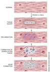
Scar Formation
Causes
A
- Regeneration not enough for repair
- Severe or chronic tissue injury
- Damage to parenchymal cells, epithelial, CT frame
- Injury of non-dividing cells
11
Q
Scar Formation
Process
A
- 3 key steps:
- Angiogenesis
- Deposition of CT ⇒ formation of granulation tissue
- Remodeling of CT
- Macrophages important
- Clear microbes/dead tissue
- Provide growth factors
- Secrete cytokines
- Repair begins within 24 hours of injury
- See granulation tissue in 3-5 days
12
Q
Scar Formation
Timeline
A

13
Q
Scar Formation
Area Preparation
A
- Immune system removes offending agent and inflammatory exudate
- Lysosomal enzymes liquefy debris
- Inflammatory response ends
- Fibroblasts recruited to begin remodeling
14
Q
Scar Formation
Angiogenesis
A
Formation of blood vessels:
-
Changes in hemodynamics
- NO ⇒ vasodilation
-
Vascular endothelial growth factor (VEGF) ⇒ ↑ vascular permeability / promote angiogenesis
- Contributes to edema in healing wounds
-
Pericytes seperate from adluminal surface
- Breaks down basement membrane
- Allows formation of vessel sprout
-
Endothelial cells
- Migrate towards injury
- Proliferate behind leading front (“tip”) of migrating cells
- Remodel into capillary beds
-
Periendothelial cells recruited to form mature vessels
- Pericytes for capillaries
- Smooth muscle cells for larger vessels
- Endothelial proliferation & migration suppressed
- Basement membrane deposited

15
Q
Scar Formation
Granulation Tissue Formation
A
- Highly vascularized CT composed of:
- New capillaries
- Infiltrating and proliferating fibroblasts
- Produces loose CT
- Pink/red and granular in gross appaerance
- Occupies tissue defect until scar can mature

16
Q
Scar Formation
Collagen Deposition
A
- Two steps:
- Fibroblasts migrate and proliferate into site of injury
- Fibroblasts produce and deposit ECM proteins
- Controlled by local cytokines
- Mostly from M2 macrophages
- TCF-β most important
- Mostly from M2 macrophages
- Further ECM deposition ⇒ more distance between capillaries and fibroblasts
- Less erythema
- Some fibroblasts become myofibroblasts

17
Q
Scar Formation
CT Remodeling
A
- Need balance between synthesis and degradation of ECM proteins
- Done through remodeling
-
Matrix Metalloproteinases (MMPs)
-
Degrade collagen and other ECM components
- Collagenases, gelatinases, stromelysins
- Produced by many cell types
- Must be activated by proteases at site of injury
- Inhibited by specific Tissue Inhibitors of Matalloproteinases (TIMPs) made by mesenchymal cells
-
Degrade collagen and other ECM components
18
Q
Healing
Influencing Factors
A
-
Infection
- Prolongs inflammation & ↑ local injury ⇒ delays healing
- DM
-
Glucocorticoids
- Anti-inflammatory
- ⊗ TGF-β production ⇒ weakens scar, ↓ fibrosis
-
Nutritional status
- Especially protein & Vit C deficiency
-
Mechanical factors
- ↑ local pressure or torsion ⇒ dehiscence
-
Perfusion
- Arteriosclerosis, DM
- Obstructed venous drainage e.g. varicose veins
- Foreign body
-
Tissue type
- Only stable and labile cells capable of restoration
-
Extent of injury
- Need intact parenchyma/BM/stem cells
-
Location of injury
- Inflammation in tissue spaces ⇒ extensive exudates
- Pleural, peritoneal, synovial
- Exudates must be digested/reabsorbed by leukocytes
- Can restore as long as there is not cellular necrosis
- Large exudate can organize ⇒ granulation ⇒ scar
- Inflammation in tissue spaces ⇒ extensive exudates
19
Q
Collagen Deposition
Effects of Nutrition
A
- Type III collagen deposited initially in granulation tissue
- Type III replaced by Type I collagen
- Hydroxylation of lysine and proline needed to crosslink
- Vit C required
- Cu2+ is a co-factor for lysyl hydroxylase
-
Collagenase required for transition from Type III ⇒ Type I
- Zn2+ is a co-factor
- Hydroxylation of lysine and proline needed to crosslink
20
Q
First Intention Healing
Overview
A
- Occurs through primary union
- Wound edges are apposed
- Primary mech ⇒ epithelial regeneration
- Limited # of dead cells
- Only minor discontinuites in BM
21
Q
First Intention Healing
Process
A
-
Day 1
- Formation of fibrin clot ⇒ coagulation
- Neutrophils arrive within 24 hours
-
Day 2
- Epithelial cells from both edges migrate and proliferate along edges
- Deposition of BM components
- Thin epithelial layer closes the wound
-
Day 3
- Neutrophils replaced by macrophages
- Wound debrided
- Fibroblasts arrive
- Start collagen production
- Epidermis covering wound now near normal thickness
- Neutrophils replaced by macrophages
-
Day 5
- Peak of neovascularization
- Granulation tissue filling incisional space
- New vessels are leaky
-
Week 2
- Fibroblast proliferation and collagen deposition continues
- See fewer neutrophils, vessels, and edema
-
End of 1st month
- Scar is cellular CT
- Nearly no inflammatory cells
- Covered by nearly normal epidermis
- Dermal appendages in line of incision are lost

22
Q
Second Intention Healing
A
- Occurs when wound edges are not in contact ⇒ tissue lost
- Repair through a combo of regeneration and scarring
- Characteristics
- Slower
- Larger fibrin clot
- More exudate and necrotic debris
- More intense inflammatory reaction
- More granulation tissue
- Accumulation of ECM and larger scar
-
Wound contraction can occur due to myofibroblasts
- ↓ size of large skin defects to 5-10% of original by 6 weeks
- More complications

23
Q
Wound Strength
A
-
7 days s/p wound
- 10% tensile strength
- Low collagen content
-
60-70 days s/p wound
- 30% tensile strength
- 100% collagen content
- High ratio of Type III : Type I collagen
-
3 months s/p wound
- 70-80% tensile strength
- 100% collagen content w/ remodeling and turnover
- Inc. Type I : type III collagen ⇒ 85% of normal skin Type I
- Never reaches 100% of tensile strength
24
Q
Fibrosis
Overview
A
Abnormal deposition of collagen in internal organs due to chronic disease.
-
Caused by persistent injurious stimuli
- Chronic infections and/or immunological reactions ⇒ loss of tissue ⇒ attempt at repair
-
Develops in space occupied by inflammatory exudate
- Ex. Organizing pneumonia
- Results in organ dysfunction and/or organ failure
-
TGF-β ⇒ major cytokine involved
- Collagen made by myofibroblasts in lung & kidney
- Collagen made by stellate cells in liver

25
Fibrosis
Examples
* Cirrhosis of the liver
* Systemic schlerosis (schleroderma)
* Fibrosing disease of the lungs
* End-stage kidney disease
* Constrictive pericarditis

26
Inadequate granulation tissue or scar formation can lead to...
**wound dehiscence** or **ulceration**
27
Wound Dehiscence
"Rupture"
* Most often seen with abdominal surgery
* Due to increased abdominal pressure from vomiting or coughing
* Creates mechanical stress on the wound
28
Wound Ulceration
* **Due to inadequate vascularization during healing**
* Ex.
* Ulceration in leg wounds with atherosclerotic peripheral vascular disease
* Areas devoid of sensation in neuropathic ulcers

29
Hypertrophic scars
* Forms due to accumulation of excess collagen
* Ex. thermal or traumatic injury
* Involves deep layers of dermis
30
Keloid
* **Scar tissue grows beyond boundaries of original wound without regression**
* Individuals can be predisposed
* More common in African Americans

31
Exuberant Granulation
* **Tissue protrudes above level of surrounding skin**
* Blocks re-epithelialization
* Called 'Proud Flesh"
* May need to remove with cautery or surgical excision
32
Desmoid
"Aggressive Fibromatosis"
* Proliferation of fibroblasts
* Can be neoplastic
33
Wound Contraction
* Part of normal healing
* If exaggerated ⇒ see contractures and deformities of wound and surrounding tissues
* Prone to develop on palms, soles, anterior thorax
* Common after serious burns
* Can compromise joint movement

