Chapter 22: The Heart Flashcards

You may prefer our related Brainscape-certified flashcards:
1
Q

Right Side of Heart (recieves blood from)

A
  • Recieves blood from the body and then pumps to lungs
2
Q

Lefts Side of heart (recieves blood from)

A

Recieves blood from lungs and then pumps to body

3
Q

The circulation between the heart and lungs is known as…

A

Pulmonary Circulation

4
Q

The _______ circulation entails blood from the heart TO the body…

A

Systemic Circulation

5
Q

A “vein” can be thought of as…

A

a blood vessel which carries blood BACK to the heart.

6
Q

Arteries can be best thought of as…

A

Blood vessels that carry blood AWAY from the heart

7
Q

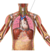
The heart is surrounded by the “pericardial sac” otherwise known as the…
A

Pericardium

8
Q

The space around the heart created by the pericardium is known as…

A

Pericardial Cavity

9
Q

Identify space “1” in the slide.

A

Pericardial Cavity

10
Q

Identify 1 and 2

A
  1. Parietal serous pericardium
  2. Visceral Serous Pericardium
11
Q

The _______ is a serous membrane that forms the outer layer of the heart wall

A

Visceral Pericardium

12
Q

The ________ is a seros membrane attached directly to the fibrous layer.

A

Parietal Paricardium

13
Q

The _________ forms a thick outer layer of connective tissue around the entire heart and pericardium.

A

Fibrous Pericardium

14
Q

Number 1 in this picture is a general structure known as the _____ of the heart?

A

Apex

15
Q

There are four chambers of the heart, what are the three shown here?

A
  1. Right Atrium
  2. Right Ventricle
  3. Left Ventricle
16
Q

Identify the view that this picture of the heart is (anterior/posterior) and the four chambers of the heart in ascending order based on the numbers in this picture.

A

Posterior View

  1. Left Atrium
  2. Left Ventricle
  3. Right Ventricle
  4. Right Atrium
17
Q

Identify the view that this picture of the heart is (anterior/posterior) and the four chambers of the heart in ascending order based on the numbers in this picture.

A

Anterior

  1. Right Atrium
  2. Right Ventricle
  3. Left Ventricle
  4. Left Atrium
18
Q

The two ventricles are seperated by the…

A

Interventricular Septum

19
Q

The blue star idicated which structure?

A

Interventricular Septum

20
Q

Which ventricle is “larger”/ “More muscular”?

A

Left Ventricle (b/c it has to pump blood to the body)

21
Q

What is the name of the structure indicated by the green star?

A

Trabeculae Carneae

22
Q

If the ventricles are seperated by the interventricular septum, the two atria are probably seperated by ______ septum.

A

“Inter”Atrial (Interatrial)

23
Q

Identify this structure highlighted in red

A

Interatrial Septum

24
Q

Identify the structure indicated by the star

A

Fossa Ovalis

25
Q

Identify (1) and (2) in the image and what view of the heart is this (anterior/posterior)

A
  1. Superor Vena Cava
  2. Inferior Vena Cava

Posterior

26
Q

The heart sounds are caused by…

A

Closing of valves

27
Q

There are two semilunar valves, what are they?

A
  1. Pulmonary Valve
  2. Aortic Valve
28
Q

There are two antriventricular (AV) valves, what are they?

A
  1. Left Atrioventricular Valve (Bicuspid/Mitral Valve)
  2. Right Atrioventricular Valve (Tricuspid Valve)
29
Q

The “Lubb” sound (S1) is associated with the _______ valves closing

A

Atrioventricular

30
Q

The “dupp” sound is associated with the ______ valves closing.

A
  1. Semilunar
31
Q

This is diagnosed as what type of heart issue?

A

Mitral stenosis

32
Q

This is diagnosed as what type of heart issue?

A

Mitral regurgitation

33
Q

This is diagnosed as what type of heart issue

A

Aortic Stenosis

34
Q

This is diagnosed as what type of heart issue

A

Aortic Regurgitation

35
Q

Number 1 is…

A

Right coronary artery

36
Q

Number 2 is…

A

Circumflex artery

37
Q

Number 3 is…

A

Left Coronary artery

38
Q

Number 4 is…

A

Anterior Interventricular Artery

39
Q

Number 1 is…

A

Posterior Left Ventricular Artery

40
Q

Number 2 is…

A

Circumflex artery

41
Q

Number 3 is…

A

Right Coronary artery

42
Q

Number 4 is…

A

Posterior Interventricular artery

43
Q

Identify number 1

A

Coronary Sinus

44
Q

Identify number 2

A

Great Cardiac vein

45
Q

Identify number 3

A

Posterior vein of left ventricle

46
Q

Number 4 is…?

A

Small cardiac vein

47
Q

Number 5 is…

A

Middle cardiac vein

48
Q

Number 1 is..

A

Anterior Cardiac Veins

49
Q

Number 2 is…

A

Small cardiac vein

50
Q

Number 3 is…

A

Great (like you… yes you :] ) Cardiac Vein

51
Q

Number 1 is…

A

Right coronary artery (in coronary sulcus)

52
Q

Number 2 is…

A

Small cardiac vein

53
Q

Number 3 is…

A

Anterior Cardiac Veins

54
Q

Number 4 is…

A

Left coronary artery (in coronary sulcus)

55
Q

Number 5 is…

A

Circumflex artery

56
Q

Number 6 is…

A

Anterior Interventricular Artery

57
Q

Number 1 is…

A

Great Cardiac Vein

58
Q

Number 2….

A

Circumflex Artery

59
Q

Number 5…

A

Coronary Sinus (in coronary sulcus)

60
Q

Identify number 3…

A

Posterior Left Ventricular Artery

61
Q

Number 8 is…

A

Posterior Vein of Left Ventricle

62
Q

Number 4 is…

A

Small cardiac vein

63
Q

Right coronary artery is which number?

A

Number 6

64
Q

The posterior interventricular artery is identified by which number?

A

7

65
Q

What is the order in which an electrical signal is sent across the heart in order beginning with the Sinoartrial Node (SA node)?

A

After the SA node discharges…

  1. Inernodal pathways
  2. Antrioventricular node
  3. Antrioventricular (AV) bundle (Bundle of His)
  4. Bundle Branches
  5. Purkinje fibers
66
Q

What do the SA Node, Internodal pathways, Atrioventricular (AV) node, AV bundle, Bundle branches, and Purkinje fibers compose?

A

The Intrinsic Conducting system of the heart

67
Q

What part of the intrinsic conducting system is the electrical impulse currently in?

A

SA Node

68
Q

What part of the intrinsic conducting system is the electrical impulse currently in?

A

Internodal pathways

69
Q

What part of the intrinsic conducting system is the electrical impulse currently in?

A

AV node

70
Q

What part of the intrinsic conducting system is the electrical impulse currently in?

A

Bundle Branches

71
Q

What part of the intrinsic conducting system is the electrical impulse currently in?

A

Purkinje Fibers

72
Q

Sir

I

Always

Act

Bad

with

Peanut Butter

A

SA Node

Internodal pathways

AV node

AV bundle

Bundle Branches

Purkinje Fibers