Week 5 - A - Urogenital imaging lecture from week 1 Flashcards
What are the layers of the kidney?
Fibrous capsule (mainly collagen and elastin) Outer cortex Inner medulla
What is the functional unit of the kidney?
This is the nephron
Which parts of the nephron are locate in the medulla?
The loop of henle and the collecting duct are located in the medulla of the kidney

What is the relationship of the renal artery, vein,ureter and hilum?
The renal vein is anterior at the hilum The renal artery is posterior The ureter is inferior leaving the hilum to go t the bladder
Describe the flow of urine from collecting duct to the prostatic urethra?
Collecting duct - exits the medulla via the renal papillae - excretes urine into the minor calyces which join to form major calyces and these join to form the renal pelvis This drains urine into the ureter which inserts posteriolaerally into the bladder which drains into the prostatic urethra

Are the kidneys retro or intraperitoneal organ and which lies higher? State the vertebral levels
Kdineys are retorperitoneal The left lies higher due to the right having the liver above pushing it downwards Left kidney - T12-L2 Right kidney - L1-L3
What is the location of the kidneys in the posterior abdominal wall?
The kidneys lie in the paravertebral gutters of the posterior abdominal wall

What are the muscles of the posterior abdominal wall that protect the kidneys?
Psoas major (medial) and quadratus lumborim (lateral)
Going from kidneys to peritoneum direction name the structures from renal capsule to the visceral peritoneum
Renal capsule Perinephric fat Renal (deep) fascia Paranephric fat Visceral peritoneum
https://s3.amazonaws.com/classconnection/403/flashcards/11907403/jpg/picture1jpggif-15B3A7FC00C6D2787FC.jpg

- Parenchyma 2. Cortex 3. Medulla 4. Perninephric fat 5. Capsule 6. Ureter 7. Renal pelvis 8. Renal vein and artery 9. Hilum 10. Minor calyx

Which structures lie anterior to the right kidney?
Anterior structures Liver and hepatorenal recess 2nd part of duodenum Ascending colon Right colic flexure - hepatic flexure
Which structures lie anterior to the left kidney?
Stomach Tail of pancreas Hilum of spleen and splenic vessels The splenic flexure
https://s3.amazonaws.com/classconnection/403/flashcards/11907403/png/picture1jpggifjpggif-15B3A8625DD6C20D2B3.png

This is the transverse colon
https://s3.amazonaws.com/classconnection/403/flashcards/11907403/png/picture1jpggifjpggifjpg-15B3A87D264593F9333.png

This is liver - sits in front of the right kidney
Which part of the duodenum lies in front of the right kidney?
The 2nd part of the duodenum lies in front of the kidney

Which renal vein is longer?
The left renal vein is longer as it has to pass over the aorta to supply the left kidney
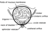
Name the two circled strcutres?

Spleen and splenic artery
What does the splenic artery arise? What does this structure also give off?
Arises from the coeliac trunk which is T12 Coeliac trunk - trifurcates into left gastric, common hepatic and splenic artery

Where on the kidneys do the suprarenal glands lie?
They lie superomedially on the kidneys
At what levels do the renal hilum arise?
Left renal hilum - - L1 Right renal hilum - L1/2
Which imagaing modality can be used to view hydronephrosis and calculi? What is hydronephrosis definition?
Use ultraound Hydronephrosis is the dilatation of the renal pelvis
https://s3.amazonaws.com/classconnection/403/flashcards/11907403/png/picture1jpggifjpggif-15B3AB4D416666EC1F2.png

A - right lobe of liver B - medulla C - Cortex
What are the 4 lobes of the liver?
Right, left, caudate, and quadrate

Even though ultrasound can detect renal stones, what is the best imaging technique to detect these?
Non contrast CT
What is a complication of contrast CT scans when used to view the kidney?
Can cause contrast induced nephropathy
What are the four phases of a CT contrast scan?
Pre-contrast - before contrast Corticomedullary - corticol enhancement Nephrographic- Renal medulla and cortex enhance equally Excretory - opacification of the renal collecting duct and urinary system
After iodinate contrast injection, when are pictures for the contrast taken?
Pre contrast - there is no contrast Corticomedullary - lasts 25-70 seconds Nephrographic - lasts from 80seconds to 180 seconds after injection Excretory - from 5 to 15 minutes after contrast injection
At what stage does the renal pelvis become enhanced?
The renal pelvis becomes enhcanced in the excretory phase - it remains dark in the nephrographic phase which only enhances the cortex and medulla Pic of excretory

Which phase is which of the CT contrast scan Also state the time after contrast injection for each stage

1 - pre contrast - good for depicting renal stones 2. corticomedullary - 25-70 seconds post iodinated contrast - good for seeing cortex 4. nephrographic - 80 - 180 - views the renal medulla and cortex but not the collecting system 3. Excretory - 5-15 mintues - views collecting system
4phases of CT – Pre-contrast, cortical medullary (mainly cortical enhancement), nephrographic (medulla and cortical) and excretory When are simple cysts best seen?
Best seen in the nephrographic phase - shows up as a clear colour against the white contrast

What are the three phases? Triple phase contrast is used in the diagnosis of a renal cell carcinoma

a - pre contrast b - corticomedullary - can see the medulla are not enhanced c - nephrographic
What is nephrocalcinosis? What is nephrolithiasis?
Nephrocalcinosis is condition where the calcium in the kidney sis increased Nephrolithiasis are renal stones
Always remember about renal function before requesting a contrast CT What is the renal function ideally wanting to be for the bare minimum?
Want a renal function of above 60 -make sure patinet is very hydrated
What are the hydration protocols for a contrast CT to try and prevent contrast induced nephropathy? 2 different things are given
Give patinet 1-1.5mls/kg/hr 0.(% saline 12 or 6 hours before and after contrast adminsitration Sodium bicarbonate adminsitered 1hour preprocedure to 6hours post - alkalnises the urine
Contrast induced nephropathy can cause acute kidney injury What is the contrast induced nephropathy seen as when measuring bloods?
rise in serum creatinine and urea – usually peaks at 72 – 96 hours post contrast
What are the three anatomical postions where the ureters narrow in diameter?
Pelviureteric junction Pelvic brim Vesicoureteric junction

strengths excellent tissue contrast resolution renal lesion characterisation vascular assessment characterises most pathologies These are for MRIs What are the weaknesses?
Lengthy acquisition time Also can cause nephrogenic systemic firbosis
Name this artery?

It is going to an organ above the left kidney and is travelling from the aort So splenic artery
What is this bifurcation?

Bifurcation of the aorta into the common iliac arteries
What is a common cause of secondary hypertension in patients under 40?
Renal arery stenosis
What drugs are contraindicated in renal artery stenosis?
ACE inhibitors - they widen the efferent arteriole and this decreases the glomerular filtraton rate due to the glomerular capillary blood (hydrostatic) pressure decreasing This decreases the overall net filtration pressure
severe systemic fibrosing disorder associated with the exposure to gadolinium-containing contrast media What is this known as when MRI is used?
Known as nephrogenic systemic fibrosis

Nephorgenic systemic fibrosis was first identifieed in 1997 It is clear both MRI (nephrogenic systemic fibrosis)and CT contrast (contrast induced nephropathy) are due to renal impairment What is the GFR values where these are epecially bad?
Contrast induced nephropathy - GFR less than 60 Nephrogenic systemic fibrosis - GFR less than 30 (chronic kidney failure stage 4)
What are the presetning signs of nephorgnic systemic fibrois due to gadolinium contrast?
Presenting symptoms are skin pruritus, erythema and pain before the tightening of the skin comes
The ureters are subdivided into the abdominal, pelvic and intravesicle ureter What muscle does the abdominal ureter coarse along?
Course along the psoas major

The three points of ureteric constriction are the PUJ, pelvic brim and VUJ What angle does the ureter change to at and where when entering the pelvic brim?
Samll kink in the ureter when it crosses the common iliac artery - can often cross at the bifurcation to enter the pelvis
What is the function of the bladder?
Simply to store the urine
workhorse for assessing the collecting system, ureters and bladder What is this imaging technique?
CT urogram
What are the two phases of a CT urogram?
Pre contrast phase and 5 -15 minute excretory phase obtained after IV contrast Take pictures throughout
The posterior surface of the ladder is also known as the base of the bladder What is the structure that is at the base of the bladder?

Trigone
What is the structure seen at the floor of the bladder?
Internal urethral orifice
What is the trigone a triangle between?
2 ureteric orificeson the posterolateral surface and the internal urethral orifice

What is the commonest technique that is used for assessing the bladder however requires a full bladder to best define the outline on the image?
This would be Ultrasound
What technique is usually used if a patient has painless hameaturia and on urinalysis, there is no signs of infection? (investigations differ depending on patient being above or below 50)
If the patient is above 50 - carry out a flexible cystocopy and then a CT urogram to image the upper renal tract If the patient is below 50 - carry out a flexible cystoscopy and then a Kidney/Bladder Ultrasound to image the upper renal tract (if they come back negative but bleeding persists, then carry out CT urogram)
gold standard for local tumour staging better tissue contrast resolution vs CT What is this technique?
MRI is better for local tumour staging but there is a lengthy acqusition
https://s3.amazonaws.com/classconnection/403/flashcards/11907403/png/picture1jpggifjpggif-15B3A97B94C7768BCAF.png

This is the rectouterine pouch - pouch of Douglas

The male urethra starts at the bladder neck What does it change from as it extends?
It becomes the prostatic urethra, membranous urethra, bulbous urethra, spongy (penile) urethra

What is the change in name of the urthra at the penoscortal junction?
The bulbous urethra becomes the penile urethra
Why is the penile urethra also known as the spongy urethra?
This is because it travels within the corpus spongiosum
What is used for local staging of prostate cancer? What is used for nodal or distant metastases?
Local staging - MRI Nodal or distant metastases - CT
Where does the ureter lympahtics drain to? Where do the testes lymphatics drain to?
Ureter - drains to the lumbar/iliac lymph nodes Testes -drain to the para-aortic lymph nodes
What is the lymphatic drainage of the bladder?
The bladder drains to the iliac nodes
ultrasound is the primary imaging of choice for: testes/scrotum uterus and ovaries What is used to check for infertiltiy in a female?
Hysterosalpingogram - looks for tubal patency and uterine anomalies
In the hysterosalpingogram, where does the dye spill into? This is normal
Dye spills into the peritoneum

What test, done in polycystic ovarian disease is done to look at the ovaries?
Transvaginal ultrasound