Ventral Muscle Identification (from 1st semester cards) Flashcards

1
Q

2

A

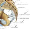
2 Bicipital aponeurosis

2
Q

3

A

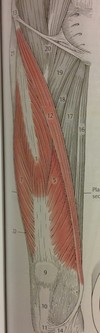
Brachioradialis muscle

origin: lat. supracondylar ridge humerus
insertion: radial styloid process
artery: radial reccurent artery
innervation: radial nerve
action: flexes elbow

3
Q

4

A

Flexor carpi radialis muscle

origin: medial epicondyle humerus (comm. flex. tendon)
insertion: base of 2nd/3rd metacarpals
artery: radial artery
innervation: median nerve
action: flexion/abduction of wrist

4
Q

5

A

5 Radial artery

5
Q

6

A

6 Flexor digitorum superficialis muscle

6
Q

7

A
  • *Median nerve**
  • only nerve to pass through carpal tunnel

innervates all of the flexors in the forearm except flexor carpi ulnaris and that part of flexor digitorum profundus that supplies the 2nd and 3rd digits

Pronator teres
Flexor carpi radialis
Palmaris longus

Flexor digitorum superficialis muscle

Flexor digitorum profundus (only the lateral half)
Flexor pollicis longus
Pronator quadratus

7
Q

8

A

Antebrachial fascia and tendon of palmaris longus muscle

8
Q

18

A

Palmaris longus muscle

origin: medial epicondyle humerus (comm. flex. tendon)
insertion: palmar aponeurosis
artery: ulnar artery
nerve: median nerve
action: flexes wrist

9
Q

20

A

20 Ulnar artery

10
Q

21 (tendon)

A

21 Tendon of flexor carpi ulnaris muscle

11
Q

23

A

23 Abductor digiti minimi muscle

12
Q

26

A

Brachialis muscle

Origin
anterior surface of the humerus, particularly the distal half
Insertion
coronoid process and the tuberosity of the ulna
Artery
radial rartery
Nerve
musculocutaneous nerve (C5, C6)
Actions

flexion at elbow joint

13
Q

28

A

28 Carpal tunnel (canalis carpi, probe)

27
Q

What are the deep flexors of the forearm in radioulnar order (based on origin)?

What are they all innervated by?

What artery supplies all of them?

A
  1. flexor pollicis longus
  2. flexor digitorum profundus
  3. pronator quadratus
  • median nerve (anterior interosseous branch)
  • anterior interosseous artery
28
Q

28

A

22 Palmaris brevis muscle

29
Q

17

(what part of the muscle)

A

17 Humeral head of pronator teres muscle

30
Q

19

A

Flexor carpi ulnaris muscle

origin:

  • humeral head: medial epicondyle humerus (comm. flex. tendon)
  • ulnar head: medial margin on olecranon

insertion: pisiform, hamulus, metacarpal V
artery: ulnar artery
innervation: ulnar nerve
action: flexion/adduction of wrist

31
Q

33

A

33 Flexor digiti minimi brevis muscle

32
Q

27

A

Flexor pollicis longus muscle

origin: middle 2/4 radial shaft and inteross. membrane
insertion: base of distal phalanx of thumb
artery: anterior interosseous artery
innervation: median nerve (ant. inteross. branch)
action: flexes thumb

33
Q

30

A

Flexor digitorum superficialis muscle

origin: medial epicondyle humerus
insertion: anterior base of middle phalanges II-V
artery: ulnar artery
innervation: median nerve
action: flexes fingers (prox. interphal. joint)

34
Q

32

A

32 Opponens digiti minimi muscle

35
Q

39

A

Pronator teres muscle

origin: medial epicondyle humerus (comm. flex. tendon)
insertion: middle lateral surface of radial shaft
artery: ulnar and radial arteries
innervation: median nerve
action: pronates forearm

36
Q

40

A

Flexor digitorum profundus muscle

origin: proximal 3/4 of anteromedial ulnar shaft, inteross. membrane, deep fascia
insertion: base of distal phalanges
artery: anterior interosseous artery
nerve: median nerve (ant. inteross. branch), ulnar nerve
action: flexes hand and interphal. joints

46
Q

What are the superficial flexors in radioulnar order (based on their proximal ends)?

What is their common origin?

What nerve innervates all of them but one?

What artery supplies all of them but one?

A
  1. Pronator teres
  2. Flexor carpi radialis
  3. Palmaris longus
  4. Flexor carpi ulnaris
  5. Flexor digitorum superficialis
  • all originate from common flexor tendon which attaches to medial epicondyle of humerus
  • median nerve innervates all but FCU
  • ulnar artery supplies all but FCR
47
25 coverings
25 Digital fibrous sheaths of tendons of flexor digitorum muscle
48
11
11 Abductor pollicis brevis muscle
49
12
12 Palmar aponeurosis
50
41
41 Lumbrical muscles
51
13
13 Superficial head of flexor pollicis brevis muscle
52
1
**Acromial part of deltoid muscle** (central fibers) ## Footnote origin: **acromion** action: **shoulder abduction**
53
2
**Scapular part of deltoid muscle** (posterior fibers) ## Footnote origin: **spine of scapula**
54
6
**Clavicular part of deltoid muscle** (anterior fibers) ## Footnote origin: **anterior border/superior surface of lateral 1/3 of clavicle** action: **shoulder abduction**, assists pec major in **shoulder flexion**
55
7 What is it? What vein passes through it?
**Deltopectoral groove** - cephalic vein passes through
56
8
**Biceps brachii muscle** origins: * short head: **coracoid process** * long head: **supraglenoid tubercle** insertion: **radial tuberosity** artery: **brachial artery** innervation: **musculocutaneous nerve** action: **flexes elbow**, supinates forearm
57
9
**Brachialis muscle** ## Footnote origin: **anterior humerus**, just below deltoid insertion insertion: **ulnar tuberosity** innervation: mostly **musculocutaneous nerve**, partly also radial nerve action: **flexes arm** at elbow joint
58
13
**Pectoralis major muscle** origins: * clavicular head: **anteromedial clavicle** * sternocostal head: **anterior sternum. costal cartilages, external oblique aponeurosis** insertion: **intertubercular sulcus**, lateral lip artery: **pectoral branch of thoracoacromial artery** innervation: **lateral and medial pectoral nerves** action: **adducts/medially rotates humerus,** **draws scapula anterior/inferior**
59
16
**Tendon of biceps brachii muscle** ## Footnote insertion: **radial tuberosity**
60
17
**Bicipital aponeurosis** - broad aponeurosis of biceps brachii located in cubital fossa - separates superficial from deep structures in fossa
61
18 | (not a muscle)
**Axillary artery**
62
24
**Brachial artery and median nerve**
63
26
**Tendon of short head of biceps bachii muscle** ## Footnote origin: **coracoid process** of scapula
64
27
**Coracobrachialis muscle** ## Footnote origin: **coracoid process** of scapula insertion: **medial humerus** artery: **brachial artery** nerve: **musculocutaneous nerve** action: **adducts humerus**
65
42
42 Tendons of flexor digitorum profundus muscle
66
34
34 Tendons of flexor digitorum superficialis muscle
67
entire colored portion
**Deltoid Muscle** origins: * scapular part: **spine of scapula** * acromial part: **acromion** * clavicular part: **anterosuperior lateral clavicle** insertion: **deltoid tuberosity** artery: **thoracoacromial, anterior and poster humeral circumflex arteries** innervation: **axillary nerve** actions: **shoulder abduction**, flexion and extension
69
What is the bottom uncolored space called? What does it contain (numbers 4 and 5)? What are its borders (numbers 6, 7 and 8)?
**Triceps Hiatus** or **Lateral Triangular Space** **-** contains **Radial Nerve** (4) and **Deep Brachial Artery** (5) Borders: * lateral: **Humerus** (6) * medial: **Long Head of Triceps** (7) * superior: **Teres Major** (8)
84
What is the proper anatomical name of the "armpit" and what are its four walls??
**Axillary Fossa** * _medial_: serratus anterior * _lateral_: humerus + arm muscles * _anterior_: pectoralis major + minor * _posterior_: latissimus doris + teres major
85
What type of joint is the shoulder? What two bone features make it up? What movements does it allow?
- ball and socket - head of humerus and glenoid cavity of scapula movements: * **medial/lateral rotation** * **abduction/adduction** * **circumduction** (combo of abd/add + antflx/retflx) * **anteflexion/retroflexion**
86
18
18 Greater sciatic foramen
87
19
19 Sacrospinous ligament base at sacrum to the apex at ischial spine
88
20
20 Sacrotuberous ligament from sacrum to ischial tuberosity
89
21
21 Lesser sciatic foramen
90
3
**Iliopsoas muscle** combination of Psoas major and Iliacus surrounded by illiac fascia and inserts on lesser trochanter • Flexes thigh at hip • Flexes vertebral column #5 below Lumbar Plexus (L1-L3)
91
18
**Vastus medialis muscle** part of quadriceps O: Linea aspera of femur, Intertrochanteric line I:Patella and tibial tuberosity via tendon of quadriceps femoris and patellar ligament * Extends leg at knee * Stabilizes patella 5 Femoral nerve (L2-L4)
92
5
**Tensor fasciae latae muscle** Origin: Anterior superior iliac spine, Iliac crest Insertion: Iliotibial tract • Flexes thigh at hip • Abducts thigh • Rotates thigh medially #21 below Superior Gluteal nerve (L5-S1)
93
6
**Sartorius muscle** O:Anterior superior, iliac spine I: Medial tibia • Flexes thigh at hip • Rotates thigh laterally • Flexes leg at knee 12 below Femoral nerve (L2-L4)
94
7
**Rectus femoris muscle** part of the quadriceps O: Anterior inferior iliac spine I: Patella and tibial tuberosity via tendon of quadriceps femoris and patellar ligament * Extends leg at knee * Flexes thigh at hip 1 below Femoral nerve (L2-L4)
95
8
**Iliotibial tract** -fibrous reinforcement of the fascia lata originates at the anterior portion of the external lip of the iliac crest inserts at the lateral condyle of the tibia/Gerdy's tubercle extend, abduct, and laterally rotate the hip. contributes to lateral knee stabilization
96
9
**Vastus lateralis muscle** part of quadriceps O:Greater trochanter of the femur, Intertrochanteric line, Linea aspera of femur I:Patella and tibial tuberosity via tendon of quadriceps femoris and patellar ligament * Extends leg at knee * Stabilizes knee 7 Femoral nerve (L2-L4)
97
15
**Pectineus muscle** O: Superior ramus of pubis I: Linea aspera of femur • Adducts thigh • Flexes thigh at hip • Rotates thigh medially 19 Obturator nerve (L2-L4)
98
16
**Adductor longus muscle** O: Pubis – near pubic symphysis I: Linea aspera of femur • Adducts thigh • Flexes thigh at hip • Rotates thigh medially 17 Obturator nerve (L2-L4)
99
17
**Gracilis muscle** O: Inferior ramus of pubis, Body of the pubis I: Medial surface of tibia * Adducts thigh * Flexes leg at knee * Rotates leg medially 16 Obturator nerve (L2-L4)
100
20
**Adductor brevis muscle** O: Inferior ramus of pubis I: Linea aspera of femur * Adducts thigh * Rotates thigh medially 18 Obturator nerve (L2-L4)
101
#2
**Vastus intermedius muscle** part of the quadriceps I: Shaft of the femur O: Patella and tibial tuberosity via tendon of quadriceps femoris and patellar ligament • Extends leg at knee 3 Femoral nerve (L2-L4)
102
29, 30, and 31 are the ?
Femoral artery Femoral vein Saphenous nerve adductor canal these blood vessels and this nerve run medially between the vastus medialis, sartorius, and adductor longus
103
There are __ adductors
7 adductors gracilis, pectineus, adductor magnus, adductor brevis, adductor minimus, adductor longus, quadratus femoris
104
5
**Obturator externus muscle** O:External surface of Obturator membrane and surrounding bone I: Trochanteric fossa of femur lateral rotator of the hip joint 9 Obturator nerve (L2-L4)
105
13
14 Adductor Minimus ## Footnote - Inferior ramus of pubis - Medial lip of Linea Aspera Obturator nerve (L2-L4) Adduction and lateral rotation of thigh
106
4, repeat
13 Soleus muscle
107
16
Medial head of gastrocnemius muscle ## Footnote O: Medial head- Medial femoral condyle O: Lateral head- Lateral femoral condyle -Calcaneal tendon of Achilles on calcaneal tuberosity Tibial nerve (S1-S2) plantar flexes foot, flexes knee
108
6
3 Peroneus Brevis - Lateral surface of Fibula (Inferior 2/3) - Tuberosity of 5th metatarsal Superficial Peroneal nerve (L5-S1) Plantarflexion, eversion
109
5
1 Peroneus Longus - Head of fibula, superior 2/3 of fibula - Tuberosity of 1st metatarsal and medial cuneiform (Crosses foot) Superficial Peroneal nerve (L5-S1) plantarflexion, eversion, supportarches
110
4
1 Tibialis Anterior (TA) - Lateral surface of tibia & interossious membrane - Plantar surface of Medial Cuneiform bone & Base of 1st metatarsal Deep Peroneal nerve (L4-S1) Dorsiflexion and Inversion of the foot
111
10
Extensor Digitorum Brevis (EDB) O: Calcaneus, near calcaneal sulcus I: Dorsal aponeurosis of 2nd – 4th digits Deep Peroneal nerve (S1-S2)
112
8
Peroneus Tertius \*\*\*\* additional tendon of extensor digitorum longus which is not always present. - distal third part of fibula & interossious membrane or from EDL - Dorsum of base of 5th metatarsal Deep Peroneal nerve (L4-S1) Pronantor and abductor of the subtalar and talocalcaneonavicular joints
113
12
**Extensor Hallucis Longus (EHL)** - Medial surface of fibula & interossious membrane - Dorsal aspect of base of distal phalanx of great toe Deep Peroneal nerve (S1-S2) Extends the big toe and assists in dorsiflexion of the foot at the ankle. Also is a weak evertor/ invertor
114
4
4 Femoral artery
115
7
**Patellar ligament** **7**
116
12
12 Aponeurosis of external abdominal oblique muscle
117
14 males only
**Spermatic cord** 13
118
14
14 Femoral vein
119
17 and 18
17 Adductor hiatus 18 Vasto-adductor membrane
120
27
27 Iliopectineal arch
121
20
20 Tendons of extensor digitorum longus muscle
122
3
3 Patellar ligament
123
6, 3 tendons from anterior to posterior
6 Tendons of deep flexor muscles from anterior to posterior: 1. tibialis posterior; 2. flexor digitorum longus; 3. flexor hallucis longus muscles
124
7
7 Flexor retinaculum
125
8
8 Tendon of tibialis anterior muscle
126
13
13 Tendon of gracilis muscle
127
14
14 Tendon of semitendinosus muscle
128
15
15 Common tendon of gracilis, semitendinosus, and sartorius muscles (pes anserinus)
129
18
18 Calcaneal or Achilles tendon
130
19
19 Calcaneus muscle
131
17
17 Superior extensor retinaculum
132
18
18 Inferior extensor retinaculum
133
19
19 Tendon of extensor hallucis longus muscle
134
5
**Extensor digitorum longus muscle** - Lateral condyle of tibia, head and superior 3/4 of medial surface of fibula and interossious membrane - Middle and distal phalanges of 2nd – 5th digits (toes) extension of toes and dorsiflexion of ankle