Respiratory conditions asthma Flashcards
Medical/Respiratory Disorders affecting the Respiratory System Name conditions
Asthma Chronic bronchitis Emphysema Pneumonia Pulmonary embolus
Asthma 4 presentations
Mild/moderate Severe Life-threatening Near fatal
Asthma can be triggered by a number of factors e.g.
allergens (pollen, dust, foodstuffs), aspirin, infection, stress, cold air or smoke
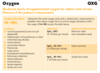
Pathology of asthma
The initial reaction involves spasmodic contraction of the bronchi and bronchioles. Inflammation of the bronchi and bronchioles. Increased mucus secretion. Oedema may follow to further complicate the condition.
Adult Asthma
Acute Severe Asthma Unable to complete a sentence in one breath Respiratory rate >25 Heart rate >110 PEF 33% - 50% of predicted normal Life Threatening Asthma Silent chest Cyanosis Poor respiratory effort Bradycardia or hypotension Exhaustion, confusion, coma PEF <33% of predicted normal SpO2 <92%
Asthma in Children
Acute Severe Asthma SpO2 <92% on air Too breathless to talk or feed Respiratory rate >50 (2 – 5 yrs), >30 (5 – 18yrs) Heart rate >130 (2 – 5 yrs), >120 (5 – 18 yrs) PEF <50% of predicted normal Life Threatening Asthma - SpO2 <85% in air Silent chest Cyanosis Poor respiratory effort Fatigue or exhaustion Agitation or reduced conscious level PEF <33% of predicted normal
Management of Asthma

Management of Asthma (1)
Ensure ABC’s
Administer Oxygen therapy if hypoxaemic (maintain SpO2 94-98%)
Consider patient position
Commence transport
Specifically consider:
Check peak flow if practicable – note the best of 3 Administer Salbutamol
In acute severe or life threatening cases Ipratropium should be administered with the salbutamol
Management of Asthma (2) In cases of hypoventilation consider
in-line nebulisation with BVM and nebuliser
Monitor using ECG and Pulse Oximeter
Repeat doses of Salbutamol in accordance with guidelines
Exclude pneumothorax
Monitor and reassess to evaluate any change in peak flow or air entry
Consider Paramedic support to administer Hydrocortisone where there is a delay getting to hospital of >30 minutes
Treatment of Life Threatening Asthma
Adrenaline 1:1000 IM/SC – repeated after 5 minutes if necessary)
Salbutamol / Ipratropium Bromide
Paramedic support for IV access [do not delay transport]
Paramedic will administer Hydrocortisone IV (if >30 minutes to hospital)
In-line nebulisation with bag, valve, mask in cases of hypoventilation.

Causes of respiratory compromise
- Obstruction of the air passages
- Chest or lung trauma
- Paralysis of respiratory nerves and muscles
- Non – oxygen atmospheres
Causes of dyspnoea

Asthma
oCharacterised by intermittent reversible airway obstruction
oBronchi become inflamed making them smaller.
oInflammation irritates the muscles around the bronchi causing spasmodic contraction
oInflammation causes mucus glands to produce excessive sputum which further blocks the airways
o
o
oCan result in a partial or complete obstruction of the airways
oAir can become trapped in the alveoli
oLeads to Hyperinflated chest
https://www.youtube.com/watch?v=EK8nzKzdnIM acute asthma video (4 mins
Peak expiratory flow rate (PEFR)

oxygen

Describe the clinical features of the 4 levels of asthma
Mild/moderate
Severe
Life-threatening
Near fatal
You should consider the possibility of COPD in patients:
- Over 35
- Smoker (or ex-smoker)
•
Patients displaying any of the following:
- Exertional breathlessness
- Chronic cough
- Regular sputum production
- Wheeze
Bronchitis
- Bronchoconstriction caused by inflammation of the airways and increased mucus production coupled with inhibition of the cilia leading to an accumulation of secretions.
- Acute bronchitis (viral or bacterial) -common in young children and the elderly -is usually short lived
- •Chronic Bronchitis is defined as where an individual has had a cough with sputum for 3 months in 2 successive years (Ross and Wilson p258)
- •Lung damage may eventually result in right heart failure, peripheral oedema and cyanosis
- Blue bloater’
- Typically overweight
- Typically cyanosed
- •Difficult inspiration & expiration
- •Productive cough
Emphysema
- Characterised by distension of the alveoli and destruction of their gas exchange membranes
- Lung tissue loses elasticity
- •The airway loses muscular integrity and collapses
- •Air is obstructed on entrance to the alveoli
‘Pink puffer’
Typically thin
Typically maintain normal skin tone
Usually maintains normal skin colour
Use of accessory muscles
Difficult often prolonged exhalation
Pursed lips
Pneumonia – signs and symptoms
- Fever with or without rigors
- Cough (often productive with green, blood-stained or rusty sputum)
- Pleuritic chest pain
- Muscle or joint pain
- Raised RR, raised HR,
- Crackles at base of lungs
Pneumonia – Management
•Oxygen therapy as required
Salbutamol if wheeze
PE – signs and symptoms
- Raised HR, Raised RR
- Lowered SpO2 (<92%)
- Signs of DVT (deep vein thrombosis)
–Unilateral swelling of lower leg, warm, red, painful, tender calf
- Dyspnoea
- Pleuritic chest pain - often specific to one place (use of finger to identify pain)
- Substernal chest pain
- Apprehension
- Cough
- Haemoptysis
- Syncope
PE – Management
- Oxygen therapy as required target SpO2 94-98%
- Time critical transport with pre-alert
- Management en-route
- Position patient for comfort, often sitting forwards
- Be prepared for arrest
- 12 lead ECG and monitor obs en-route
- Consider assisting ventilation
Hyperventilation syndrome (HVS)
A DIAGNOSIS OF EXCLUSION – don’t miss a life threatening cause
Excessive breathing causes little change to oxygen levels
BUT a significant decrease in CO2 raising blood pH (alkalosis)
Alkalosis affects calcium binding and can cause tetany (commonly carpopedal spasm)
- Tingling / numbness in hands, feet & mouth
- Chest pain
- Tetany / carpopedal spasm
- Severe anxiety – feeling of suffocation
- Syncope
HVS – Management
- Exclude life threating causes
- Oxygen therapy as required target SpO2 94-98%
- Coach breathing
–Remove bystanders – including friends and family
–Ensure calm quiet environment
–Eye contact and at eye level
–Calm and confident approach
–Demonstration
Continue to consider medical causes for hyperventilation
Do not leave patient until symptoms have subsided completely and remained absent for approx. 10 minutes
–Clear instructions – include hand gestures
–Explain that it will take time to adjust body chemistry
–Stay with patient
––Use CO2 rebreathing device if necessary
